English日本語简体中文
Skip to main content
Dr. Peter C. Kang's Rhinoplasty Stories
PrologueBlogMy thoughtsNasal obstructionSurgical journalSurgical photos
About the surgeon

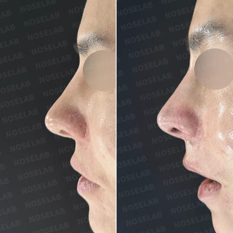
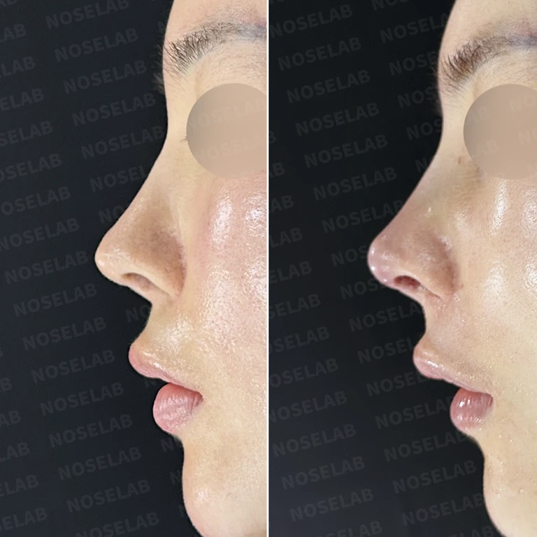
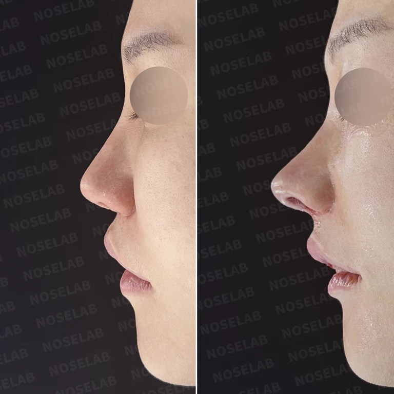
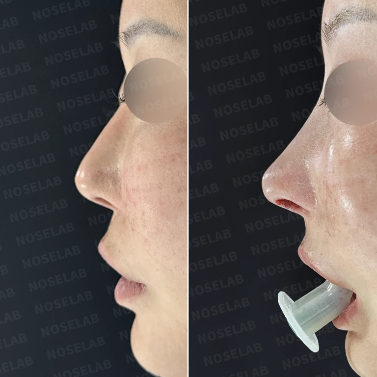
Thin tip skin and soft tissue reinforcement

In skin reinforcement cases, the internal cartilage framework of the nasal tip and the overlying soft tissue can be evaluated separately. Records document the use of artificial dermis and scar tissue to address cartilage show-through in thin skin, as well as reinforcing the nasal tip with autologous dermis harvested from behind the ear. Even within the same term "reinforcement," the actual tissue source varies.

Cases also include reinforcing nasal tip skin with dermis alongside repositioning the lower lateral cartilages and placing ear cartilage grafts. Because lateral osteotomy or columella correction may also be performed, the overall before-and-after changes cannot be attributed solely to skin reinforcement. We recommend distinguishing the thinned areas, visible structures, and additional cartilage work described in the original records.

View before-and-after cases ↓Comprehensive rhinoplasty guide

Searching for surgical cases matching your criteria.
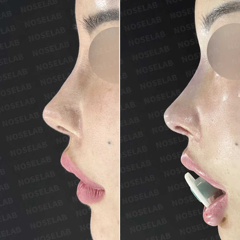
Why an L-shaped silicone implant gradually causes the nasal tip to become upturned: removal and reconstruction — Before-and-after comparison
Before-and-after comparison

Why an L-shaped silicone implant gradually causes the nasal tip to become upturned: removal and reconstruction

A case where an L-shaped implant placed about 10 years ago caused skin thinning and an upturned nasal tip. The L-shaped silicone implant was removed, and nasal tip reconstruction with autologous rib cartilage, septal correction, and dermal tissue reinforcement were performed.

Performed or used in this surgery
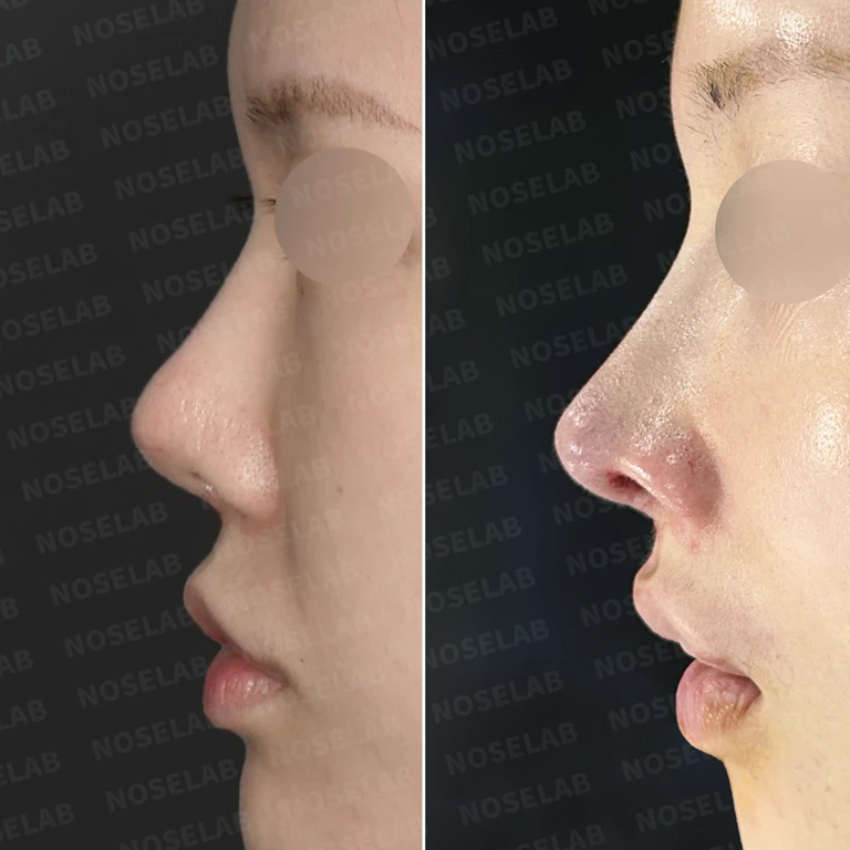
Why an implant is palpable and visible through the nasal tip: rib cartilage reconstruction and dermis reinforcement — Before-and-after comparison
Before-and-after comparison

Why an implant is palpable and visible through the nasal tip: rib cartilage reconstruction and dermis reinforcement

A case where a silicone implant became palpable, the nasal tip skin thinned, and asymmetry developed after a previous rhinoplasty. The implant was removed, the nasal tip and septum were reconstructed using sliced autologous rib cartilage, and artificial dermis was used for reinforcement.

Performed or used in this surgery

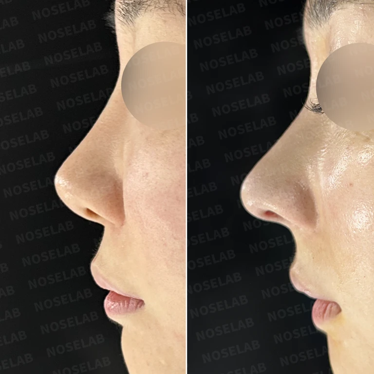
Why implants alone should not be placed in a small, low nose and nasal tip support structure — Before-and-after comparison
Before-and-after comparison

Why implants alone should not be placed in a small, low nose and nasal tip support structure

A primary rhinoplasty case involving a small, low nose and an upturned tip causing excessive nostril show. Using a closed approach, septal reconstruction with autologous rib cartilage, nasolabial angle correction, artificial dermis skin reinforcement, and a silicone implant were performed.

Performed or used in this surgery
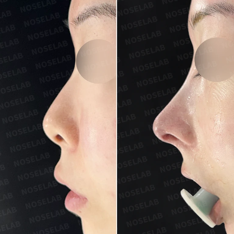
Why a high radix makes the nose look long and overly prominent — Before-and-after comparison
Before-and-after comparison

Why a high radix makes the nose look long and overly prominent

Following prior surgeries, a bulbous and drooping long nose with an excessively high radix and nasal obstruction was corrected using a closed approach. Scar tissue debridement, osteotomy, nasolabial angle correction, and septoplasty were performed concurrently.

Performed or used in this surgery

Why mesh remaining in the septum causes an upturned nose, and reconstruction after removal — Before-and-after comparison
Before-and-after comparison

Why mesh remaining in the septum causes an upturned nose, and reconstruction after removal

This is a case where mesh left in the septum after previous surgeries resulted in an upturned nose and excessive nostril show. All foreign material was removed, followed by septal reconstruction using autologous rib cartilage, replacement of the silicone implant, and reinforcement of the skin with acellular dermal matrix.

Performed or used in this surgery

Why noses become short and upturned after multiple revision surgeries, and methods for reconstruction — Before-and-after comparison
Before-and-after comparison

Why noses become short and upturned after multiple revision surgeries, and methods for reconstruction

A case correcting an upturned nasal tip caused by loss of the septum from repeated revision surgeries. Autologous rib cartilage septal reconstruction, nasal mucosal advancement flap, silicone implant adjustment, and rhinitis surgery were performed concurrently.

Performed or used in this surgery

Why nasal tip skin depresses and implants show through after inflammation, and how to reconstruct it — Before-and-after comparison
Before-and-after comparison

Why nasal tip skin depresses and implants show through after inflammation, and how to reconstruct it

A case where repeated surgeries and inflammation led to depressed nasal tip skin and implant show-through. The damaged skin was revised, dermal and cartilage grafts were placed, the septum was reinforced with autologous rib cartilage, and the implant was replaced.

Performed or used in this surgery
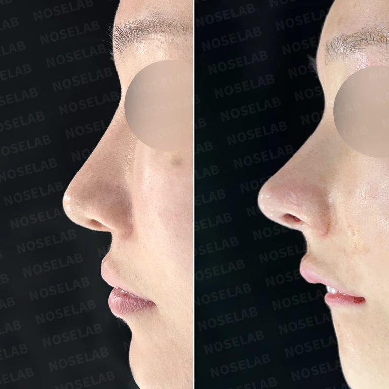
Why implants show through and nostrils look M-shaped, and how to reconstruct them — Before-and-after comparison
Before-and-after comparison

Why implants show through and nostrils look M-shaped, and how to reconstruct them

Implant show-through at the nasal tip and thinned skin were reinforced with donor dermis, and foreign substances were removed. The nasal tip was reconstructed using autologous rib cartilage, along with dragon nose correction and lateral osteotomy.

Performed or used in this surgery

Why contracture remains in multiply operated noses: Reconstructing the columella and support framework — Before-and-after comparison
Before-and-after comparison

Why contracture remains in multiply operated noses: Reconstructing the columella and support framework

Contracture and septal defect resulting from repeated surgeries were reconstructed using autologous rib cartilage. For the retracted columella, retroauricular free skin flap grafting and perichondrium grafting were performed to restore its shape.

Performed or used in this surgery

Nasal obstruction from a severely deviated septum: why cartilage reconstruction is necessary — Before-and-after comparison
Before-and-after comparison

Nasal obstruction from a severely deviated septum: why cartilage reconstruction is necessary

In a primary rhinoplasty patient, a severely deviated septum was reconstructed using autologous rib cartilage, and hypertrophied inferior turbinates were reduced. Dermis was grafted into the depressed alar region, and the nasolabial angle was adjusted.

Performed or used in this surgery
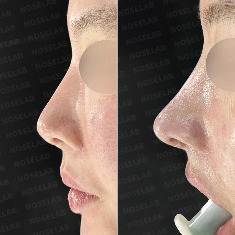
Why a nose operated on multiple times continues to appear upturned, and how septal reconstruction is performed — Before-and-after comparison
Before-and-after comparison

Why a nose operated on multiple times continues to appear upturned, and how septal reconstruction is performed

Following previous surgeries and double-jaw surgery, an upturned nasal tip and nostril asymmetry were precisely corrected using donor rib cartilage septal reconstruction, lower lateral cartilage restoration, and a composite dermis-fat graft to the soft triangle.

Performed or used in this surgery
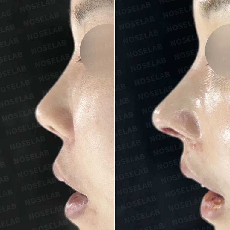
Why nasal tip skin thins after multiple surgeries: autologous dermis reinforcement and reconstruction methods — Before-and-after comparison
Before-and-after comparison

Why nasal tip skin thins after multiple surgeries: autologous dermis reinforcement and reconstruction methods

We removed previous foreign materials and implants, reconstructed the nasal tip using autologous rib cartilage, reinforced the thinned skin with autologous dermis, recarved a silicone implant, and performed fat grafting between the eyebrows.

Performed or used in this surgery
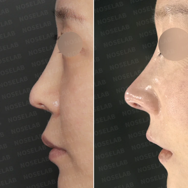
Why the columella retracts and the alar rim looks pinched after rhinoplasty, and how autologous rib cartilage reconstruction is performed — Before-and-after comparison
Before-and-after comparison

Why the columella retracts and the alar rim looks pinched after rhinoplasty, and how autologous rib cartilage reconstruction is performed

In a patient presenting with columellar retraction and a pinched right alar rim following rhinoplasty with silicone and donor rib cartilage, the existing materials were removed and reconstruction of the septum and alar cartilage was performed using autologous rib cartilage.

Performed or used in this surgery
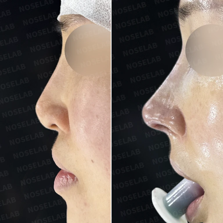
Why nasal obstruction persists despite multiple surgeries: Correcting nasal valve stenosis and nostril asymmetry — Before-and-after comparison
Before-and-after comparison

Why nasal obstruction persists despite multiple surgeries: Correcting nasal valve stenosis and nostril asymmetry

A case of persistent nostril asymmetry and nasal valve stenosis after multiple surgeries, addressed by removing existing implants, performing septoplasty and turbinoplasty, and reconstructing the structure with rib cartilage and a skin graft.

Performed or used in this surgery
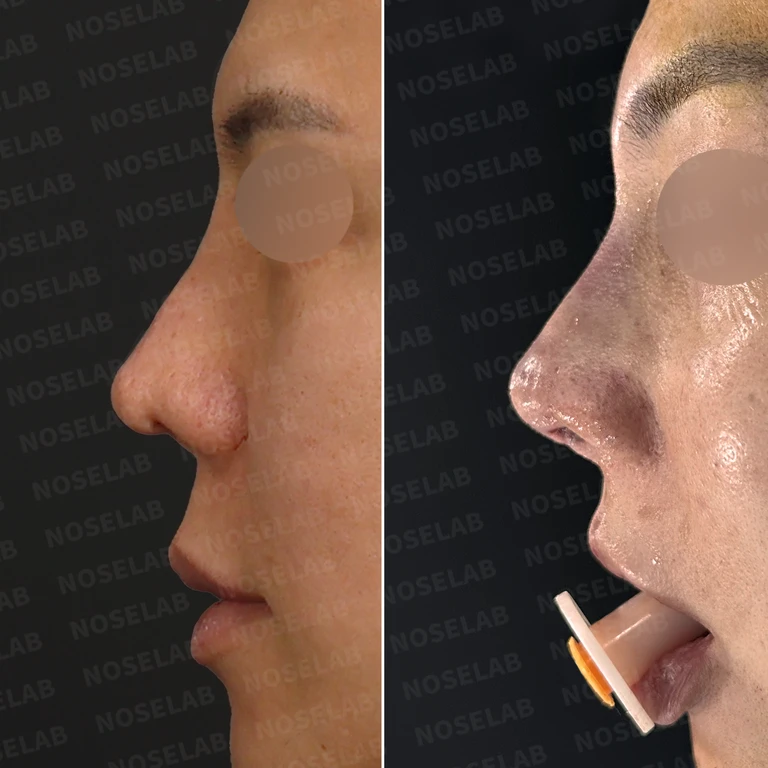
Why nasal obstruction persists after multiple surgeries: How to reconstruct the septum and nasal valve — Before-and-after comparison
Before-and-after comparison

Why nasal obstruction persists after multiple surgeries: How to reconstruct the septum and nasal valve

For a patient whose septum had collapsed after repeated surgeries, causing internal nasal valve stenosis and inferior turbinate adhesions, we performed septal reconstruction with autologous rib cartilage, osteotomy reversal, and adhesiolysis.

Performed or used in this surgery
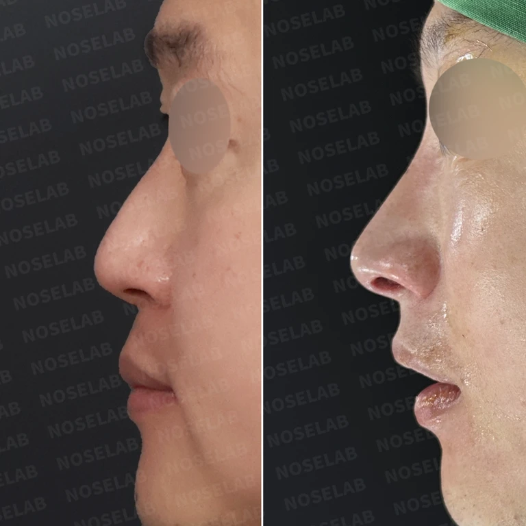
Why the nostrils look severely uneven on both sides: cartilage reconstruction and skin graft correction — Before-and-after comparison
Before-and-after comparison

Why the nostrils look severely uneven on both sides: cartilage reconstruction and skin graft correction

Congenital asymmetry, along with a residual crooked nose and nostril asymmetry from a previous rhinoplasty, was stabilized using lower lateral cartilage reconstruction, internal nostril skin grafting (free composite graft), and septal reconstruction.

Performed or used in this surgery
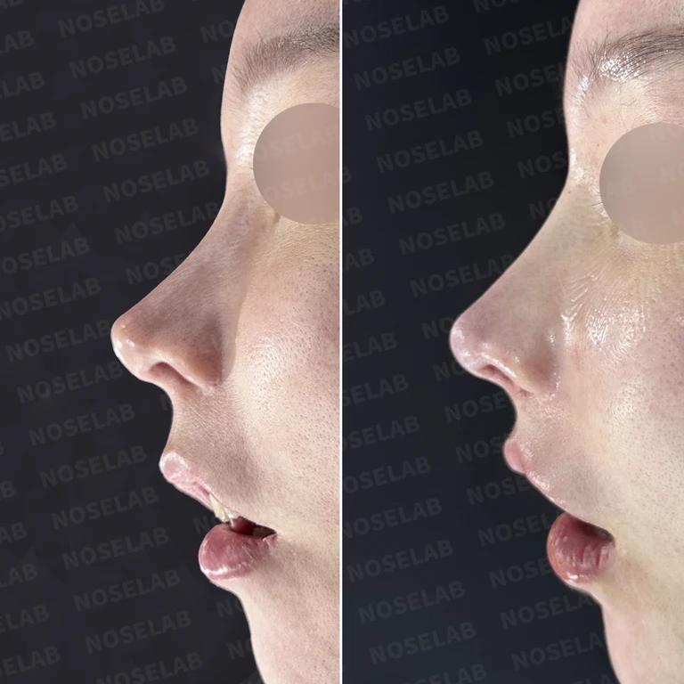
Why nasal obstruction worsens after rhinoplasty: How septal reconstruction and osteotomy reversal are performed — Before-and-after comparison
Before-and-after comparison

Why nasal obstruction worsens after rhinoplasty: How septal reconstruction and osteotomy reversal are performed

Nostril asymmetry, over-osteotomy, and a collapsed septum resulting from prior alar reduction were corrected using a skin graft, osteotomy reversal, septal reinforcement with autologous rib cartilage, and rhinitis surgery.

Performed or used in this surgery
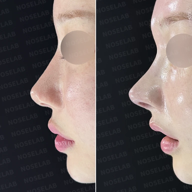
Why internal nasal structures collapse after multiple rhinoplasties and total reconstruction with rib cartilage — Before-and-after comparison
Before-and-after comparison

Why internal nasal structures collapse after multiple rhinoplasties and total reconstruction with rib cartilage

Following four prior rhinoplasties, septal and lower lateral cartilage defects and nasal bone erosion were addressed by treating a chronic foreign body reaction, performing total reconstruction with autologous rib cartilage, and placing a nostril skin graft.

Performed or used in this surgery
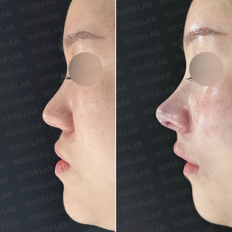
No search results found

Please try searching with different keywords.

Scroll to keep reading

1 / 4Next case

Common questions before reading the cases

What types of tissue are used in nasal tip skin reinforcement cases?

The records document surgeries using a combination of artificial dermis and scar tissue, as well as surgeries using autologous dermis harvested from behind the ear. In some cases, only "dermis" is noted, so they cannot all be assumed to be the same material. Checking whether the original text specifies the exact source and placement site helps clarify the differences among skin reinforcement methods.

Is cartilage also corrected during surgery for a nose with thin skin?

One case describes reinforcing the nasal tip skin with dermis while repositioning the lower lateral cartilages and placing an ear cartilage graft at the nasal tip. This is a record of addressing both the overlying soft tissue and the underlying cartilage framework together. Other cases also involve columella correction and osteotomy, so it is helpful to distinguish each surgical step associated with the changes seen in the photos.

Related topics to explore

Thin nasal skinImplant show-through and palpabilityAutologous dermisArtificial and allogeneic dermis

Surgical records referenced in this guide

  • Correcting asymmetry and function in a small, upturned nose with thin skin
  • The structure and correction methods for a bifid (cleft) nasal tip
  • Why repeat rhinoplasty thins the nasal tip skin causing cartilage show-through, and how autologous dermis reinforcement helps
Original article

Read more records

Planning a visit to Seoul?Prepare your consultation with the NOSELAB team.

International patient guideWhatsApp ↗
Dr. Peter C. Kang's Rhinoplasty Stories
Rhinoplasty guideCases by topicDr. Peter C. KangNaver Blog ↗
View original photo
Enlarged photo
WhatsAppChat with NOSELAB↗