Hello, I am Dr. Peter C. Kang.
Today, I would like to share a revision rhinoplasty case of a patient who visited our clinic concerned about columellar retraction and a pinched alar appearance following a previous rhinoplasty.
🔍 Surgical history
The patient had undergone rhinoplasty about 5 years ago.
About 5 years ago: Nasal bridge augmentation with a silicone implant, nasal tip surgery with donor rib cartilage, and unilateral osteotomy to correct nasal bone asymmetry. After that previous surgery, retraction in the columellar area and a pinched deformity of the right alar rim developed, leading the patient to decide on revision rhinoplasty.
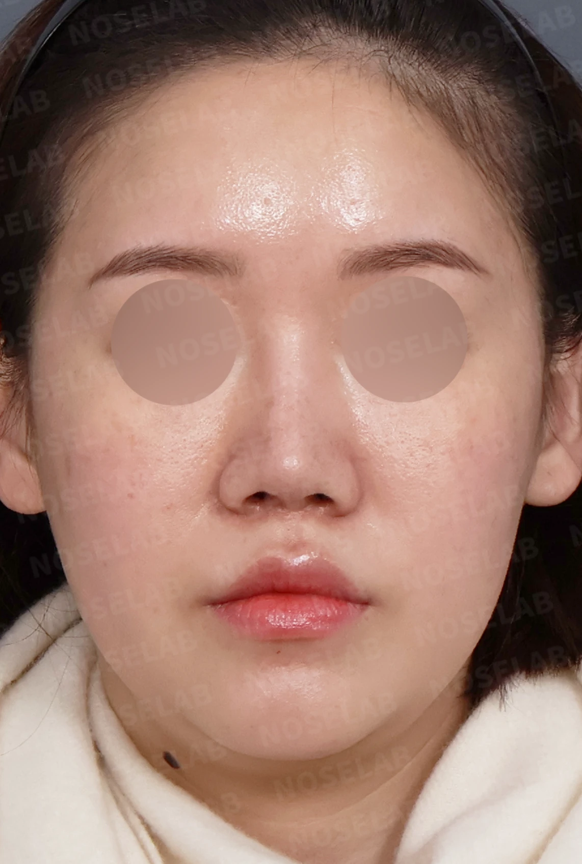
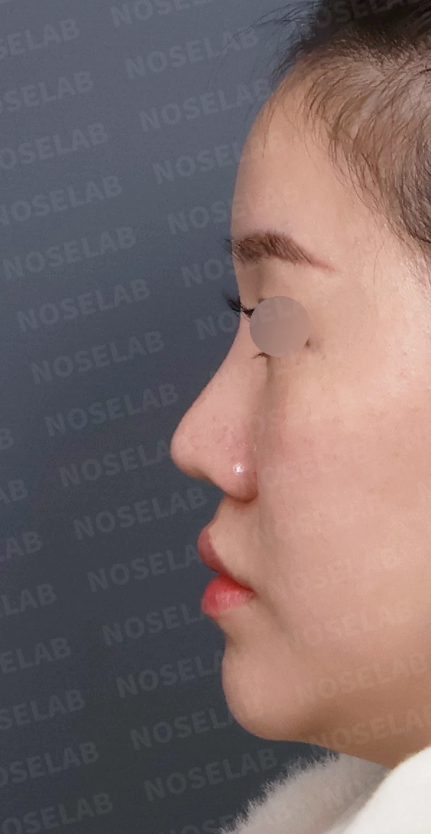
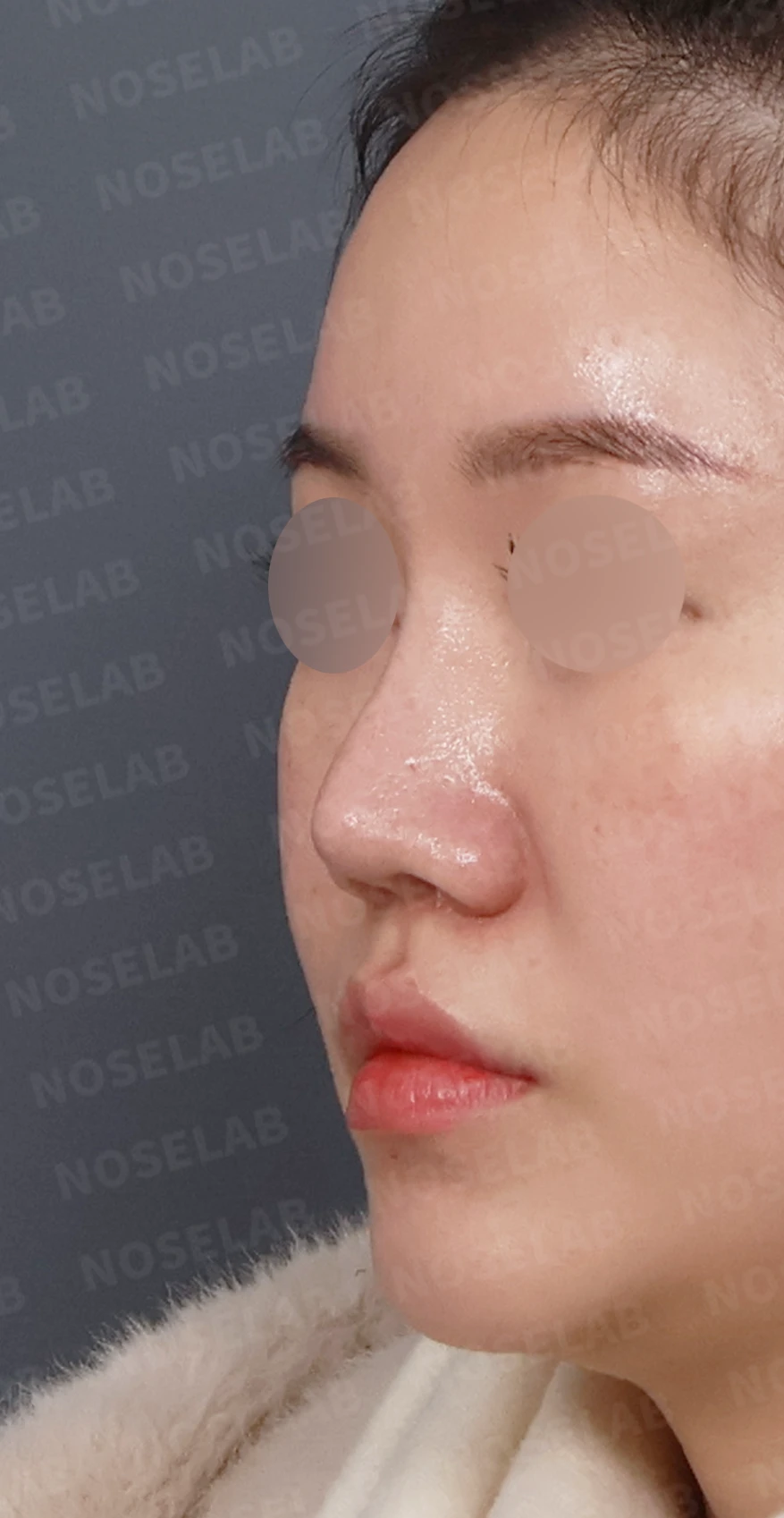
Front, left profile, and oblique views during design consultation.
💉 Patient requests
The patient's specific requests were as follows:
Improvement of the retracted columellar area
Correction of the pinched appearance of the right alar rim
Natural refinement of the overall nasal shape to balance the face
Relief of nasal obstruction (rhinitis symptoms)
Correction of the sunken appearance around both nasolabial folds (protruding mouth correction)
The patient expressed trust in Dr. Kang's recommendations for the overall nasal shape, and after considering replacing the donor rib cartilage from the previous surgery with autologous rib cartilage, ultimately chose autologous rib cartilage.
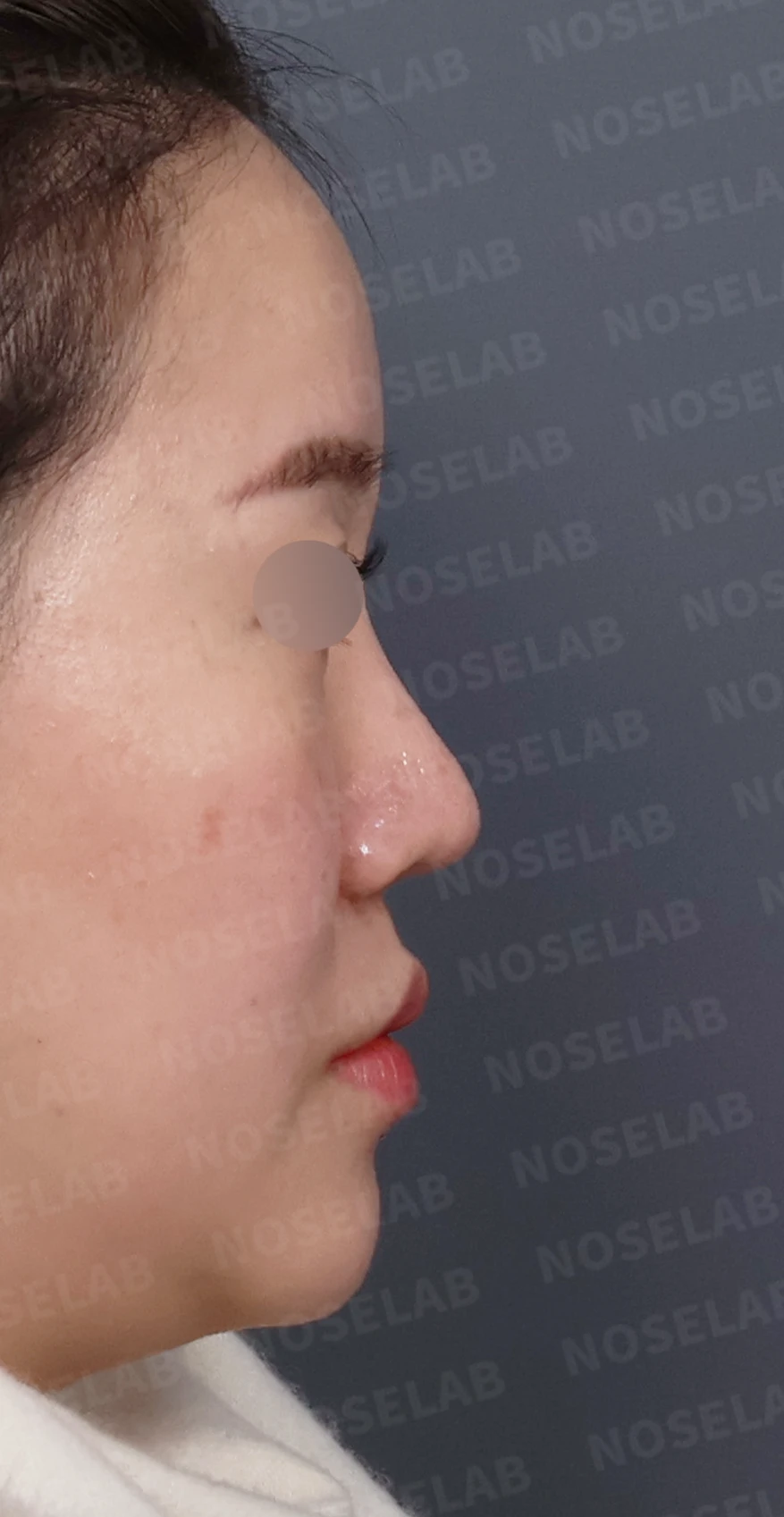
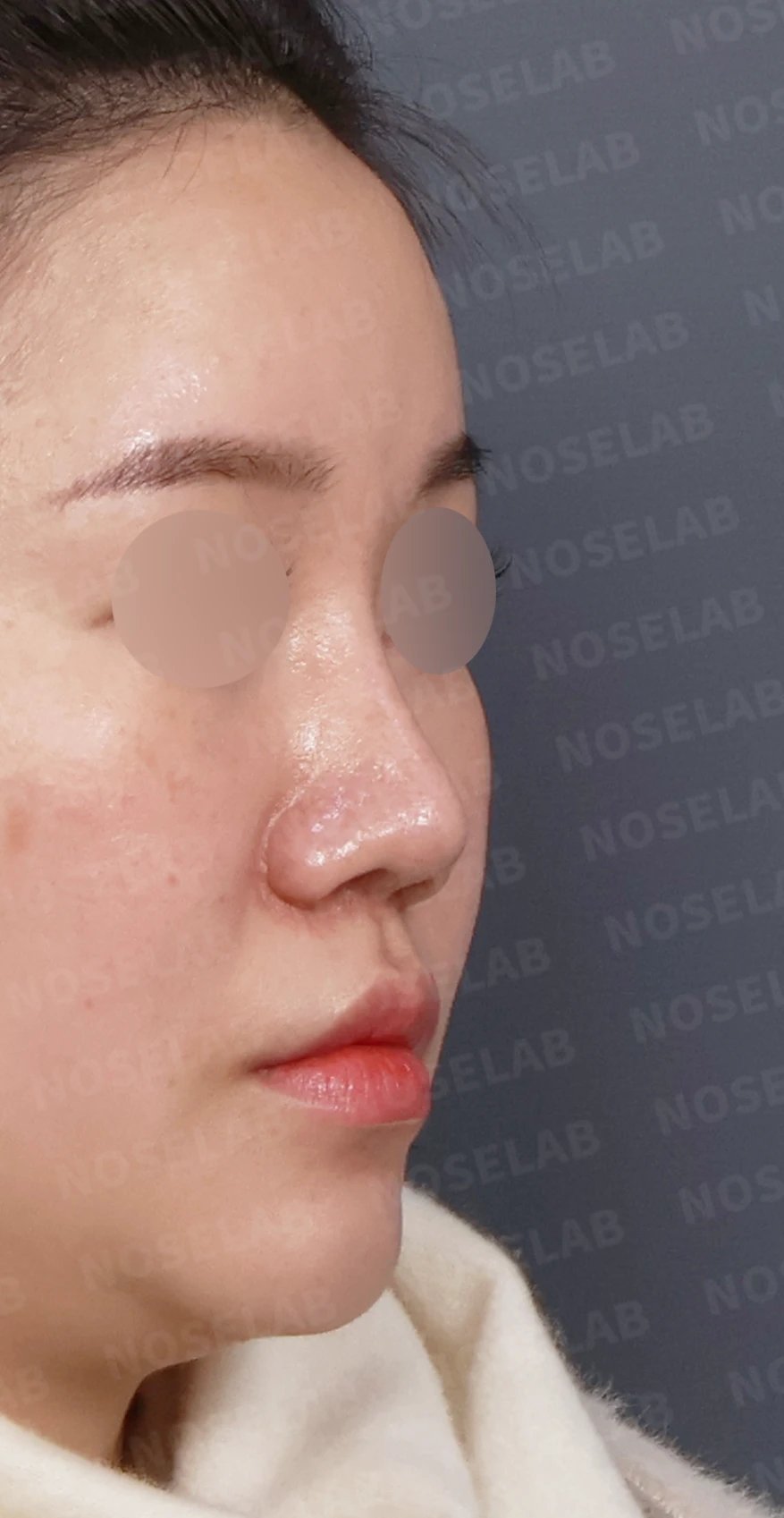
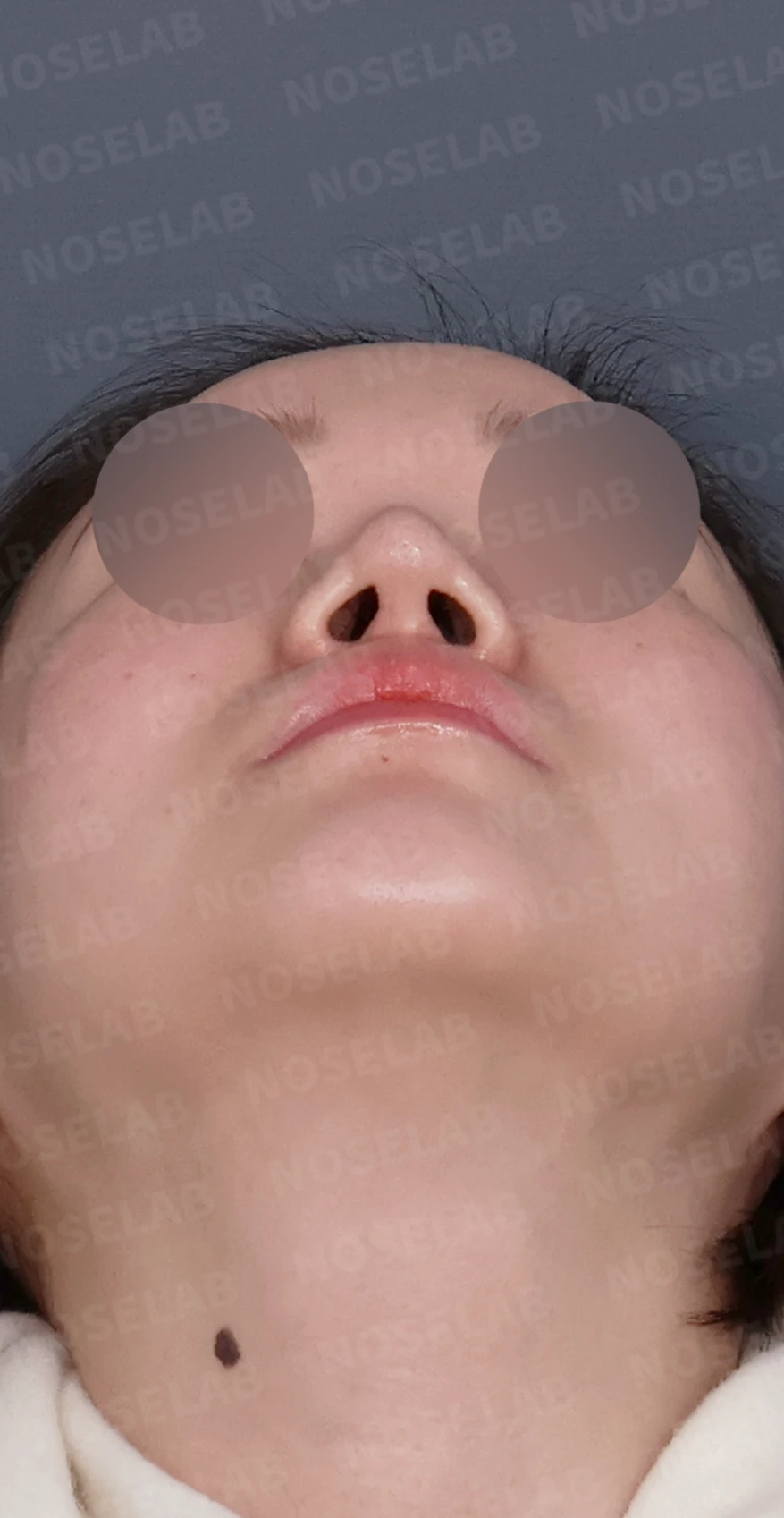
Right profile, oblique, and nostril views during design consultation.
🔧 Surgical plan and procedure
1. Removal of existing implant and graft material
Removal of existing silicone implant
Removal of the donor rib cartilage used in the previous surgery
Detailed evaluation of deformed tissues
2. Anterior septal reconstruction (correction of columellar retraction)
Identification of the damaged anterior septal cartilage causing the columellar retraction
Sturdy reconstruction of the damaged anterior septum using autologous rib cartilage
Restoration of columellar height and support
3. Reconstruction of the right alar rim
Reconstruction of the pinched right alar rim using autologous rib cartilage
Natural refinement of the alar contour through soft-tissue grafting
Restoration of symmetry between both alae
4. Septoplasty and inferior turbinate reduction
Internal nasal structures realigned by correcting the deviated septum
Rhinitis symptoms improved by reducing hypertrophied inferior turbinates
Restoration of breathing function
5. Paranasal augmentation (improvement of protruding mouth)
Implant placement in both nasolabial fold areas
Restoration of midface volume in sunken areas
Improvement in protruding mouth appearance
🌟 Surgical results
Front view changes
Natural restoration of the retracted columella
Symmetric correction of the previously pinched right alar rim
Overall balanced nasal shape
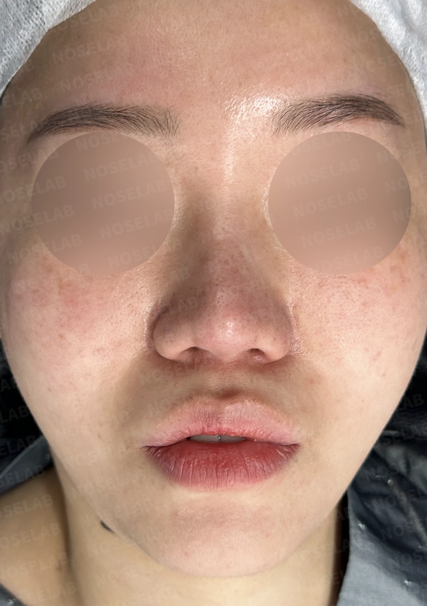
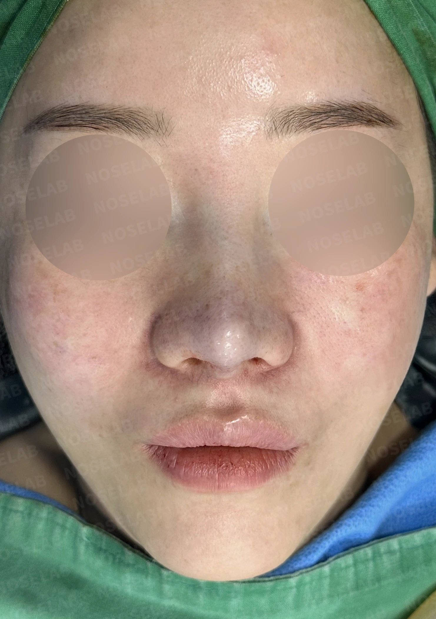
Before surgery / Day 0 immediately after surgery Photos taken immediately after surgery show swelling and may differ from the final result.
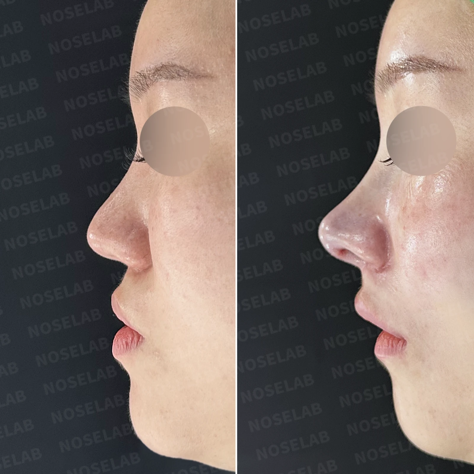
Profile view changes
Restored columellar height creating a natural tip line
Proper adjustment of the nasolabial angle
Improvement of the protruding mouth as midface volume is restored with paranasal augmentation
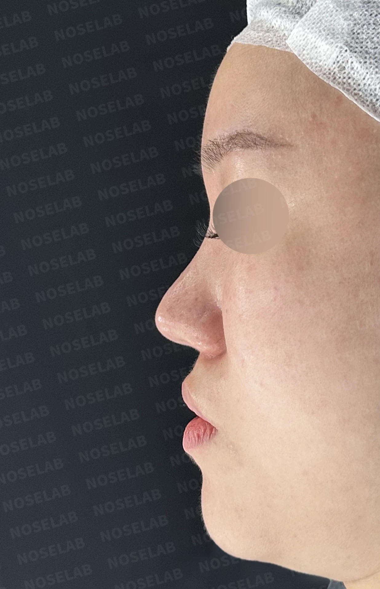
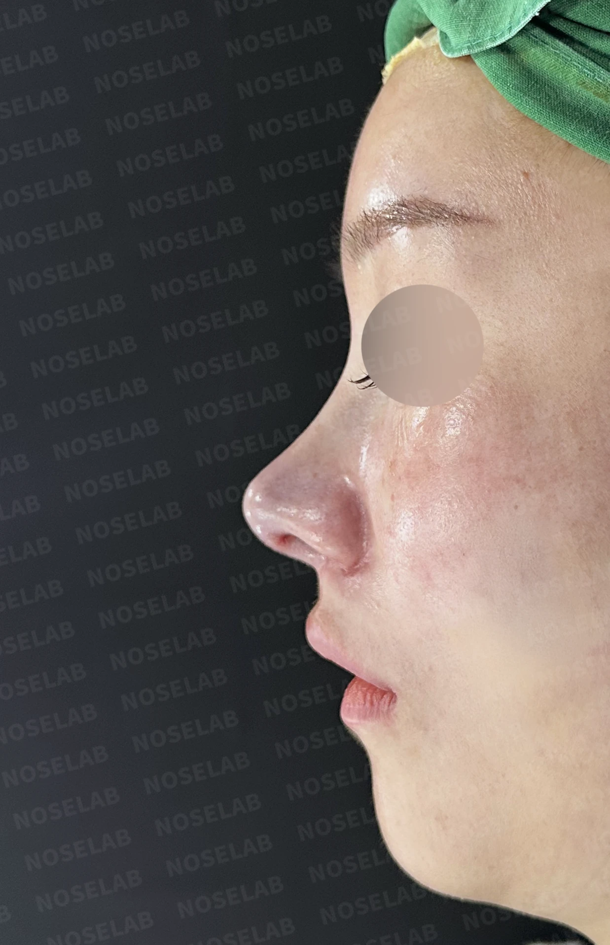
Before surgery / Day 0 immediately after surgery Photos taken immediately after surgery show swelling and may differ from the final result.
Changes on the oblique view
Natural nasal contour visible even at a 45-degree angle
Clean nasal lines following correction of the alar deformity
Improved overall facial balance through midface volume restoration
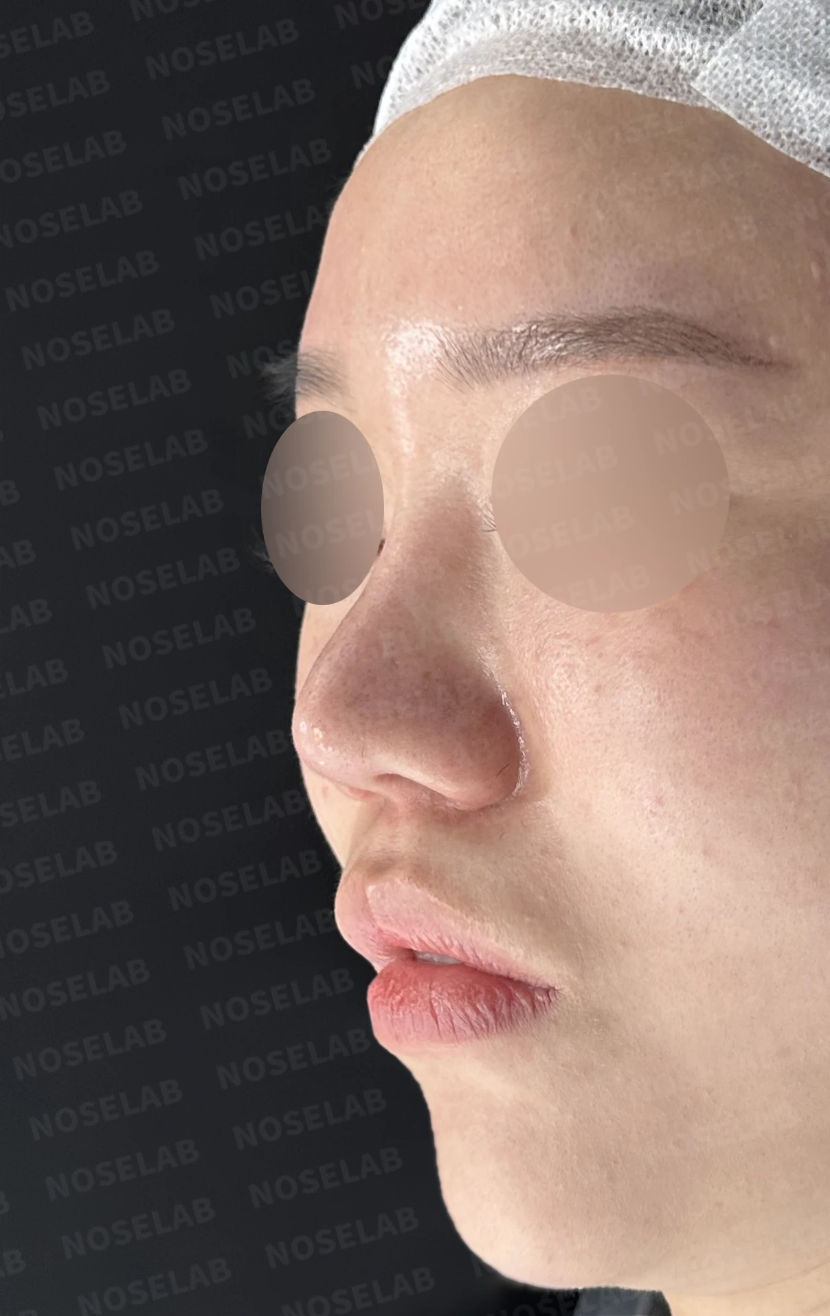
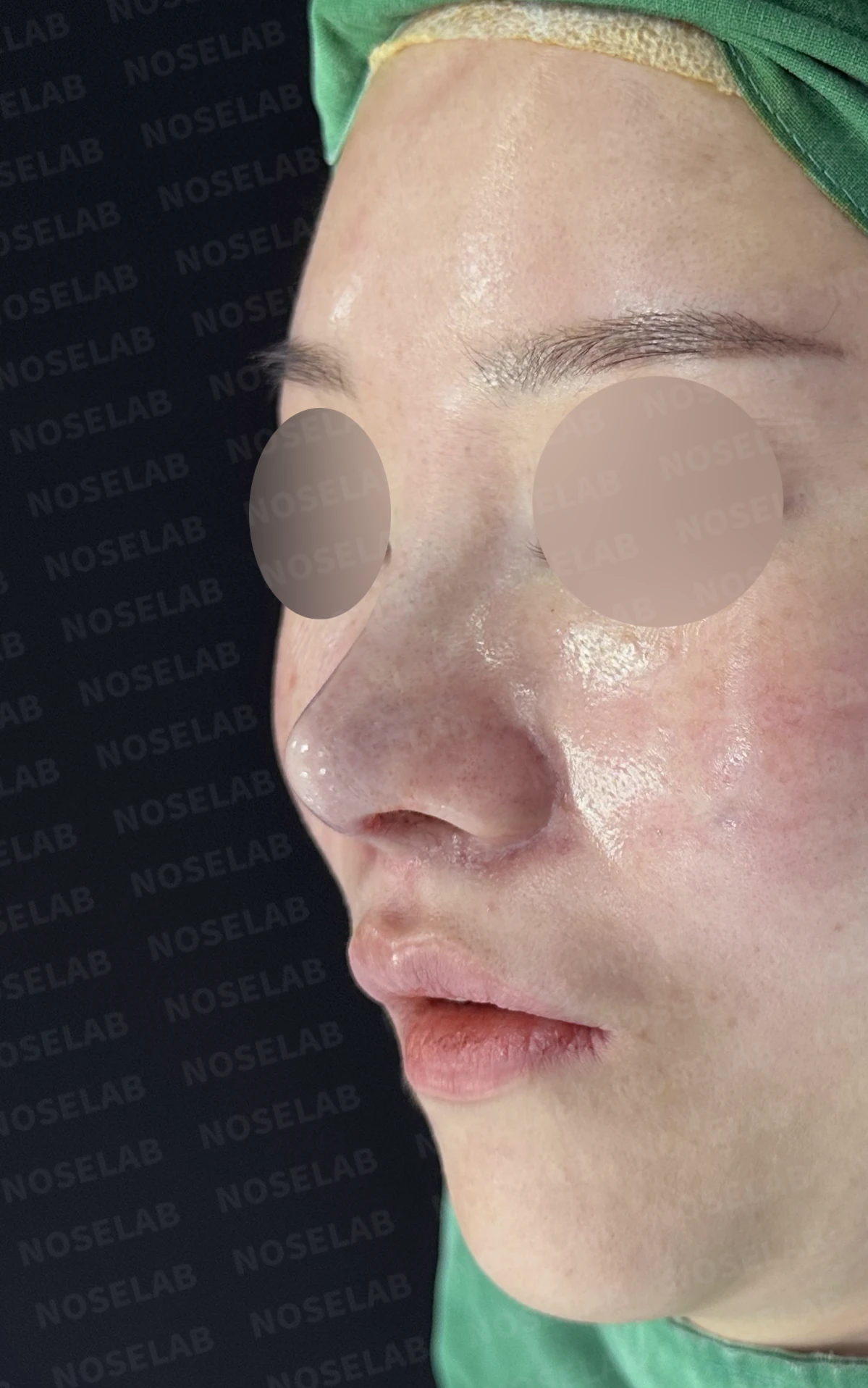
Before surgery / Day 0 immediately after surgery Photos taken immediately after surgery show swelling and may differ from the final result.
Changes in nostril shape
Natural improvement of nostril shape following restoration of the retracted columella
Restoration of symmetry between both nostrils after reconstructing the pinched right alar rim
Overall neat and refined nasal base line
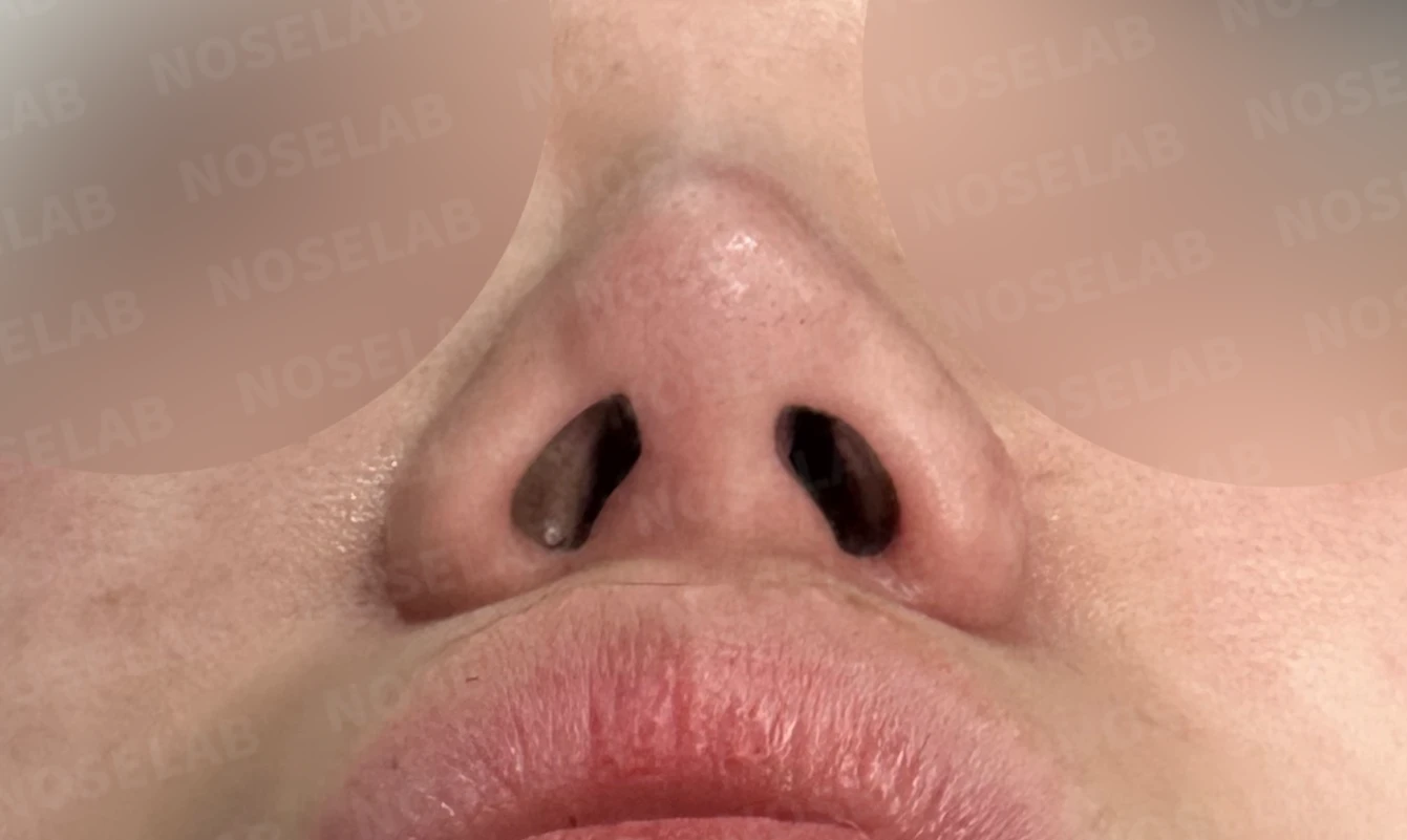
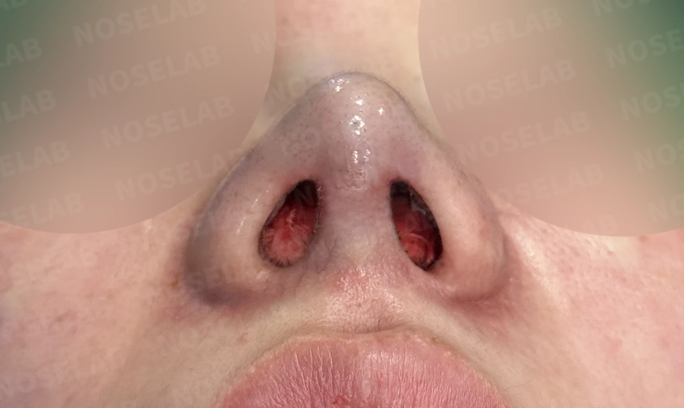
Before surgery / Day 0 immediately after surgery Photos taken immediately after surgery show swelling and may differ from the final result.
💡 Specialist opinion
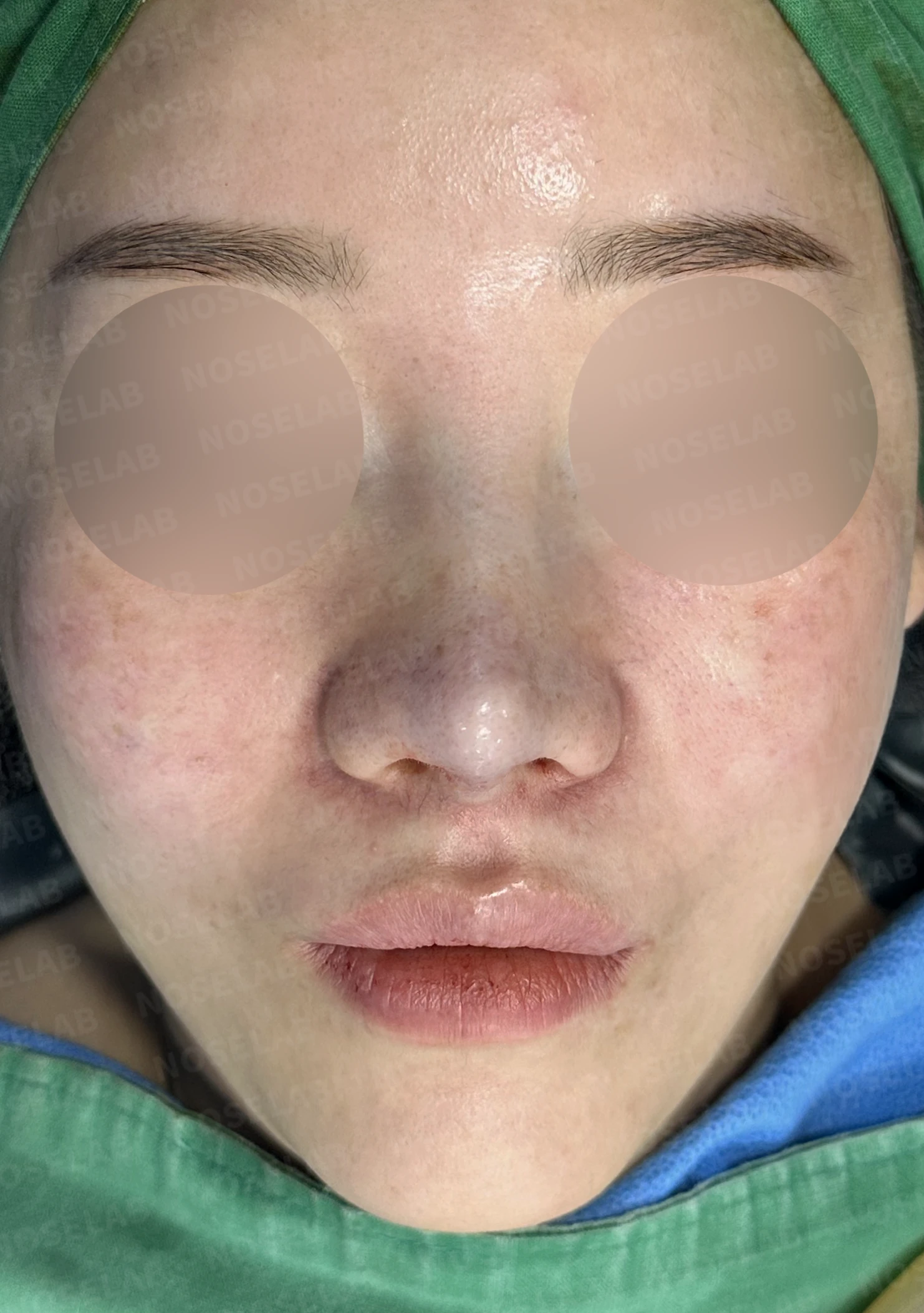
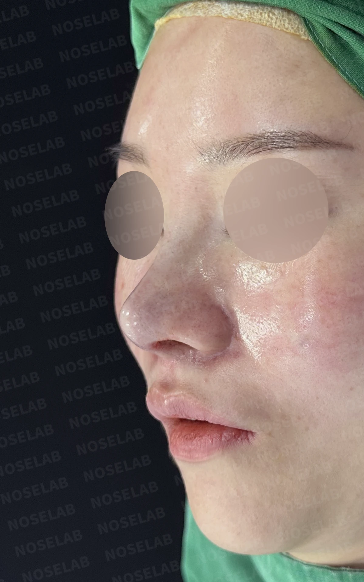
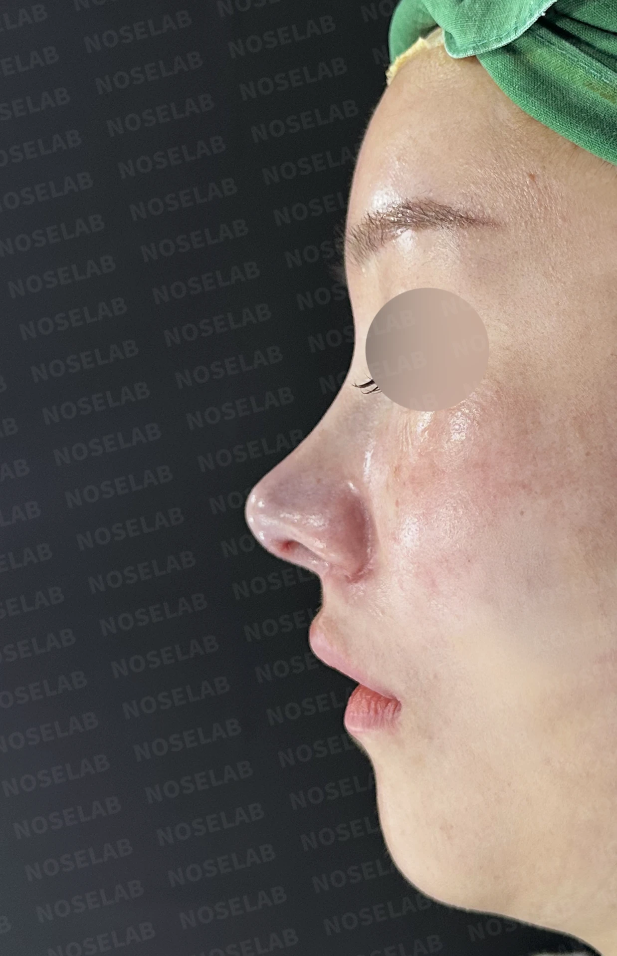
Front, oblique, and profile views on day 0 immediately after surgery. Photos taken immediately after surgery show swelling and may differ from the final result.
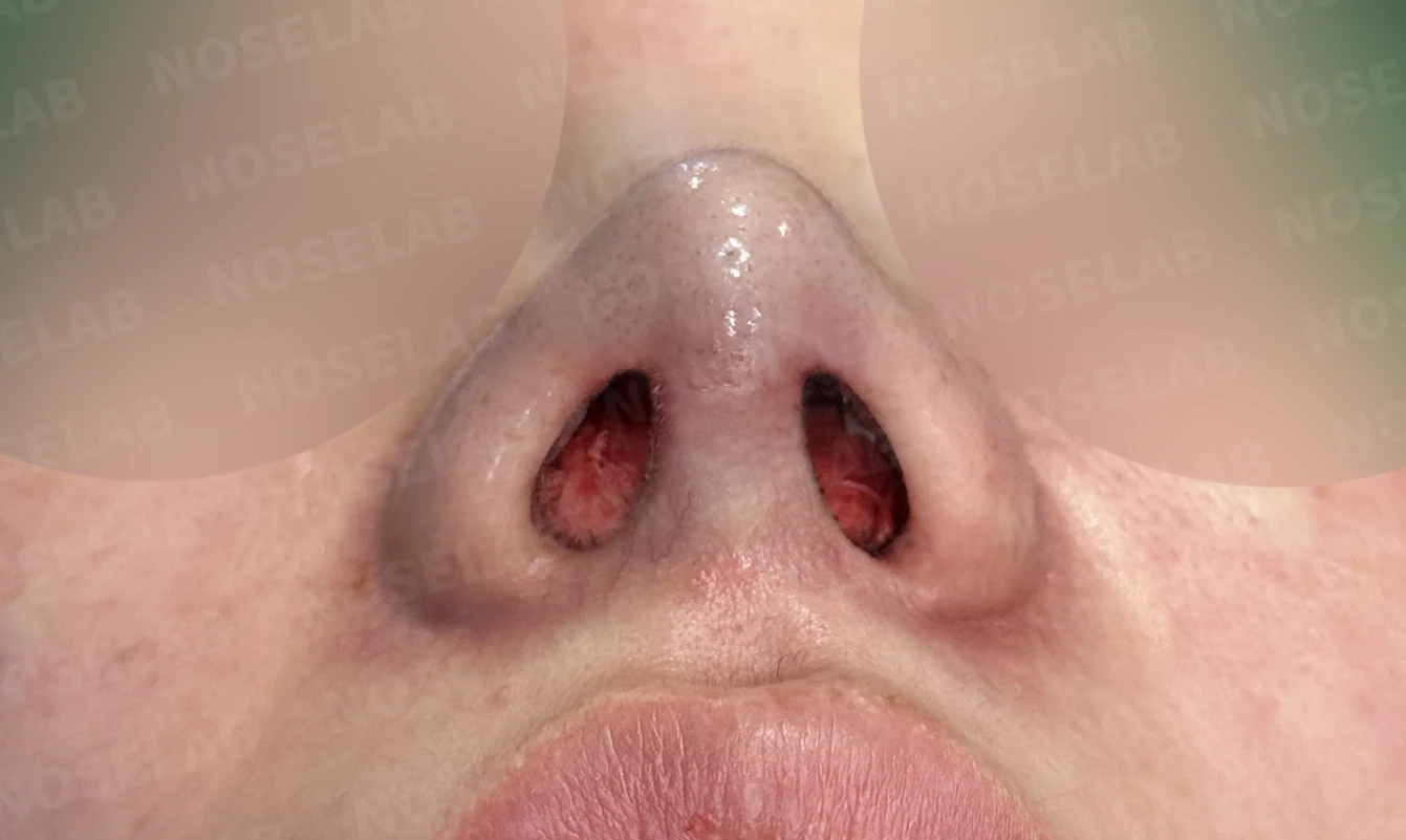
Nostril view on day 0 immediately after surgery. Photos taken immediately after surgery show swelling and may differ from the final result.
This is a revision rhinoplasty case involving columellar retraction and alar deformity following a previous surgery. A detailed analysis of why the columella had retracted revealed that damage to the anterior portion of the septal cartilage had weakened the structural support of the nasal tip as the root cause.
In such cases, simply raising the nasal tip is not sufficient; the core priority is first sturdily reconstructing the damaged anterior septum. In this surgery, autologous rib cartilage was used to reinforce the supporting framework of the anterior septum and naturally restore columellar height.
In addition, the pinched appearance of the right alar rim was corrected through alar reconstruction with autologous rib cartilage combined with soft-tissue grafting. Alar deformities are among the common issues encountered in revision surgery, and both cartilage and soft tissue must be supplemented simultaneously to achieve a natural, stable contour.
Septoplasty and inferior turbinate reduction were performed concurrently to relieve nasal obstruction, and paranasal augmentation was performed to supplement midface volume and address the sunken appearance around the nasolabial folds and the protruding mouth. In this way, during revision rhinoplasty, it is important to consider not only the nose itself but also its overall harmony with surrounding facial structures.
Because revision rhinoplasty must account for scar tissue, altered anatomy, and weakened structural support from previous operations, it demands far higher levels of diagnostic insight and surgical skill than primary surgery. Even in such complex revision cases, NOSELAB strives to accurately identify the root causes and provide fundamental solutions to achieve the intended result for every patient.
This was Dr. Peter C. Kang. Thank you.
#closedrhinoplasty #closedrevisionrhinoplasty #revisionrhinoplasty #columellartraction #alarreconstruction #autologousribrhinoplasty #septalreconstruction #paranasalaugmentation #protrudingmouthcorrection #deviatedseptum #nasalobstructionsurgery

Limitations and risks
When autologous rib cartilage is used, scarring and pain may remain at the donor site on the chest, and pneumothorax (pleural injury) can occur rarely. Over time, the grafted cartilage may warp or undergo partial resorption.
When using donor rib cartilage, long-term follow-up is necessary because the grafted cartilage may resorb or warp.
When revising after implant removal, contracture may progress again after removal, the nasal height and length may decrease, and staged surgery may be necessary depending on the skin condition.
When the septum is corrected at the same time, nasal obstruction may persist or recur, and in rare cases, septal perforation or changes in smell may occur.
Common potential risks include infection, bleeding, asymmetry, scarring, skin necrosis, and insufficient improvement; results vary depending on individual skin condition and previous surgical history.