Hello, I am Dr. Peter C. Kang.
Today, I would like to share the case of a patient who visited our clinic suffering from severe nasal obstruction after three prior rhinoplasties. For this revision, improving nasal breathing was the most important goal.
🔍 Surgical history
Approximately 2 to 3 years ago: Tip plasty using ear cartilage and dorsal hump reduction (at another clinic) → Nostril appearance did not improve
Approximately 2 years ago: Revision rhinoplasty using ear cartilage (at another clinic) → Developed a crooked nose; nostril appearance did not improve
Approximately 8 months ago: Revision rhinoplasty using autologous rib cartilage and ear cartilage (at another clinic)
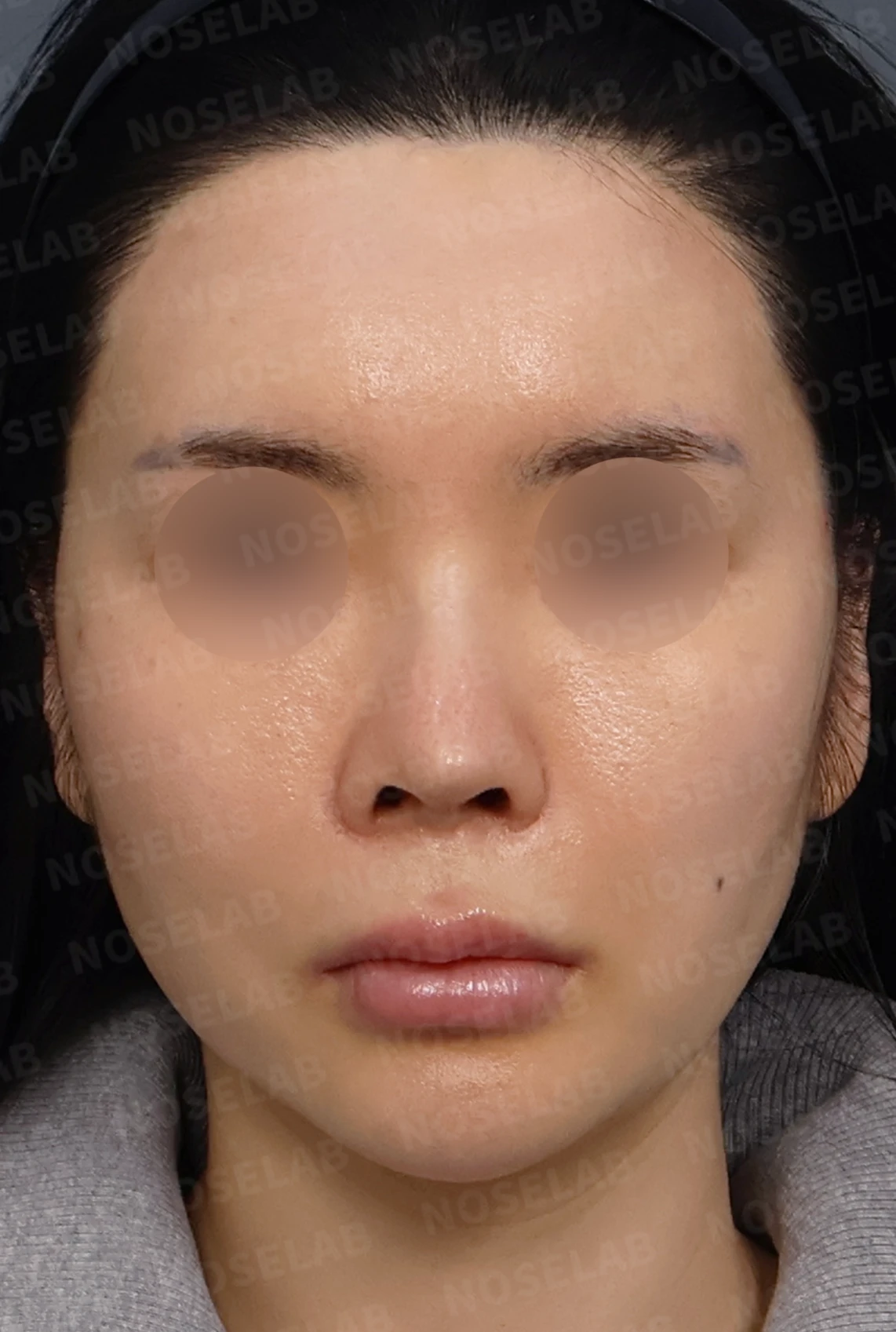
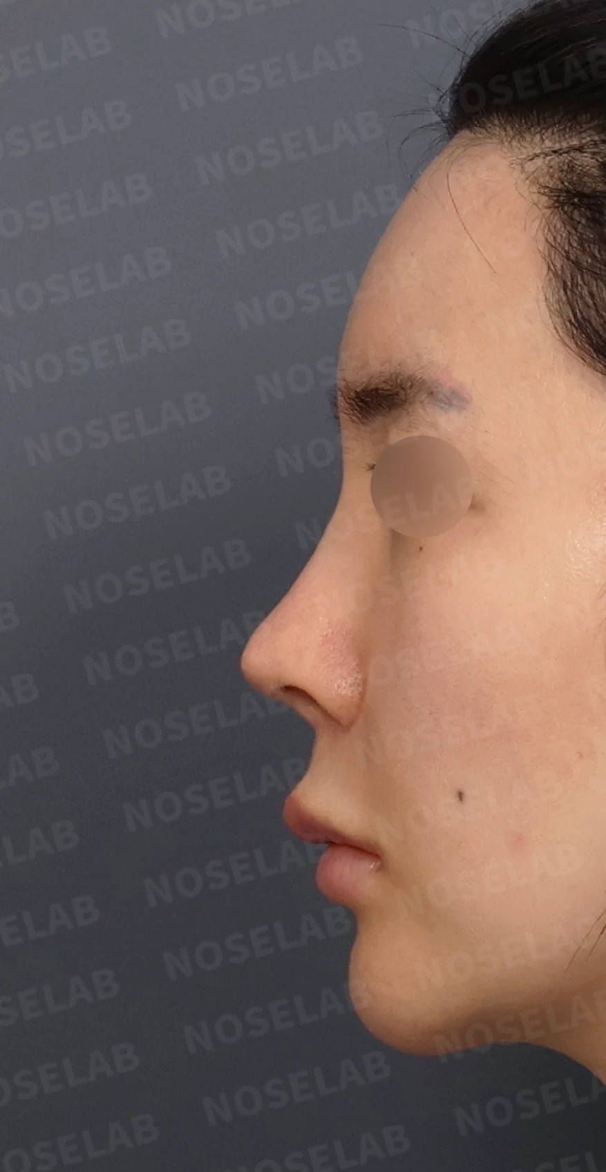
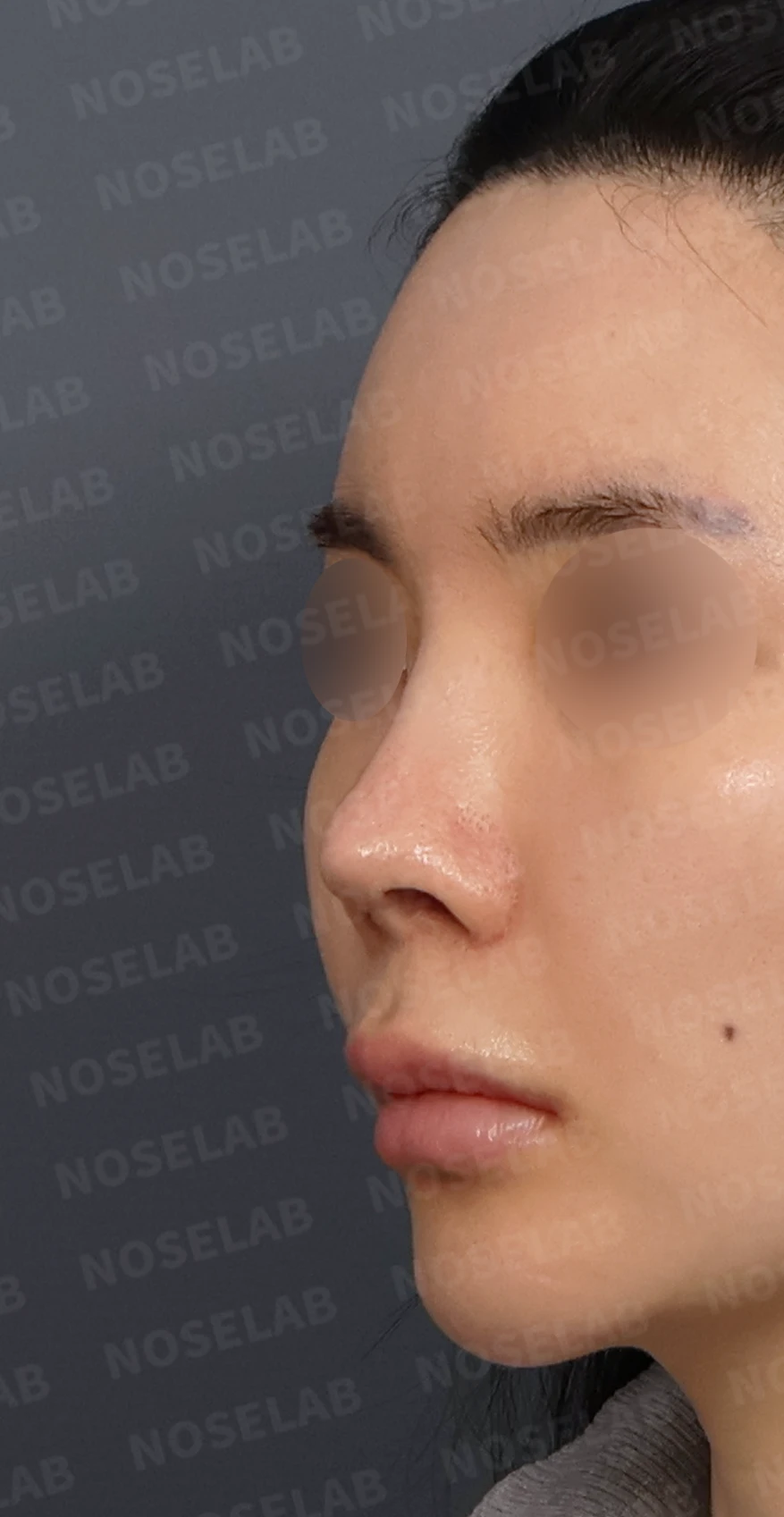
Front, left profile, and oblique views during design consultation.
💉 Patient requests
Difficulty breathing was the most distressing concern
Relieving nasal obstruction was the highest priority
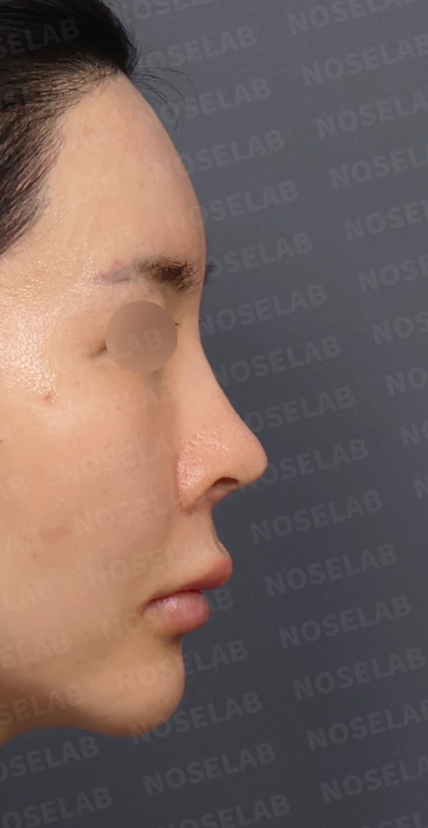
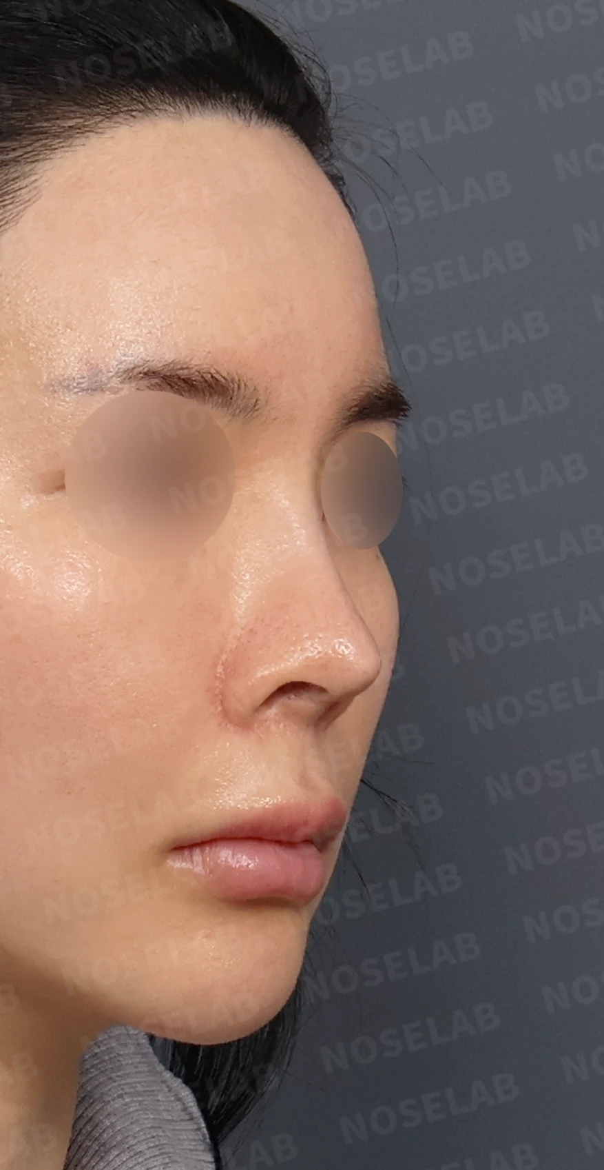
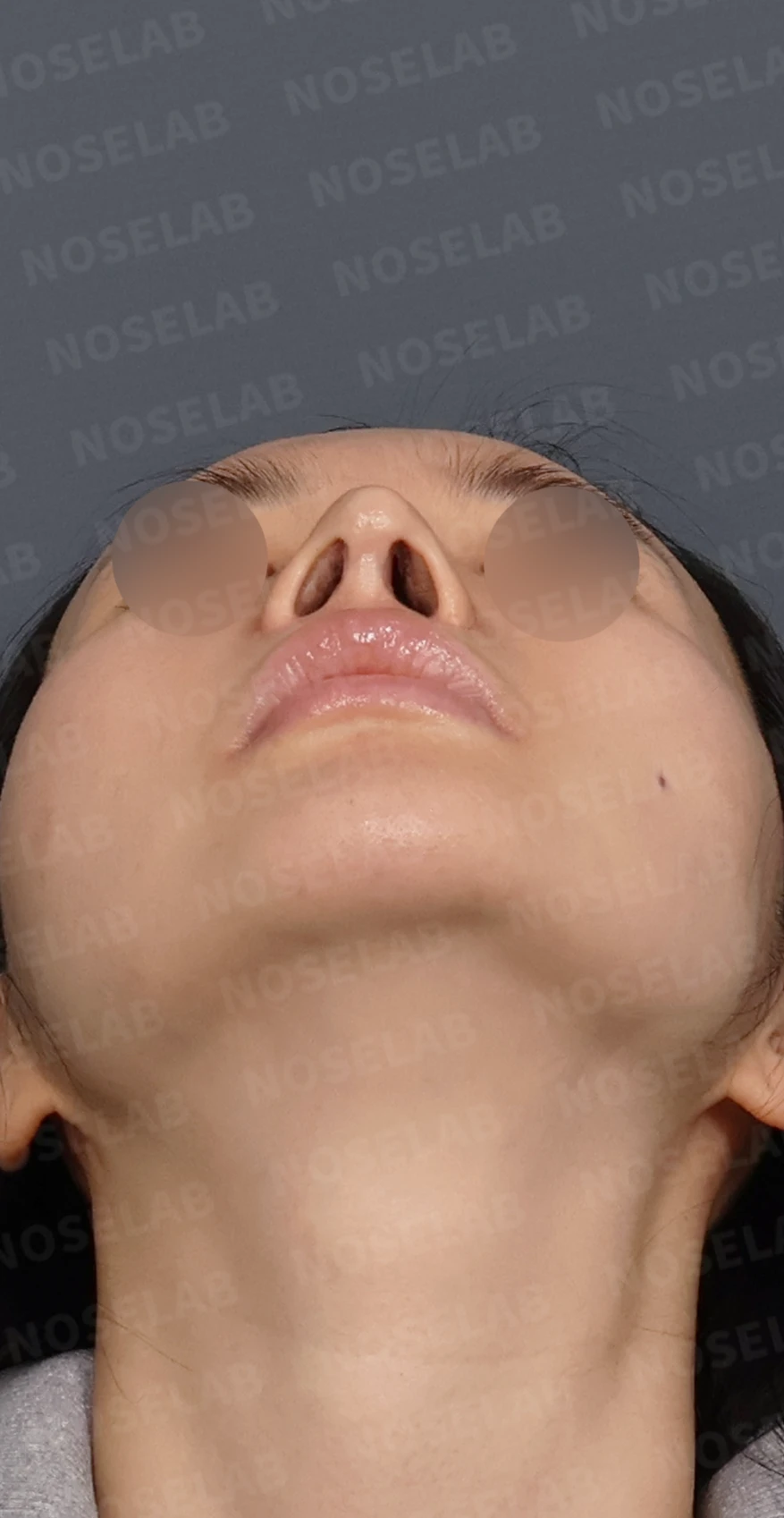
Front, left profile, and oblique views during design consultation.
🔧 Surgical plan and procedure
1. Septal reconstruction
From previous surgeries, the septum had collapsed to the right and the nasal roof had caved in, causing marked stenosis of the internal nasal valve
Because the septum was in very poor condition, it was reconstructed using autologous rib cartilage
2. Restoration of previous osteotomy
The nasal cavity space was narrowed due to overly aggressive lateral osteotomy performed during previous cosmetic surgery
Internal nasal volume was restored through osteotomy reversal surgery
Because a significant portion of the central nasal bone had been rasped away, restoring the bone caused a depression in the mid-dorsum
The depressed mid-dorsal area was corrected using autologous rib cartilage
A slight overcorrection of the nasal bone height was performed, anticipating that it would gradually settle over time
3. Tip skin reinforcement and alar cartilage reconstruction
Because the nasal tip skin was extremely thin, it was reinforced using dermis and costal perichondrium
With almost no native alar cartilage remaining, the lower lateral cartilage was reconstructed using autologous rib cartilage
4. Inferior turbinate surgery
Potential empty nose syndrome was noted, resulting from excessive inferior turbinate reduction during previous rhinitis surgery
Severe outfracture of the inferior turbinate had occurred, and the anterior part of the inferior turbinate had adhered to the lateral nasal wall
Airway space was restored by infracturing the inferior turbinate and releasing the adhesions
Anti-adhesion techniques were also applied to prevent recurrence
5. Nasolabial angle correction
The nasolabial angle between the nose and upper lip was naturally adjusted
🌟 Surgical results
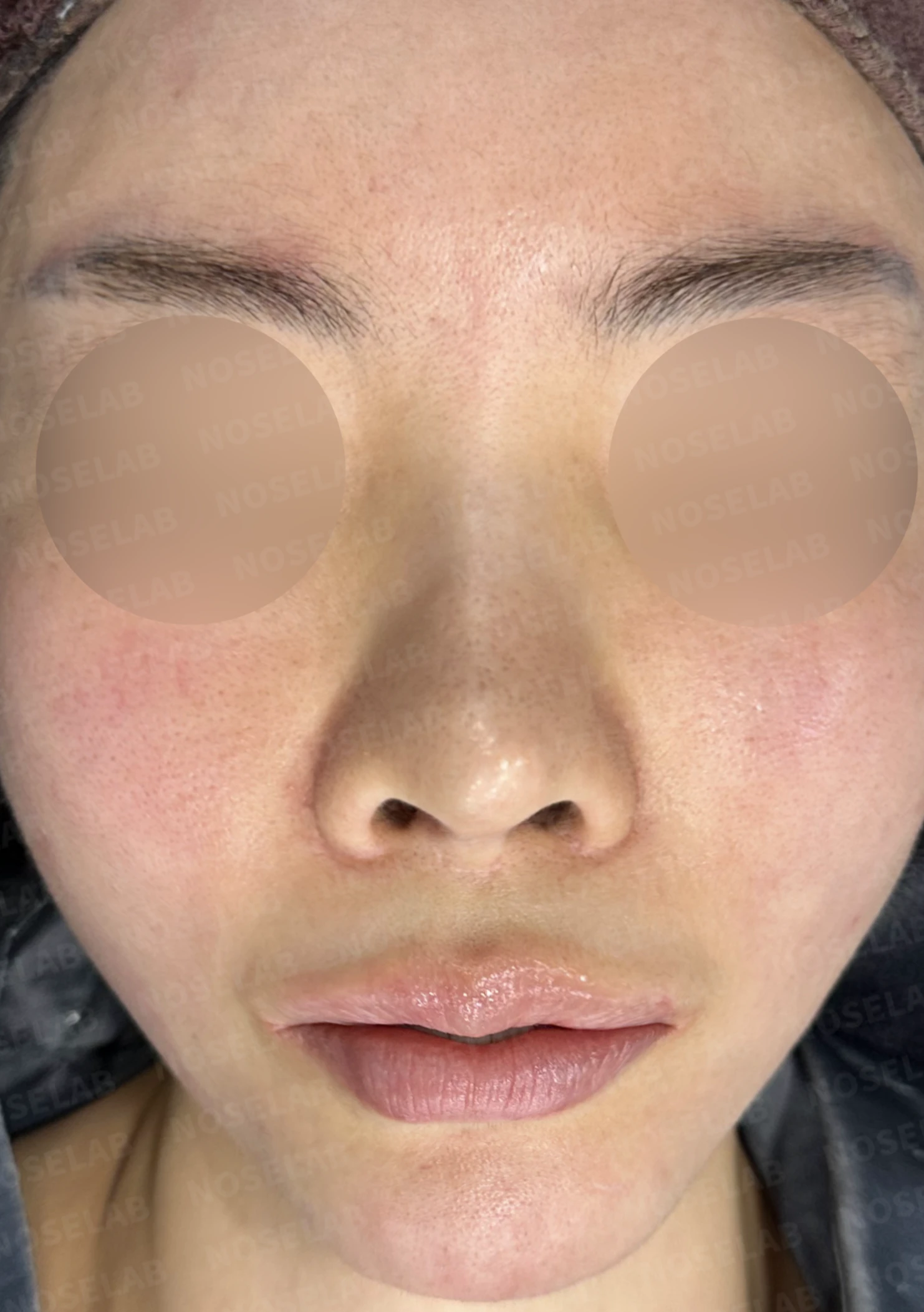
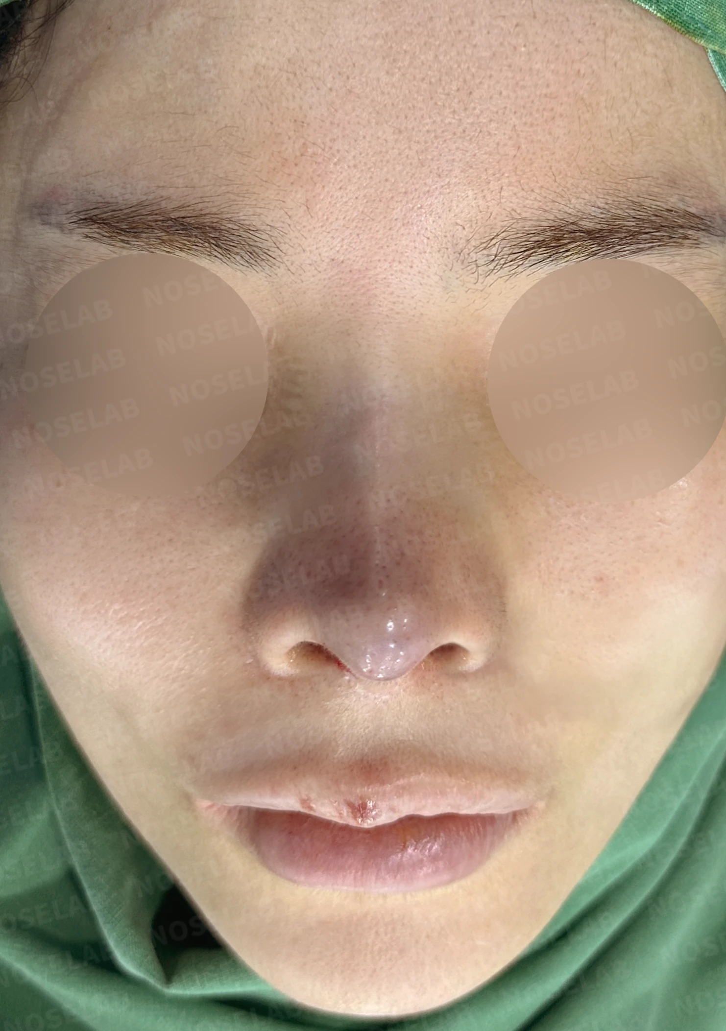
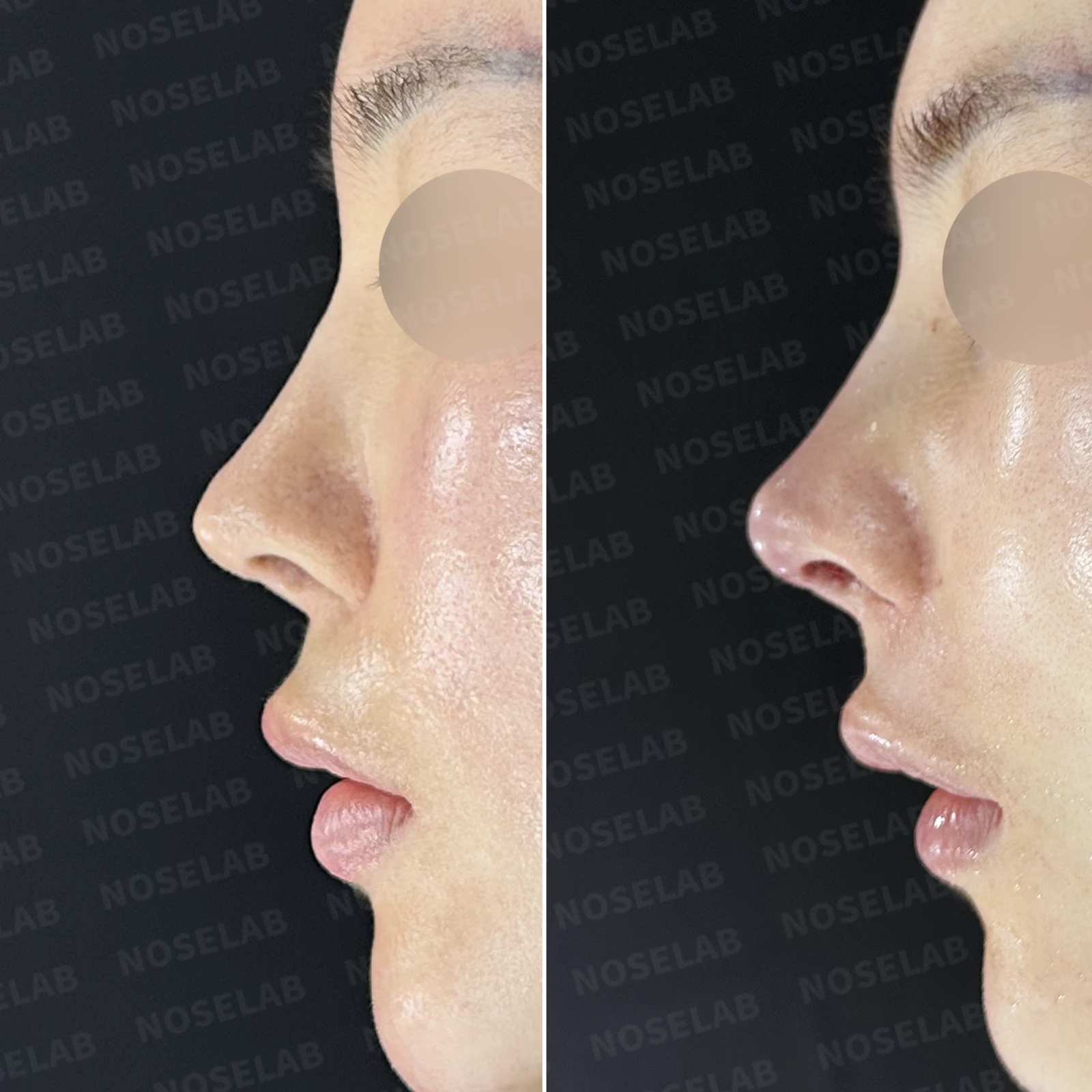
Before surgery / Day 0 immediately after surgery Photos taken immediately after surgery show swelling and may differ from the final result.
Functional improvement:
Internal nasal structures normalized through septal reconstruction
Breathing airway secured through internal nasal valve reconstruction
Internal nasal volume restored through osteotomy reversal
Right-sided nasal obstruction improved through inferior turbinate infracture and adhesiolysis
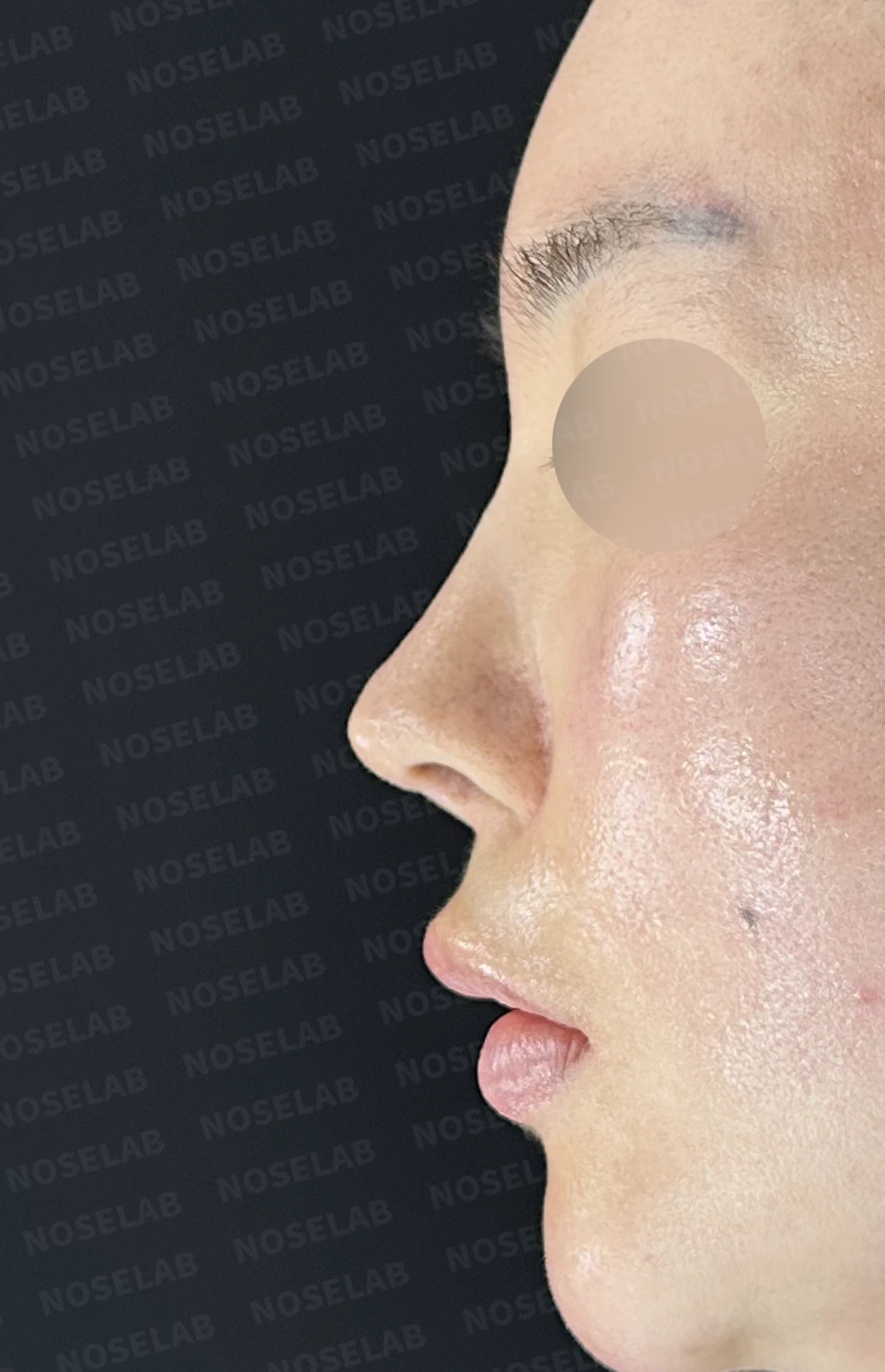
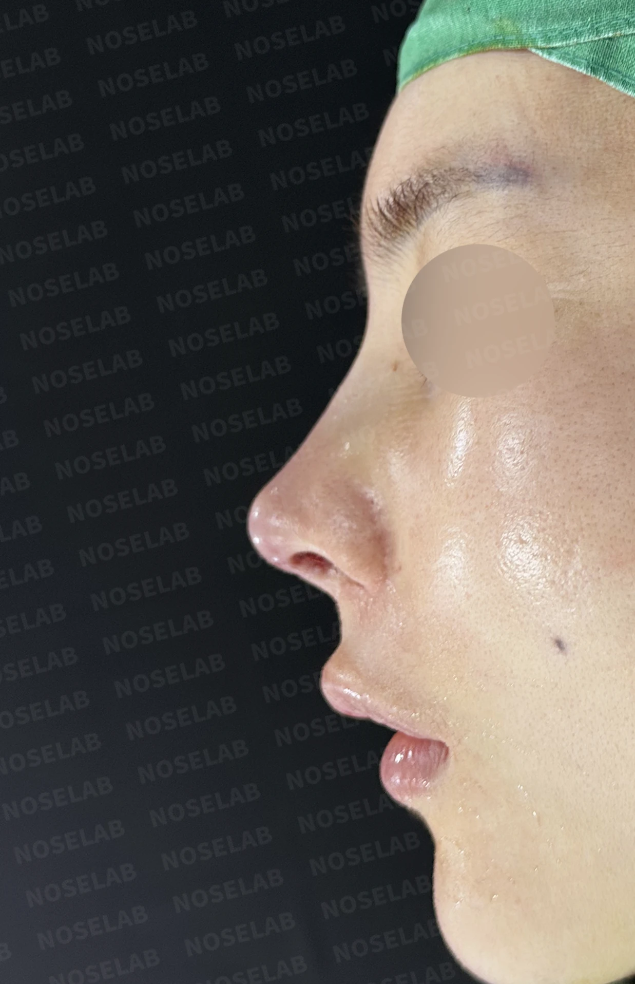
Before surgery / Day 0 immediately after surgery Photos taken immediately after surgery show swelling and may differ from the final result.
Cosmetic improvements:
Depressed mid-dorsal area corrected (it may appear slightly high initially, but will look more natural over time)
Tip skin reinforced to support a stable tip shape
A refined profile line through nasolabial angle correction
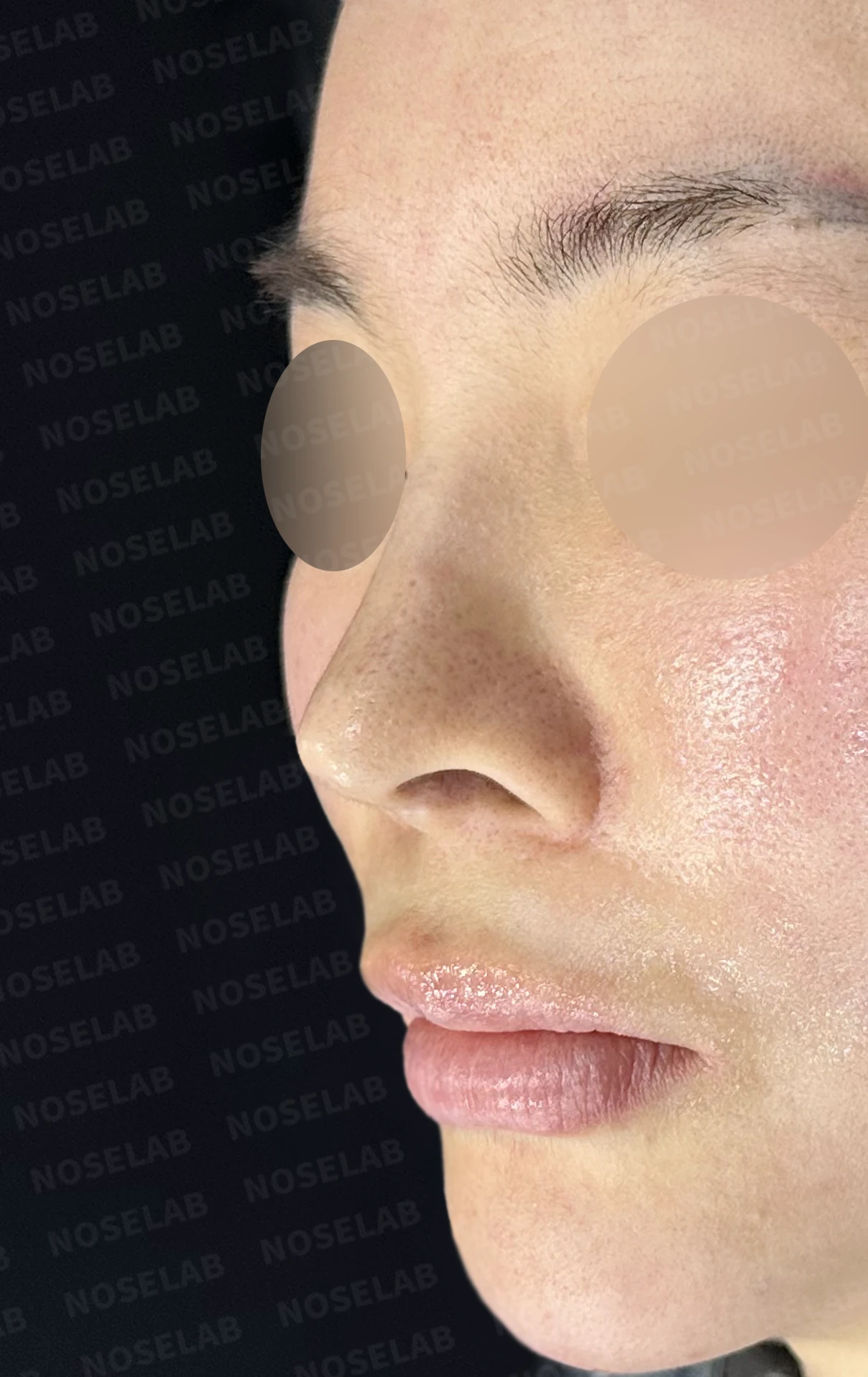
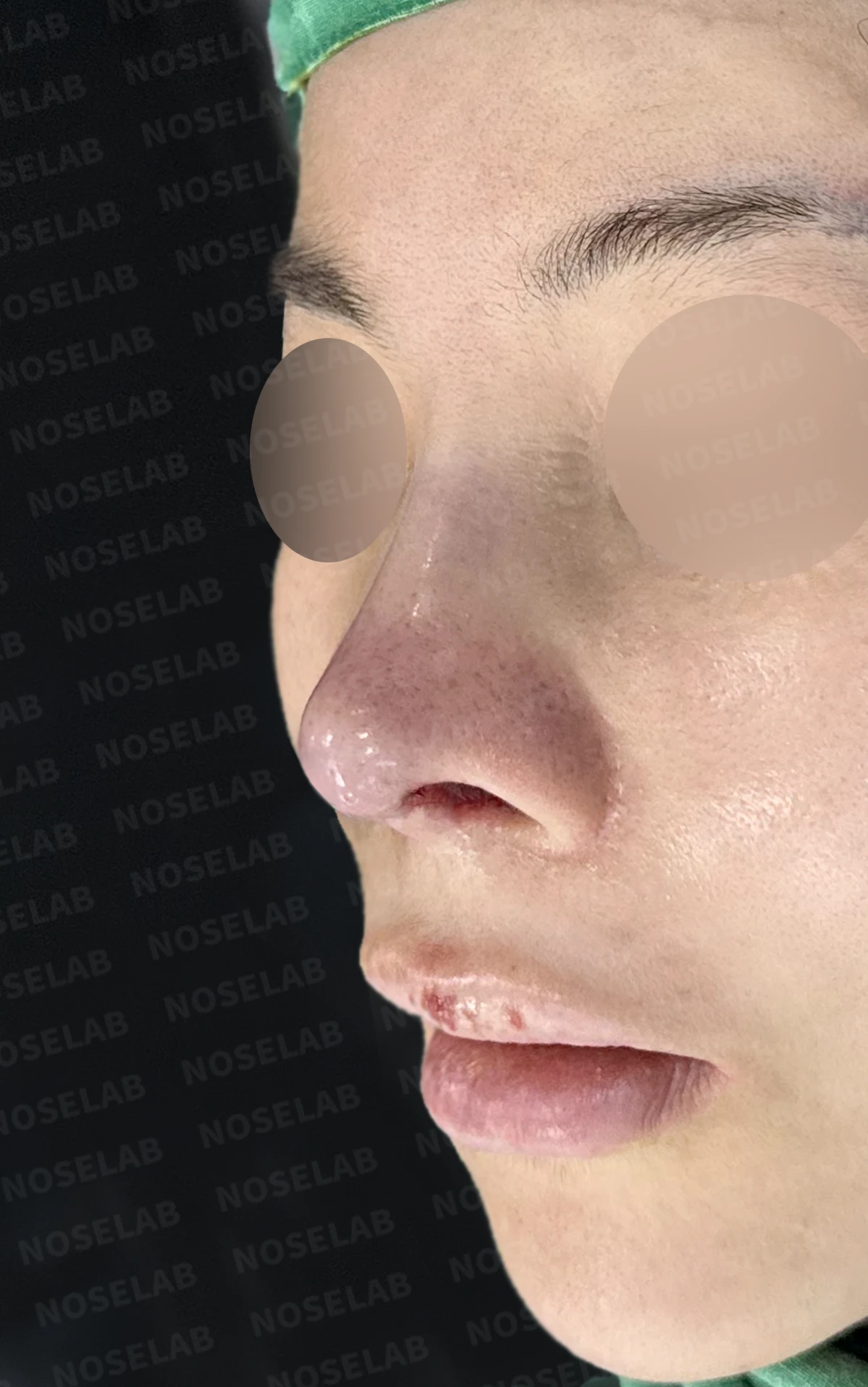
Before surgery / Day 0 immediately after surgery Photos taken immediately after surgery show swelling and may differ from the final result.
Structural stability:
Long-term structural stability supported through septal reconstruction with autologous rib cartilage
Tip support framework restored through alar cartilage reconstruction
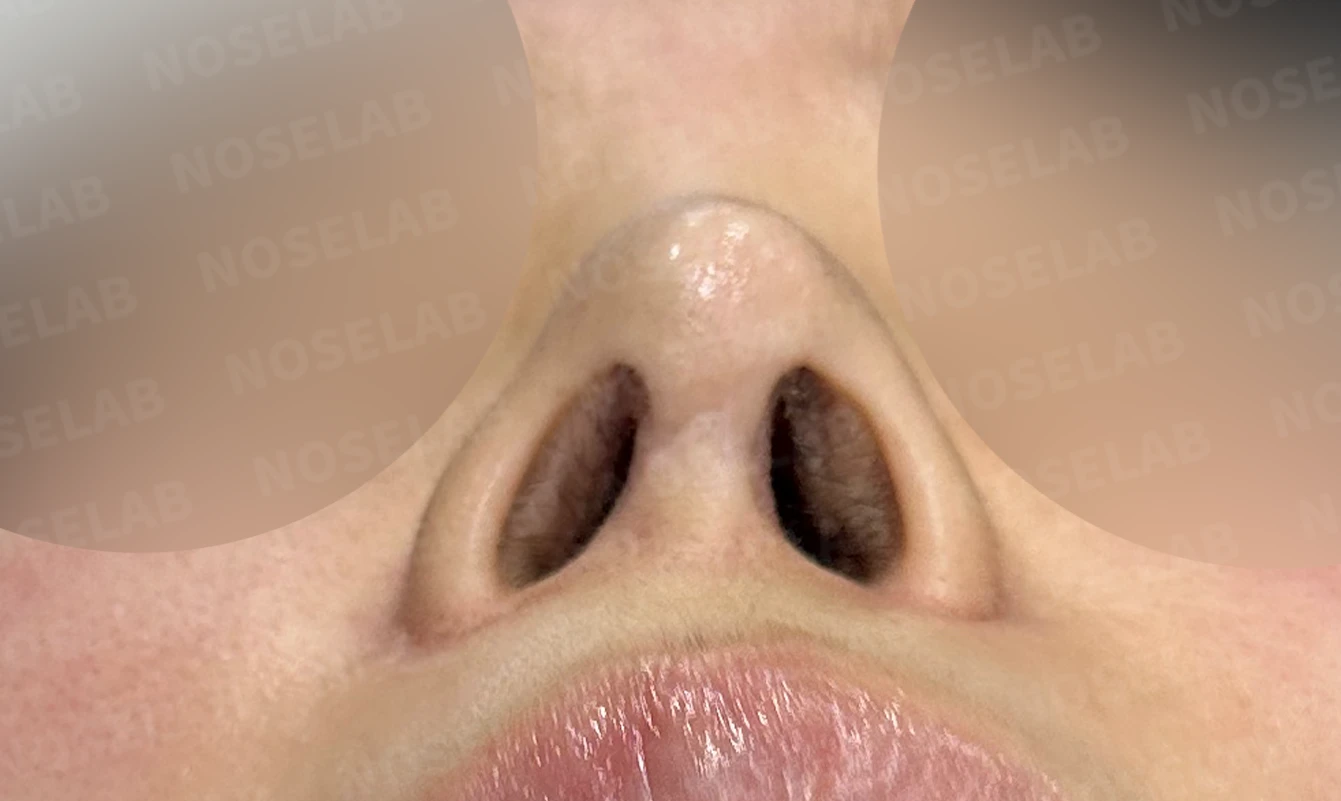
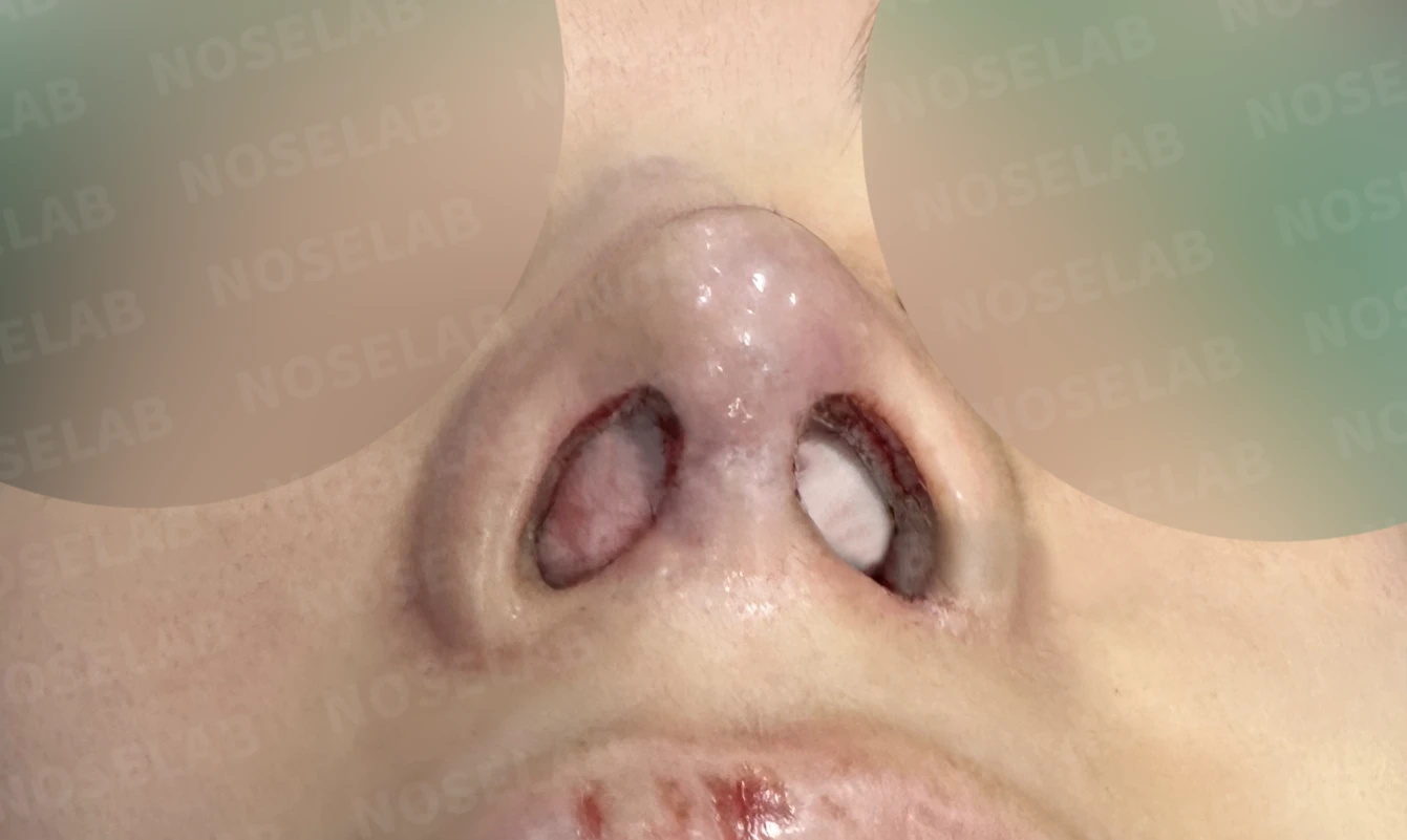
Before surgery / Day 0 immediately after surgery Photos taken immediately after surgery show swelling and may differ from the final result.
💡 Specialist opinion
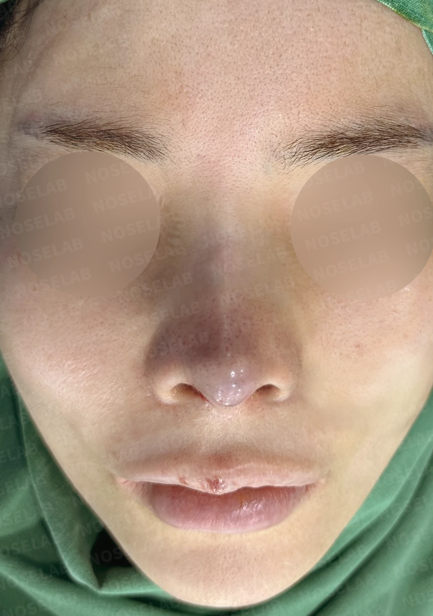
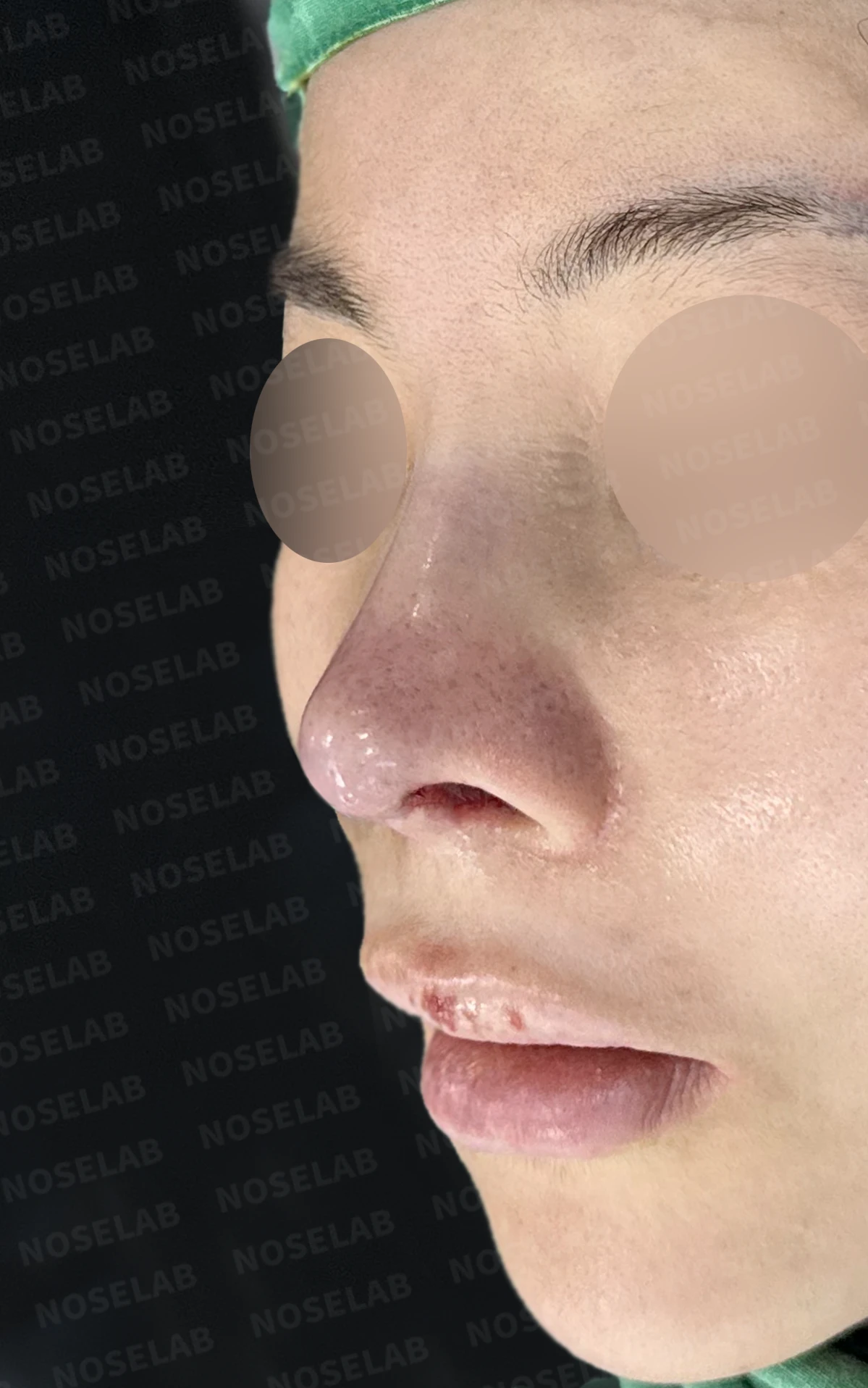
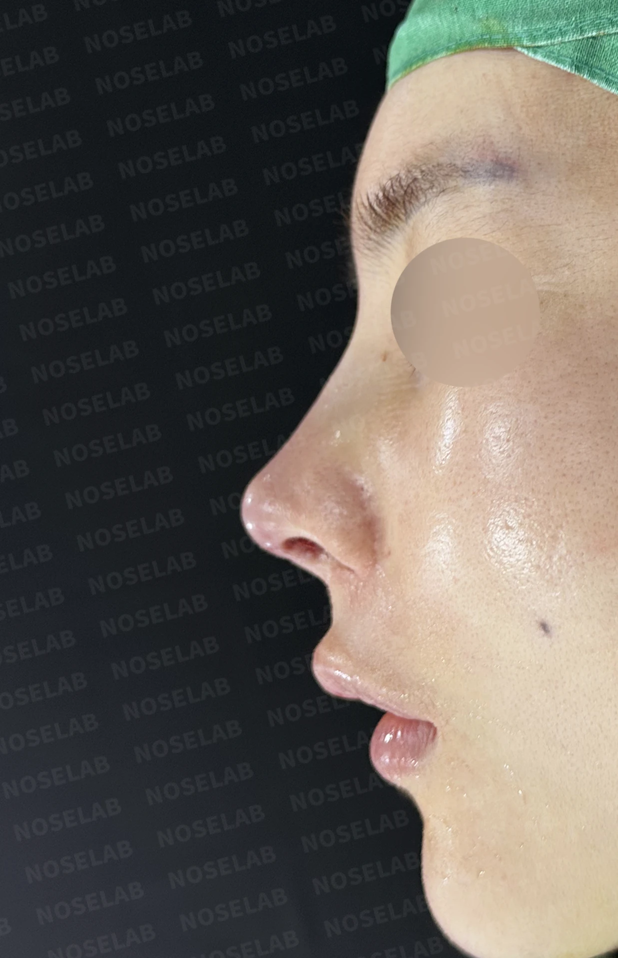
Front, oblique, and profile views on day 0 immediately after surgery. Photos taken immediately after surgery show swelling and may differ from the final result.
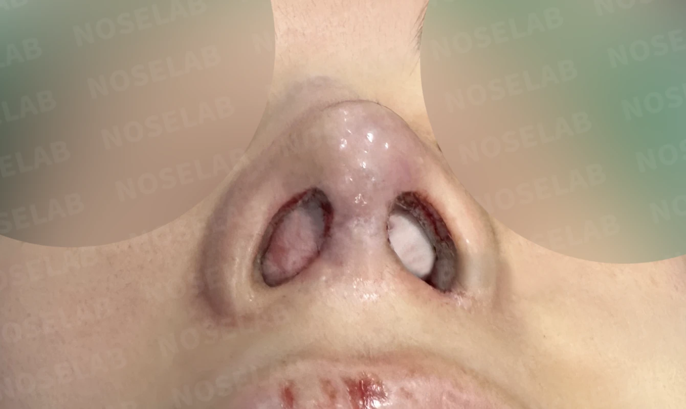
Nostril view on day 0 immediately after surgery. Photos taken immediately after surgery show swelling and may differ from the final result.
This was a complex case where the internal structures of the nose were substantially compromised after three prior surgeries. The nasal obstruction that troubled the patient most was not caused by a single issue, but by multiple contributing factors.
The internal nasal valve had narrowed from the septal collapse, internal nasal space had decreased due to overly aggressive lateral osteotomy, and complications had arisen from excessive inferior turbinate resection during prior rhinitis surgery—all coexisting simultaneously.
To address these combined issues comprehensively, we performed septal reconstruction, internal nasal valve reconstruction, right alar cartilage reconstruction, and inferior turbinate infracture with adhesiolysis. Autologous rib cartilage was chosen for every reconstruction step because it offers the most reliable stability when internal tissues have already been compromised by multiple surgeries.
I hope this case is helpful to those with similar concerns. This has been Dr. Peter C. Kang, Medical Director. Thank you.
#nasalobstructionrelief #septalreconstruction #osteotomyreversal #emptynosesyndrome #nasalvalvereconstruction #revisionrhinoplasty #autologousribcartilage #inferiorturbinatesurgery #alarcartilagereconstruction #nasolabialanglecorrection #closedrhinoplasty #closedrevisionrhinoplasty

Limitations and risks
When autologous rib cartilage is used, scarring and pain may remain at the donor site on the chest, and pneumothorax (pleural injury) can occur rarely. Over time, the grafted cartilage may warp or undergo partial resorption.
When the septum is corrected at the same time, nasal obstruction may persist or recur, and in rare cases, septal perforation or changes in smell may occur.
Common potential risks include infection, bleeding, asymmetry, scarring, skin necrosis, and insufficient improvement; results vary depending on individual skin condition and previous surgical history.