Hello, I am Dr. Peter C. Kang.
Today, I would like to share the case of a patient who resolved complex issues stemming from multiple surgeries and inflammation over 14 years through autologous rib cartilage reconstruction.
📍 Surgical history
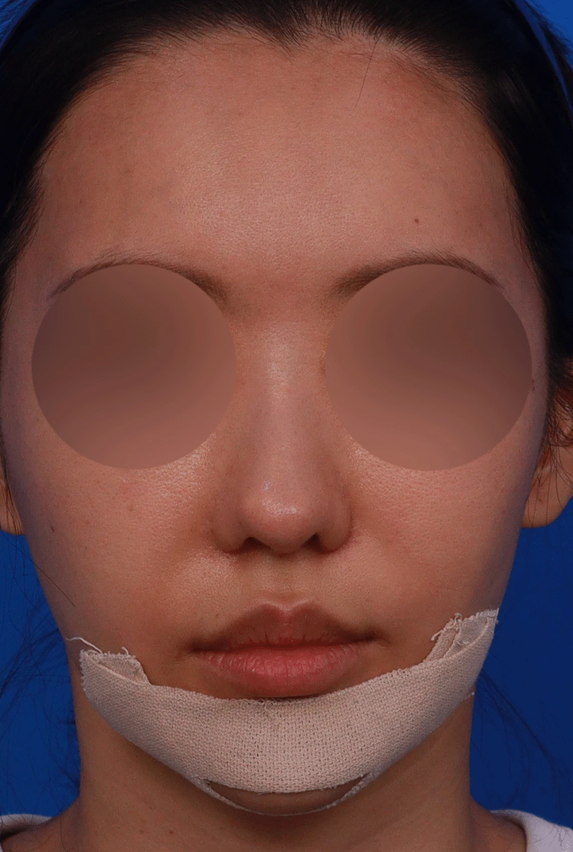
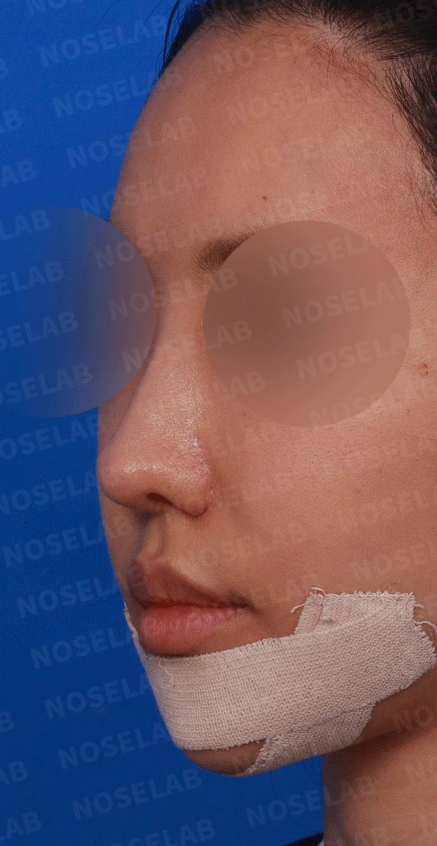
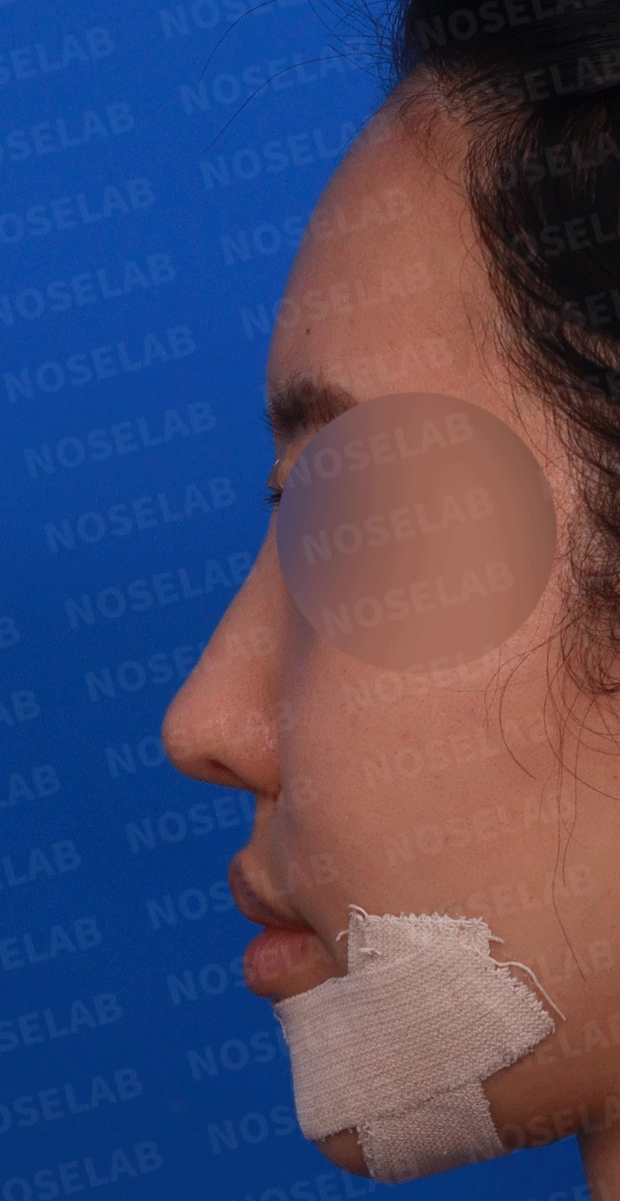
These are design consultation photos: front, oblique, and left profile views.
14 years ago
Gore-Tex + ear cartilage
13 years ago
Silicone implant + alar reduction + osteotomy
12 years ago
Removal at the glabella due to inflammation
10 years ago
After implant removal, filler treatment along the entire nasal bridge and nasal tip
9 years ago
After filler removal, silicone implant + ear cartilage + alar reduction
Recently
Silicone implant + ear cartilage
Dermis procedure (buttock dermis) on the nasal tip only
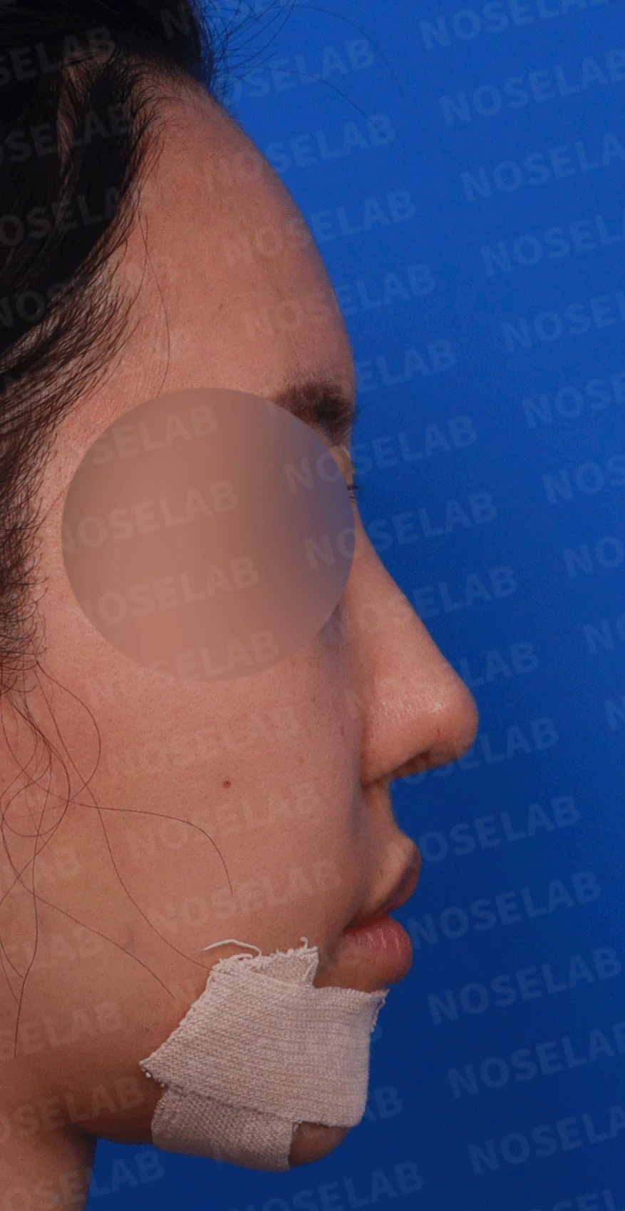
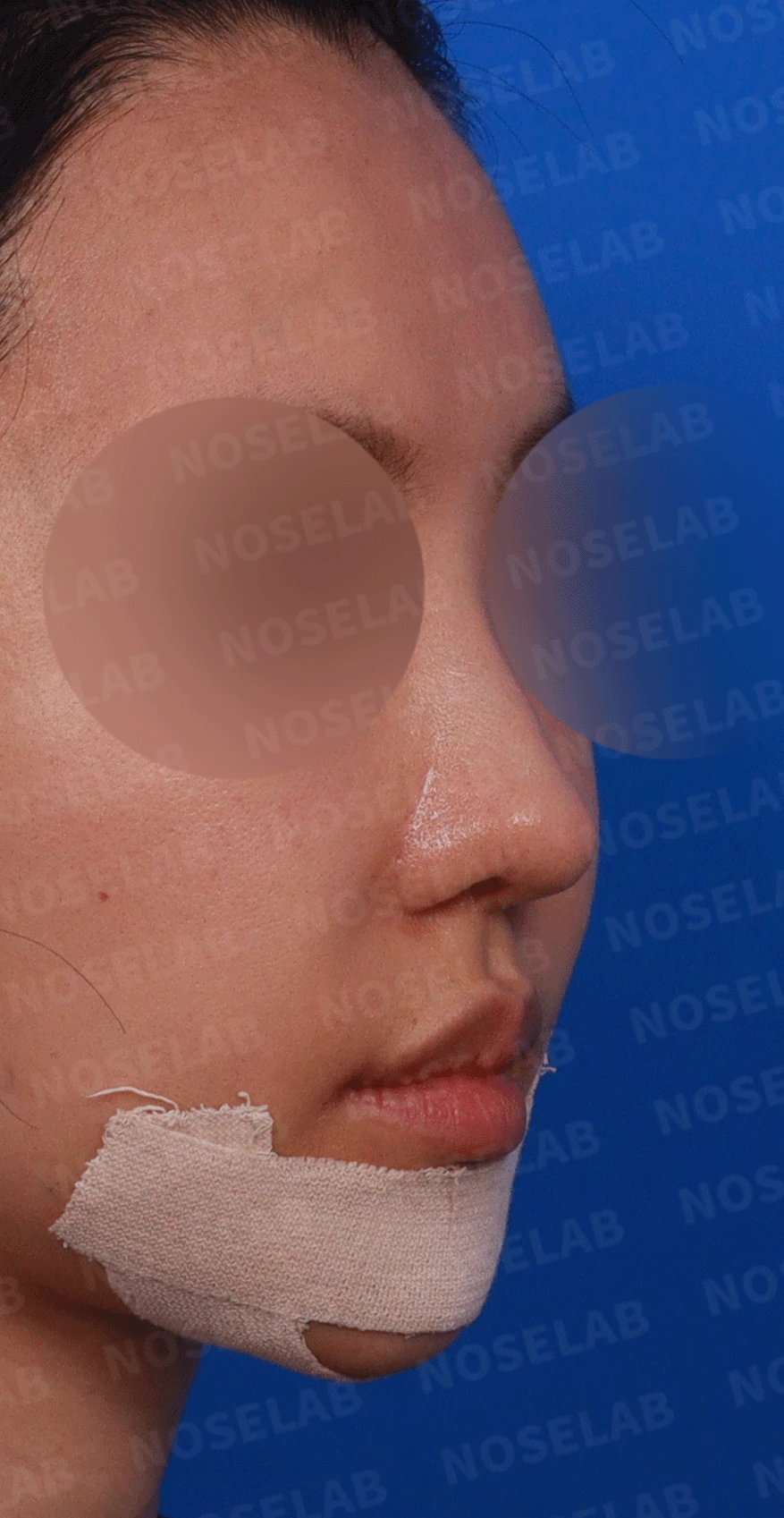
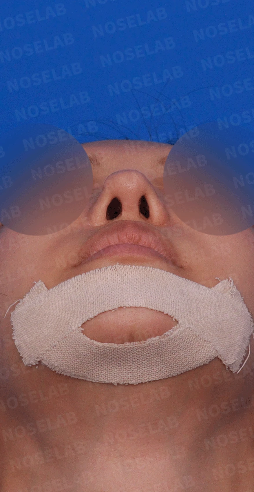
Design consultation right profile, oblique view, and nostril photos.
🔎 Initial consultation findings
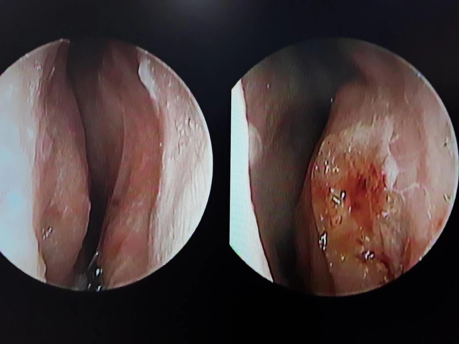
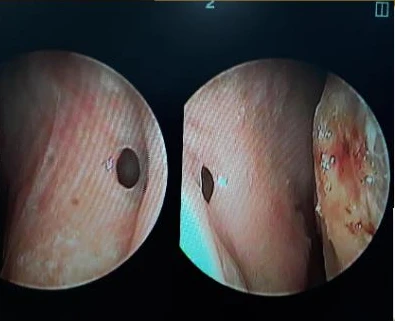
Nasal endoscopy photos
Functional issues
7x9 mm septal perforation
Severe damage to the septal cartilage
Chronic rhinitis
Impaired respiratory function
Aesthetic concerns
Cartilage show-through due to thinned nasal tip skin
Protruding mouth findings
Nostril asymmetry
Special notes
7 repeated surgeries over 14 years
Tissue damage due to inflammation
Nasal tip skin thinned as packed cartilage pieces became tangled together
Limitations in correction due to previous alar reduction
✨ Patient's requests
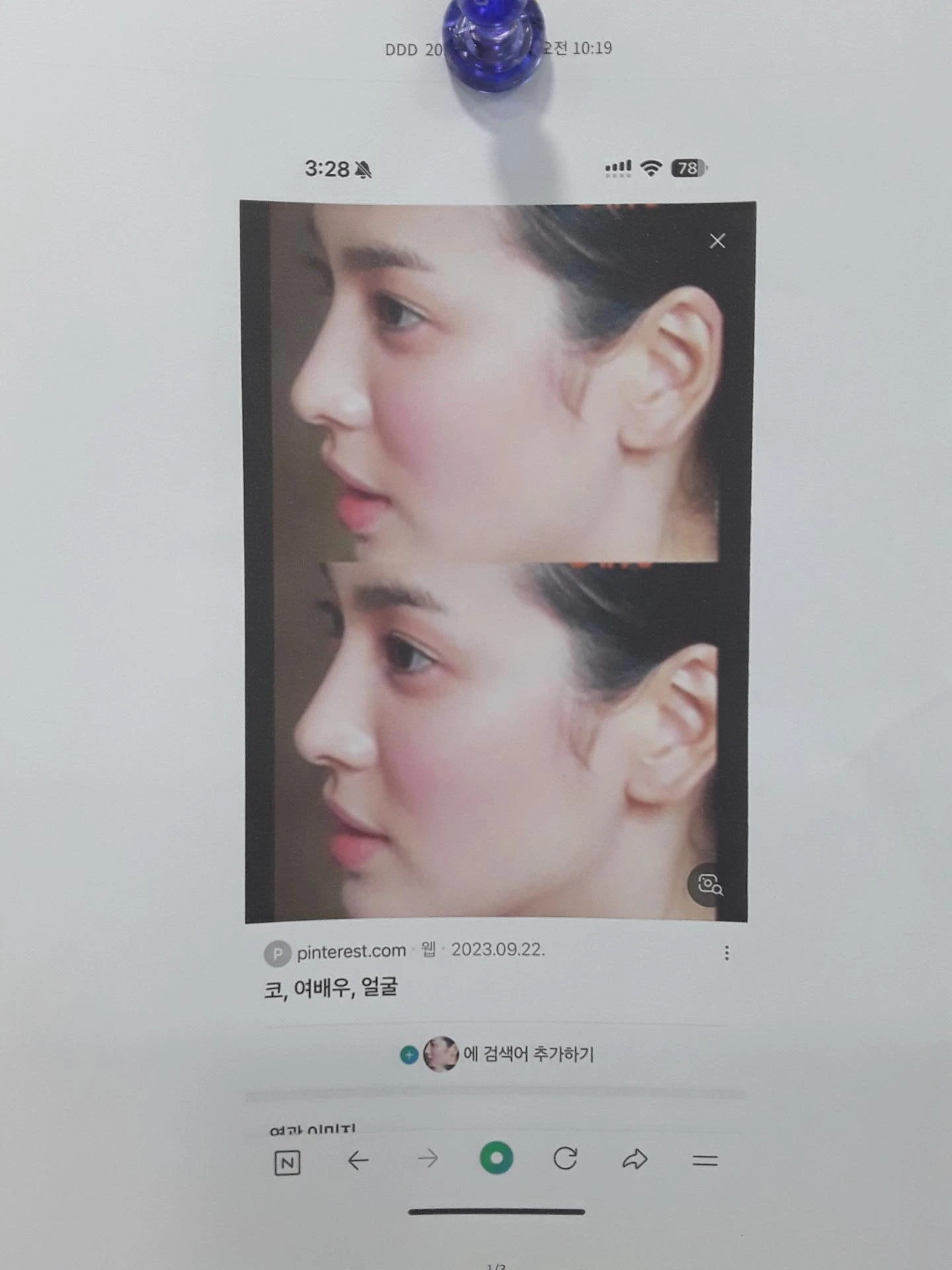

The patient's desired look
Improvement of awkwardness caused by a protruding mouth
Natural nasal shape
Resolution of functional problems
Stable results
Surgical procedure details
Septal reconstruction
Septal reconstruction using autologous rib cartilage for severely damaged septal cartilage
Correction of the 7x9 mm septal perforation
Structural reconstruction
Full reconstruction using autologous rib cartilage
Trimming and restructuring tangled existing cartilage
Dermal graft to restore nasal tip skin thickness
Improvement in shape
Improvement of protruding mouth through nasolabial angle correction
Restoration of a natural philtrum angle
Creating a natural overall nasal contour
Functional improvement
Restoring respiratory function with septoplasty
Improving rhinitis with inferior turbinate surgery
Managing limiting factors
Explanation of limitations in correcting nostril asymmetry due to previous alar reduction
Maximizing improvement within the feasible range
✅ Surgical results
Functional improvement
Complete closure of the septal perforation to restore respiratory function
Marked relief of rhinitis symptoms
Stable internal nasal framework
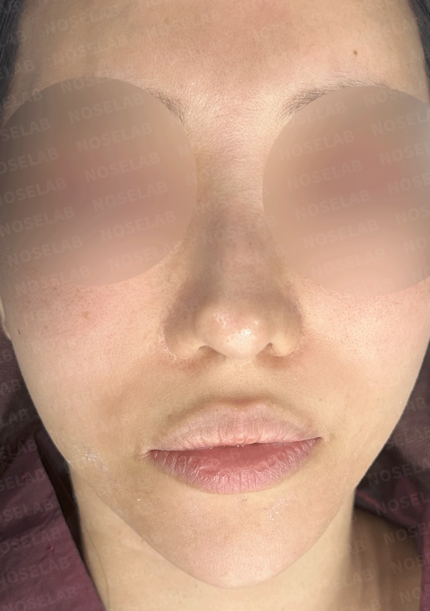
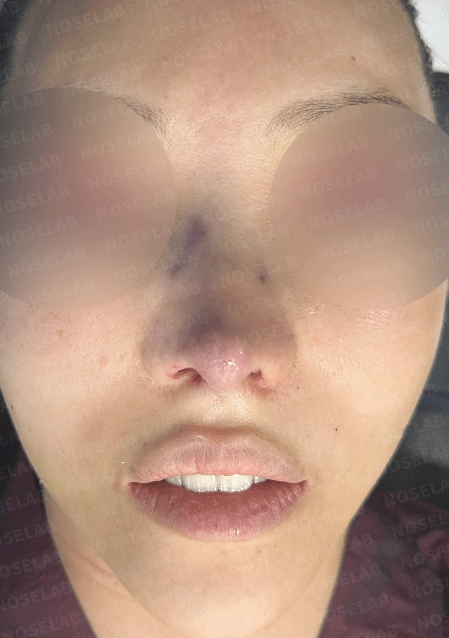
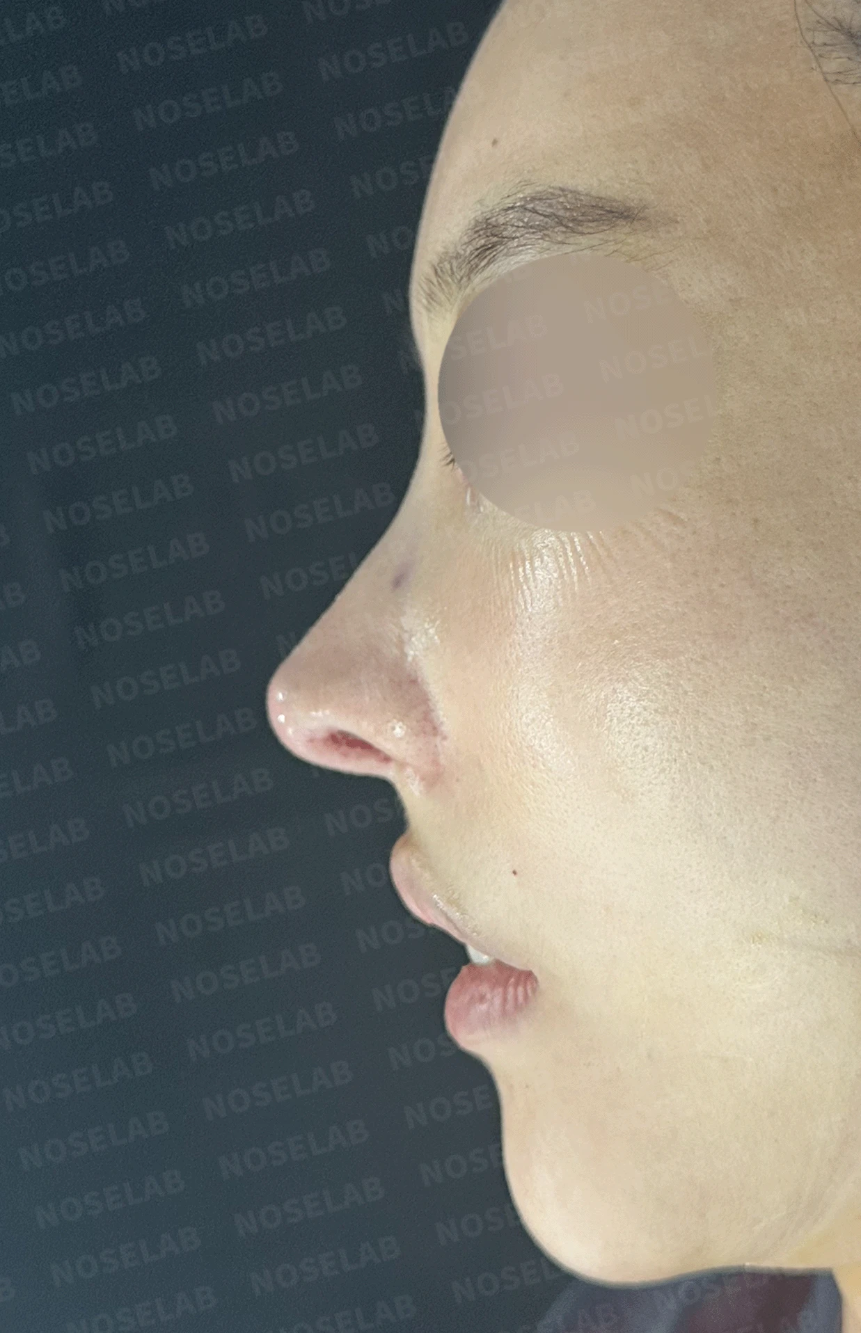
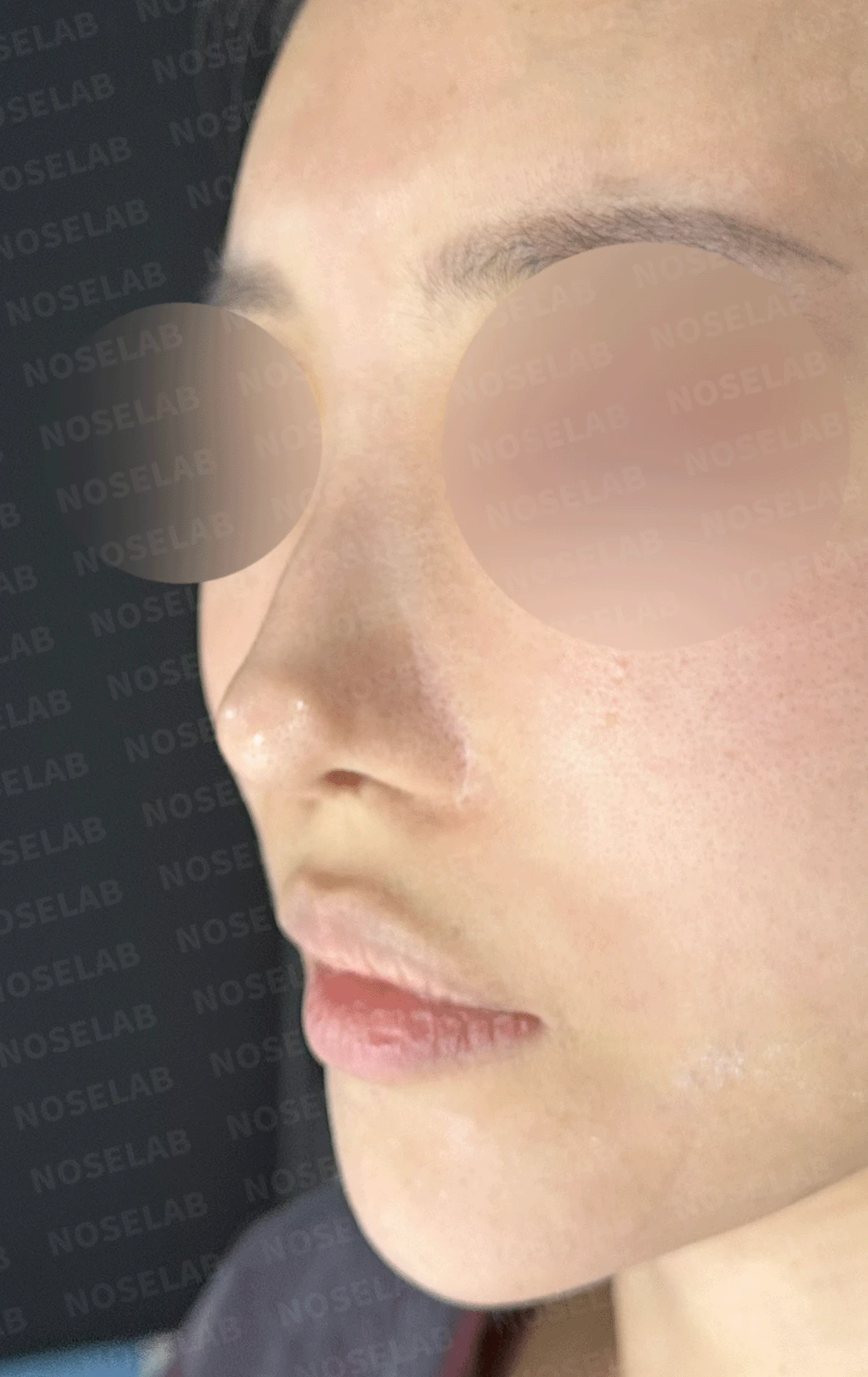
Before surgery / Day 0 immediately after surgery Photos taken immediately after surgery show swelling and may differ from the final result.
Front view
Natural overall nasal contour
Balanced facial proportions
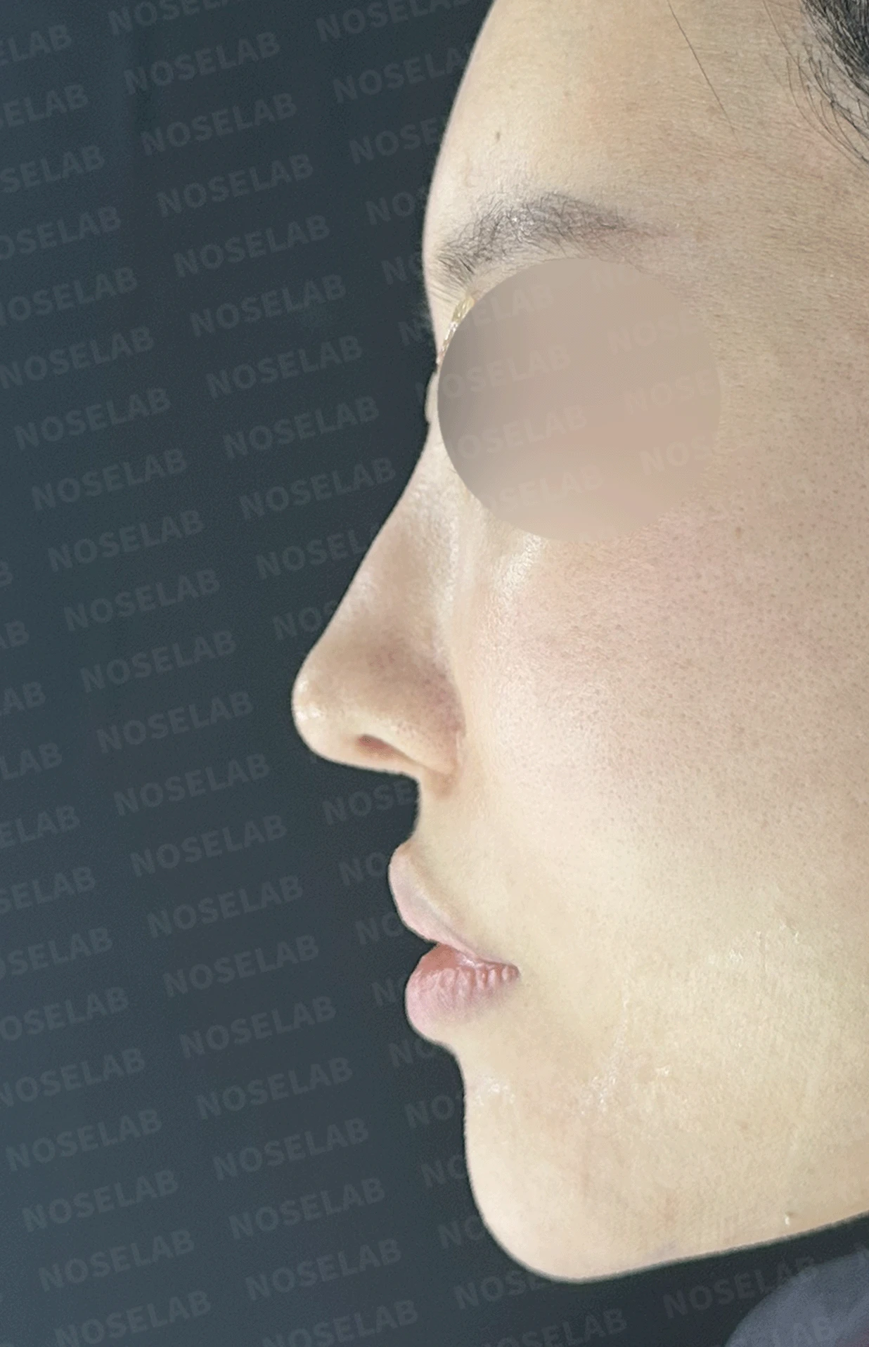
Before surgery / Day 0 immediately after surgery Photos taken immediately after surgery show swelling and may differ from the final result.
Profile view
Improved protruding mouth
Natural nasolabial angle
Natural transition between the philtrum and columella
Harmonious facial profile
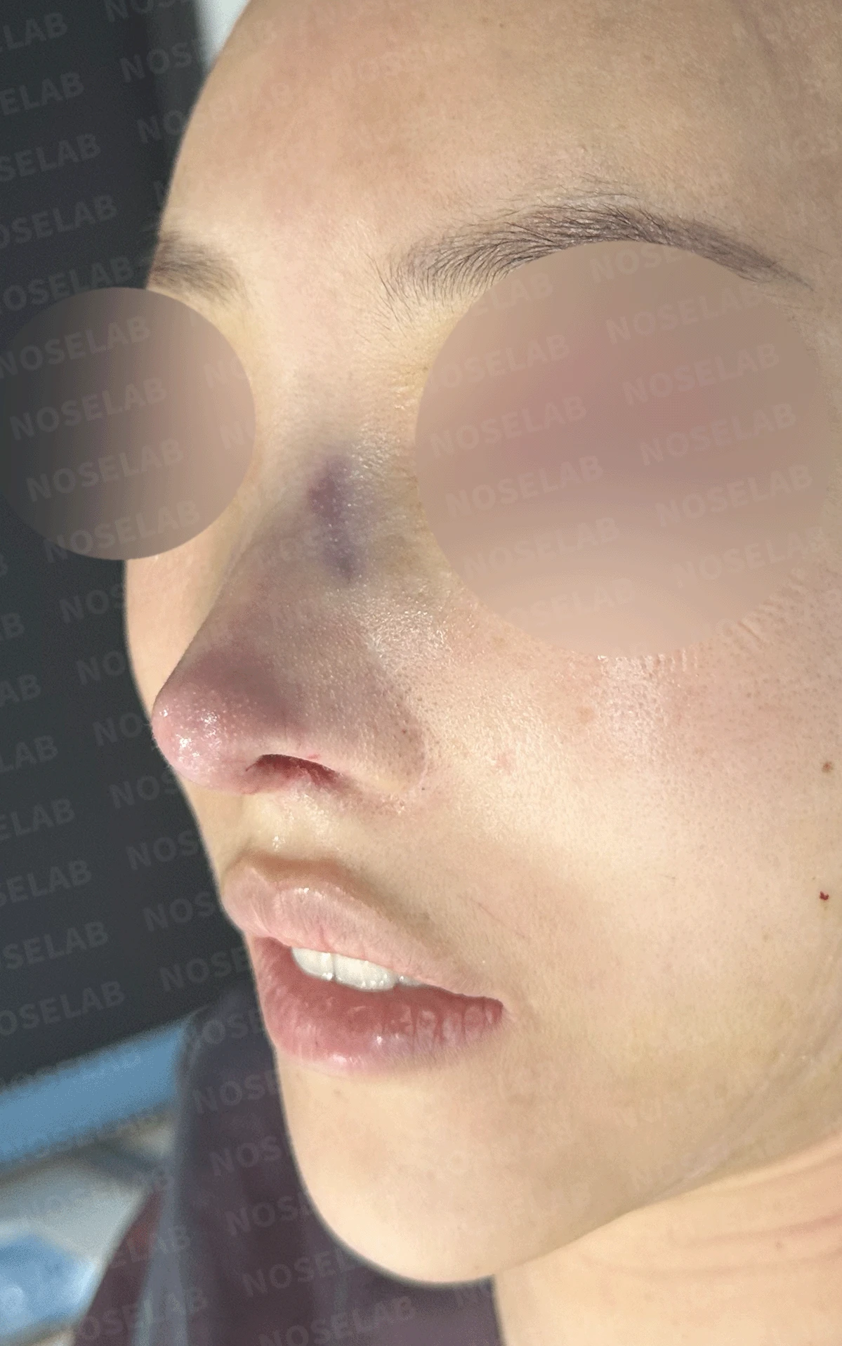
Before surgery / Day 0 immediately after surgery Photos taken immediately after surgery show swelling and may differ from the final result.
Nostrils
Improved nostril symmetry (within structural limits)
Nasal tip condition
Improvement of cartilage show-through
Restoration of adequate skin thickness
Natural nasal tip shape
Long-term stability
Long-term stability using autologous rib cartilage
Minimizing the risk of revision surgery
👨⚕️Specialist's assessment
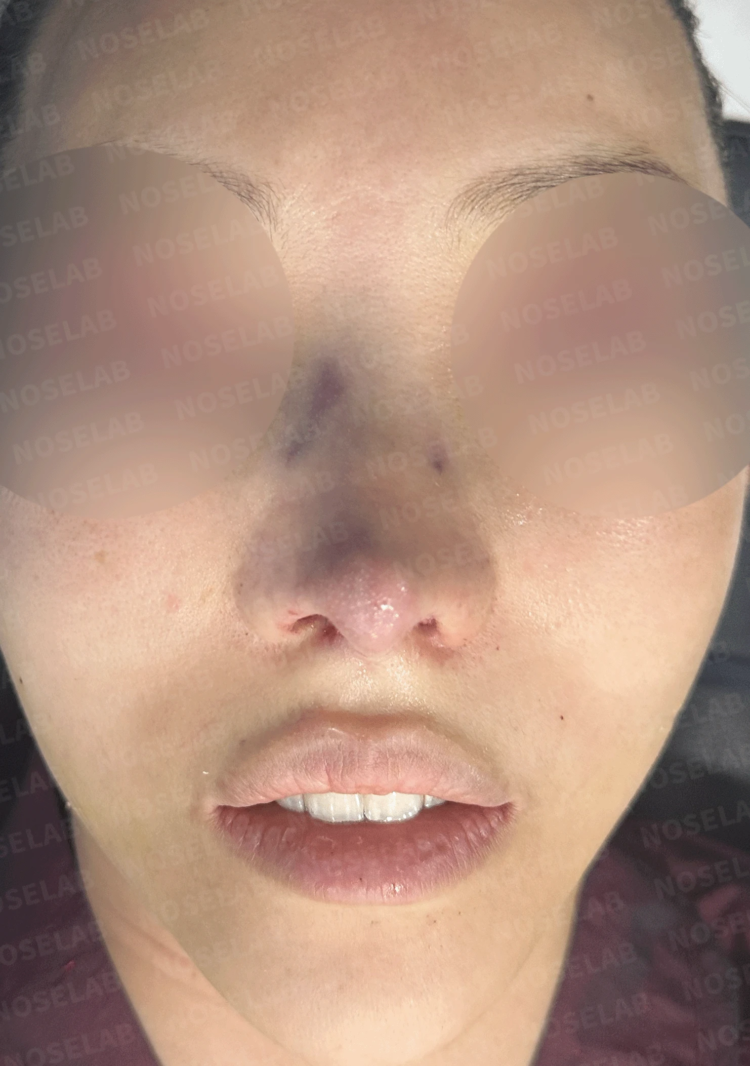
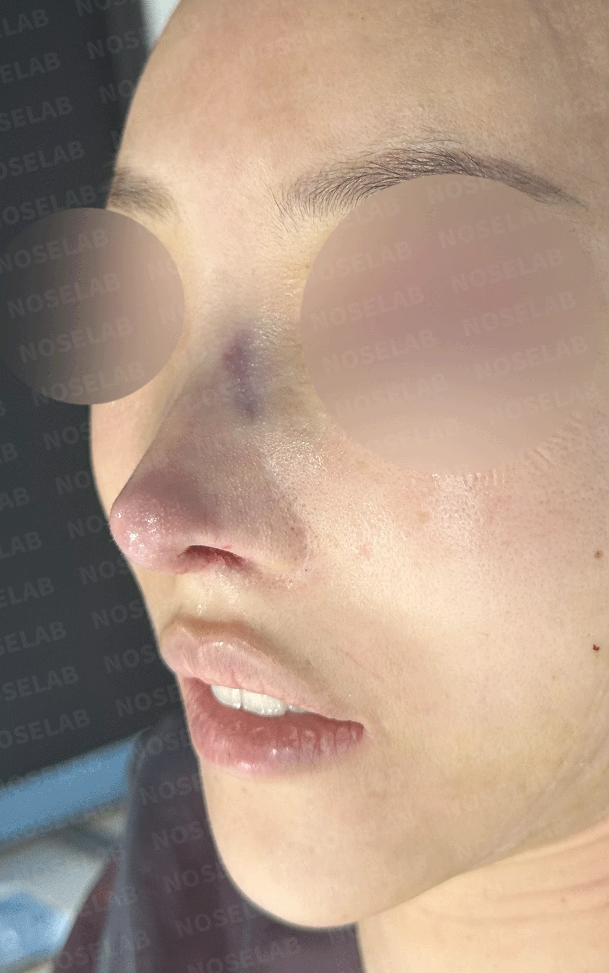
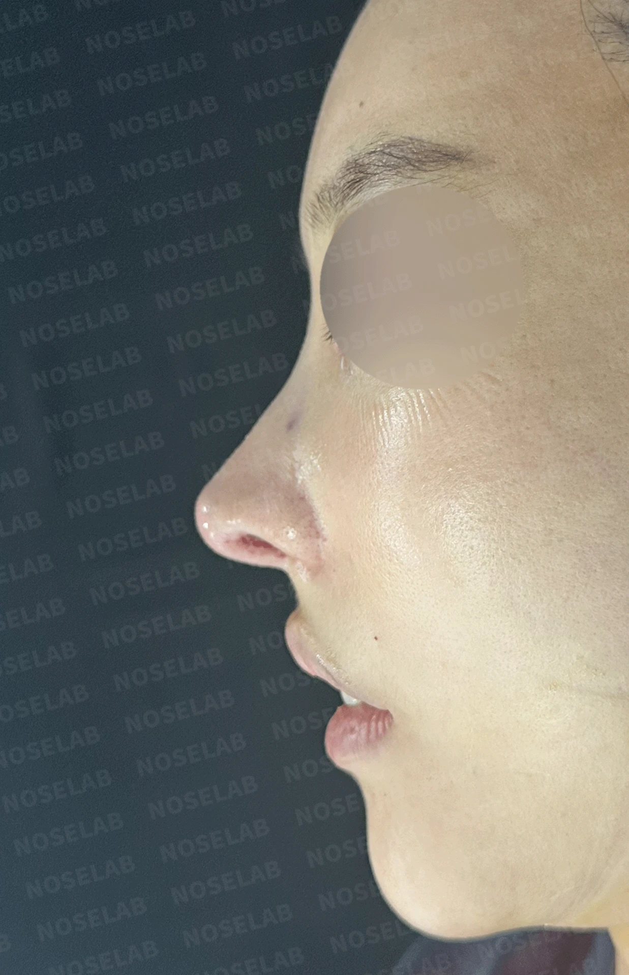
Front, oblique, and profile views on day 0 immediately after surgery. Photos taken immediately after surgery show swelling and may differ from the final result.
This case involved a patient whose nasal condition became highly complex due to 7 repeated surgeries and bouts of inflammation over 14 years. In particular, it was a challenging case compounded by a 7x9 mm septal perforation, severely damaged septal cartilage, and complex structural deformities in the nasal tip.
The highest priority was correcting the 7x9 mm septal perforation. A perforation of this size significantly impairs breathing, and because the existing septal cartilage was severely damaged, complete septal reconstruction using autologous rib cartilage was necessary. Fortunately, the septal perforation was closed entirely, restoring normal respiratory function.
At the nasal tip, cartilage pieces packed in during previous procedures had become tangled together, thinning the skin and causing visible cartilage show-through. This was not only an aesthetic concern, but it also posed a long-term risk of skin breakdown that needed to be addressed. The problematic existing cartilage was trimmed and reorganized, a new, stable framework was constructed using autologous rib cartilage, and a dermis graft was placed to restore skin thickness.
The awkward appearance from a protruding mouth, which was a major concern for the patient, was improved by correcting the nasolabial angle. Correcting the nasolabial angle created a natural connection between the philtrum and the columella, enhancing overall facial harmony.
Regarding nostril asymmetry, I explained thoroughly before surgery that previous alar reduction procedures placed limitations on achieving complete symmetry. Nevertheless, we achieved significant improvement within the feasible range to deliver a natural outcome.
Although the patient had suffered through 7 surgeries over 14 years, systematic reconstruction with autologous rib cartilage resolved both the functional and cosmetic concerns. In particular, the septal perforation was closed completely to restore normal breathing function, and the natural contour from the philtrum to the columella was achieved, leaving the patient very satisfied.
This case meaningfully demonstrates that even the most complex and demanding revision rhinoplasties can achieve successful results through accurate diagnosis, systematic planning, and the appropriate selection of graft materials.
This was Dr. Peter C. Kang. Thank you.
Thank you.
#closedrhinoplasty #revisionrhinoplasty #autologousribcartilage #septalperforationrepair #protrudingmouthcorrection #customrhinoplasty

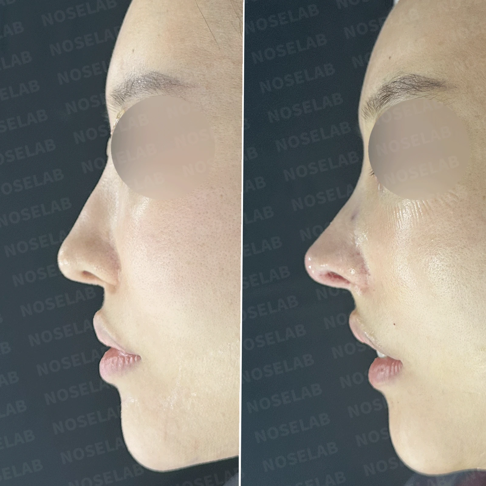
Limitations and risks
When autologous rib cartilage is used, scarring and pain may remain at the donor site on the chest, and pneumothorax (pleural injury) can occur rarely. Over time, the grafted cartilage may warp or undergo partial resorption.
When revising after implant removal, contracture may progress again after removal, the nasal height and length may decrease, and staged surgery may be necessary depending on the skin condition.
When foreign materials such as injected fillers are removed and reconstructed, contracture may recur, nasal height and length may decrease, and staged surgery may be necessary depending on the skin condition.
When the septum is corrected at the same time, nasal obstruction may persist or recur, and in rare cases, septal perforation or changes in smell may occur.
Common potential risks include infection, bleeding, asymmetry, scarring, skin necrosis, and insufficient improvement; results vary depending on individual skin condition and previous surgical history.