Hello, I am Dr. Peter C. Kang.
Today, I would like to share a revision rhinoplasty case of a patient whose internal nasal structures were severely damaged from multiple previous nose surgeries. In cases like this, abnormal findings around the implant on the preoperative CT scan required an even more meticulous approach.
🔍 Surgical history
The patient had undergone four prior nose surgeries over the course of about 10 years.
About 10 years ago: First rhinoplasty using ear cartilage and a silicone implant
About 7 years ago: Revision rhinoplasty using ear cartilage and a silicone implant
About 5 years ago: Revision rhinoplasty using donor rib cartilage and a silicone implant
About 1.5 years ago: autologous rib cartilage, silicone implant, and alar base elevation
Due to multiple surgeries, damage to the normal internal cartilage framework was suspected.
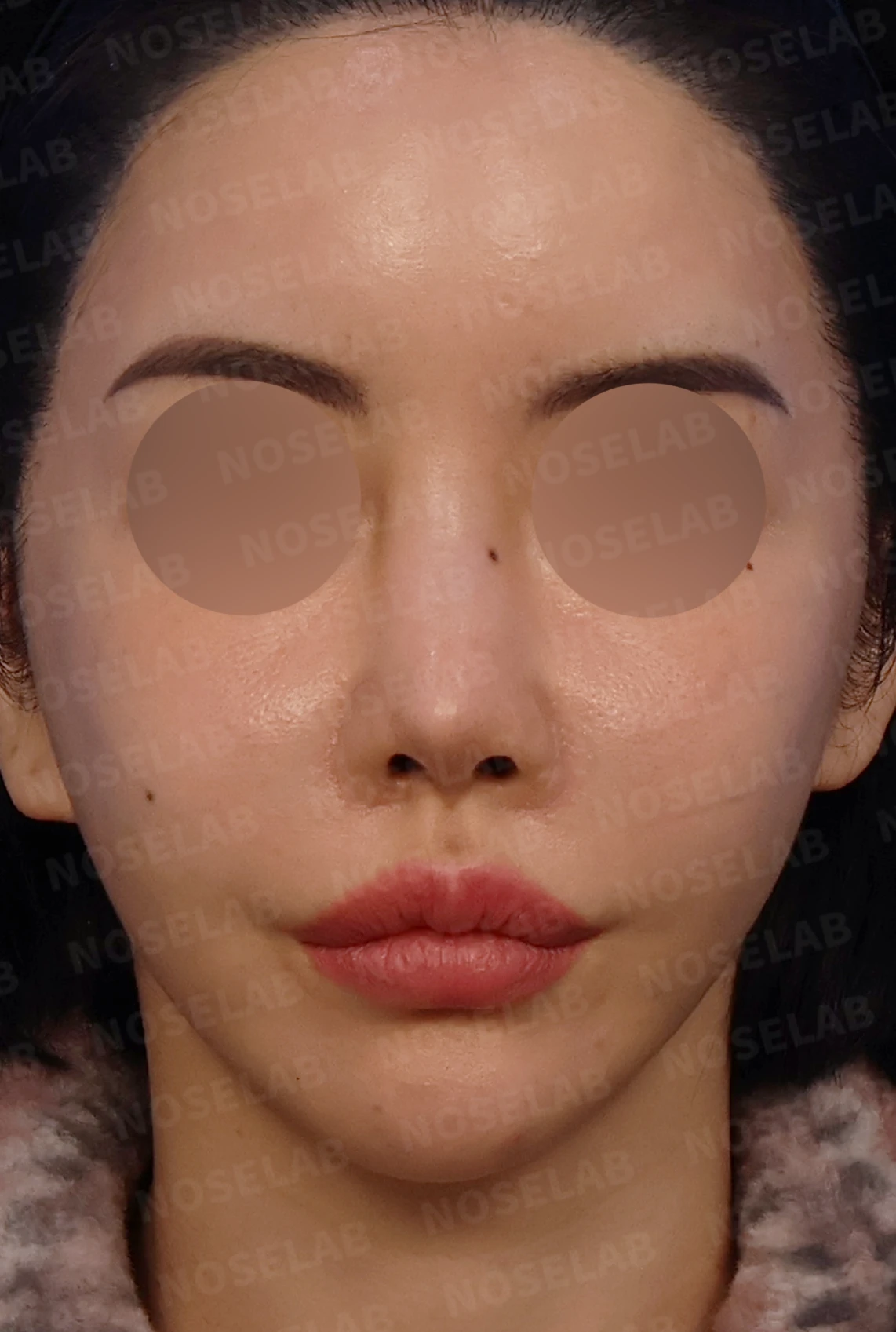
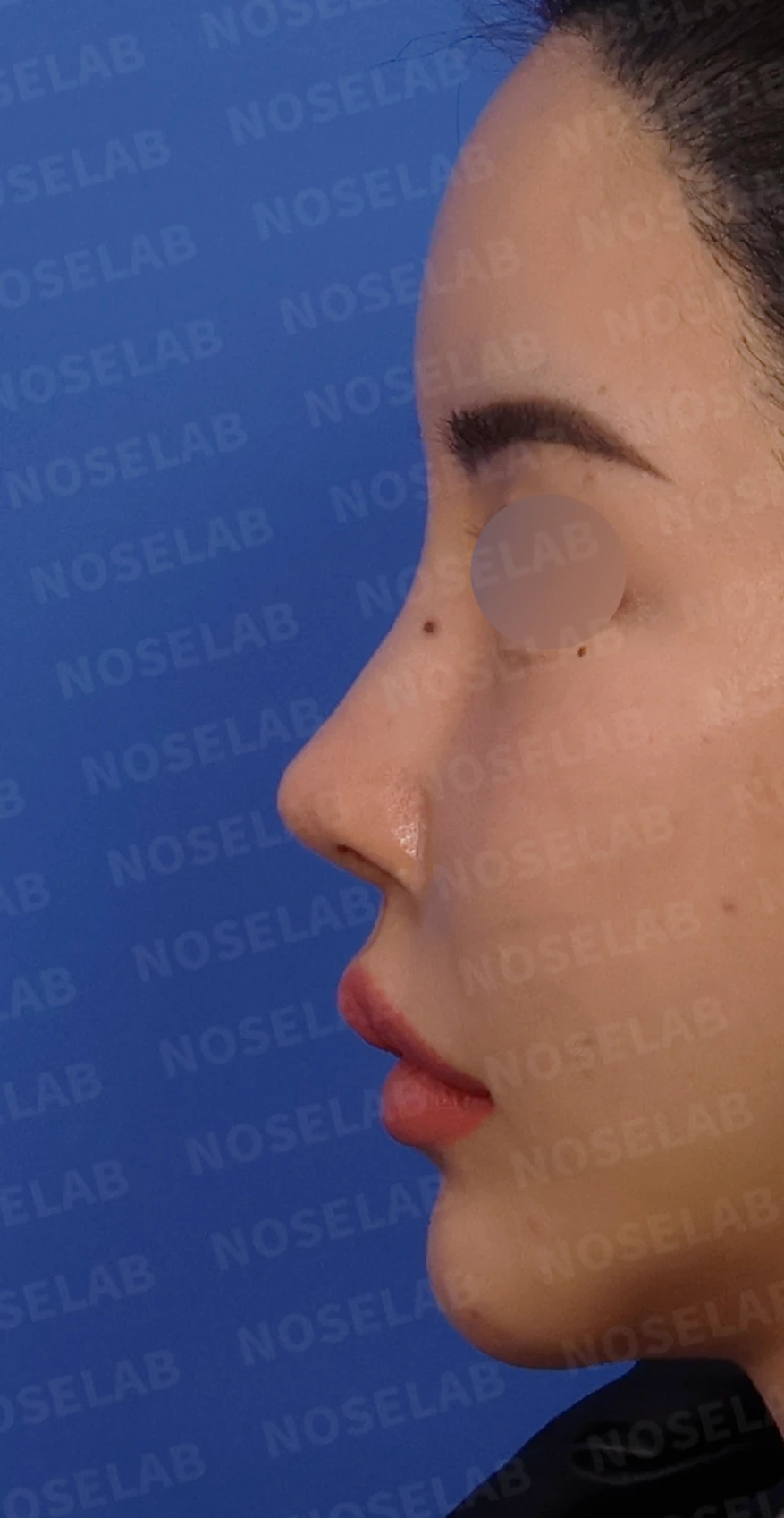
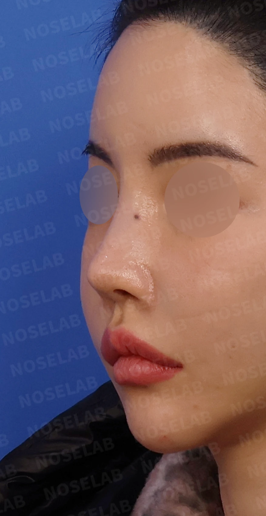
Design consultation photos: front, left profile, and oblique views
💉 Patient requests
When the patient visited our clinic for consultation, the requests were as follows:
Correction of deviated nose
Desired an increase in overall nasal height
Desired lowering the radix (starting point)
Improvement of nostril asymmetry
Desired a nasal tip that is not sharp or pointy
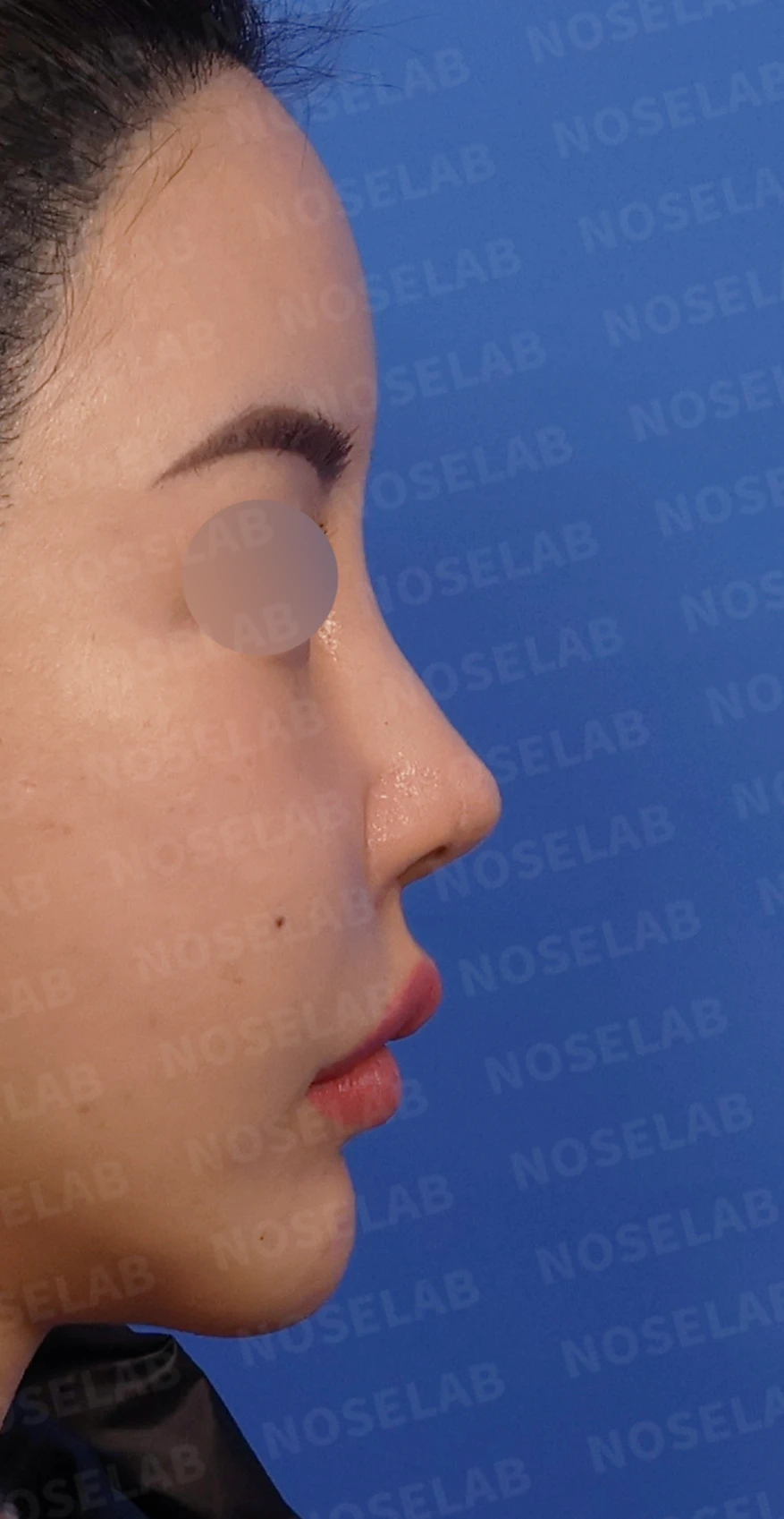
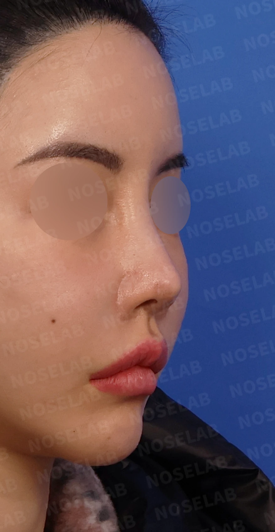
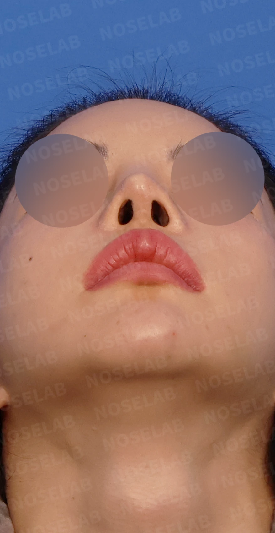
Design consultation photos: right profile, oblique, and nostril views
🔧 Surgical plan and procedure
On the preoperative CT scan taken at our clinic, fluid retention suspicious for inflammation was observed around the implant. It was necessary to determine intraoperatively whether this was simple foreign material or inflammatory fluid.
Surgical findings and procedure
During the actual surgery, serous fluid was identified, and bacterial culture testing was ordered. Fortunately, it was not purulent discharge, so it was diagnosed as a chronic foreign body reaction. After antibiotic irrigation and complete capsule removal, reconstruction proceeded using autologous rib cartilage.
1. Foreign material removal and antibiotic irrigation
Complete removal of foreign material surrounding the implant
Capsule removal caused by chronic foreign body reaction
Antibiotic irrigation to prevent infection
2. Septal reconstruction using autologous rib cartilage
Confirmed that almost no septal cartilage remained, as anticipated
Complete reconstruction of the septum using autologous rib cartilage
Secured a stable structural foundation for nasal tip support
3. Lower lateral cartilage reconstruction using autologous rib cartilage
Almost no lower lateral cartilage remained due to multiple surgeries
Complete reconstruction of the lower lateral cartilages using autologous rib cartilage
Established a structural foundation to shape the nasal tip
4. Nasal bridge reconstruction
Identified erosion of part of the nasal bone after removing the foreign material
The height of the nasal bridge had become significantly diminished
Secured bridge height within a safe margin, considering the compromised condition of the tissues
Reconstructed into a safe and aesthetically pleasing structure
5. Correction of nostril asymmetry via skin graft
The right nostril had become narrowed and smaller
Performed a skin graft inside the nostril
Slightly enlarged the nostril size to correct asymmetry as much as possible
🌟 Surgical results
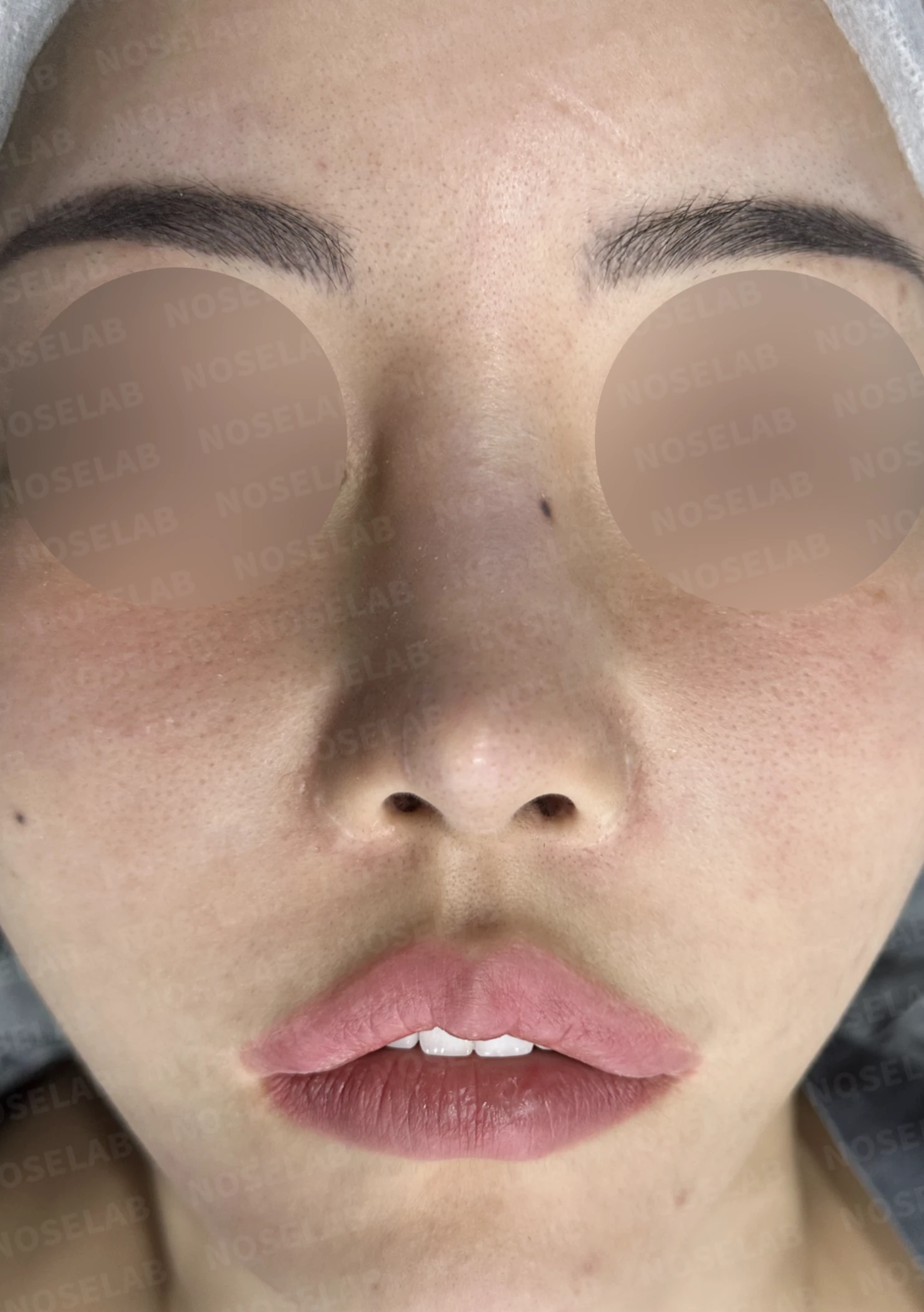
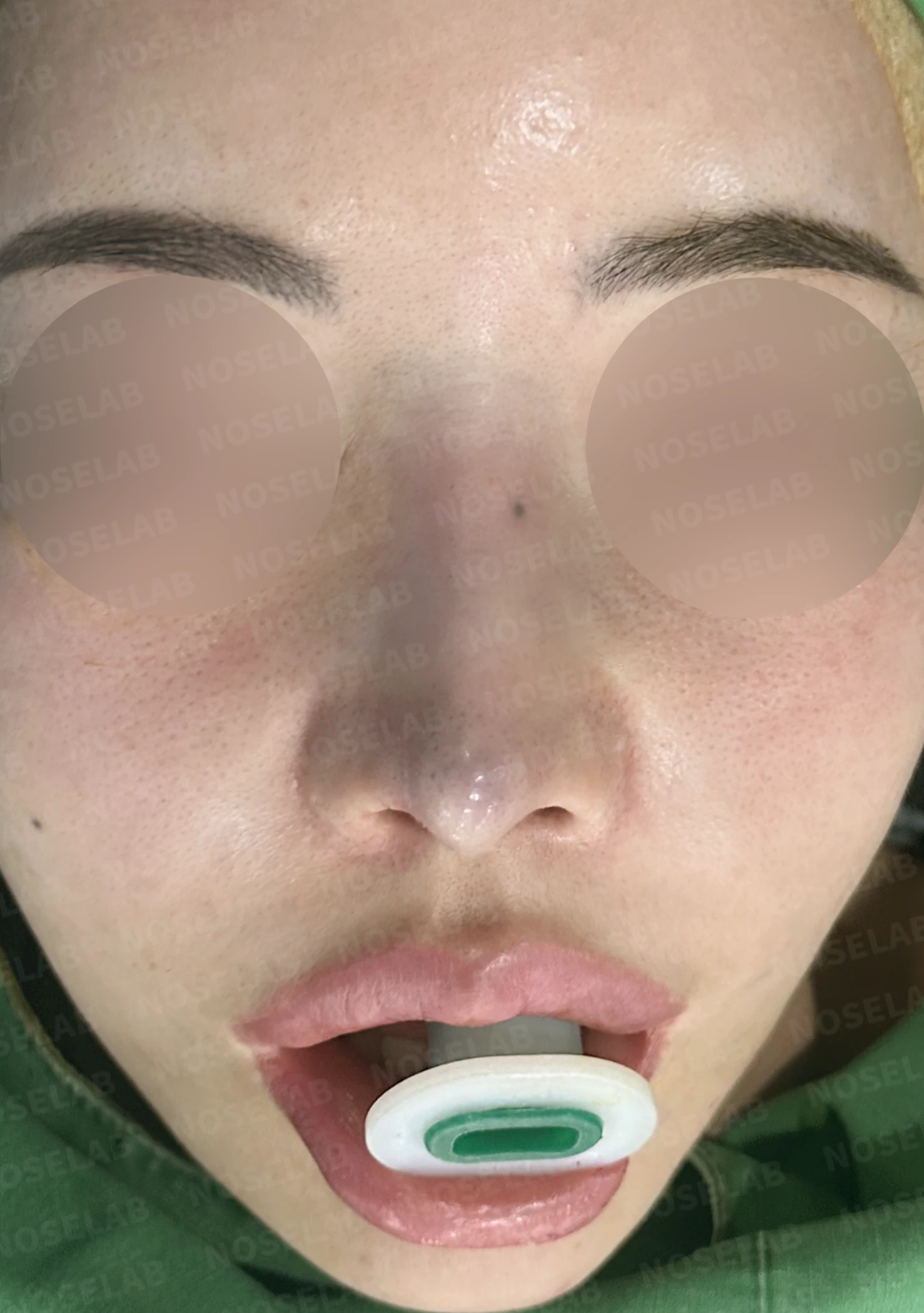
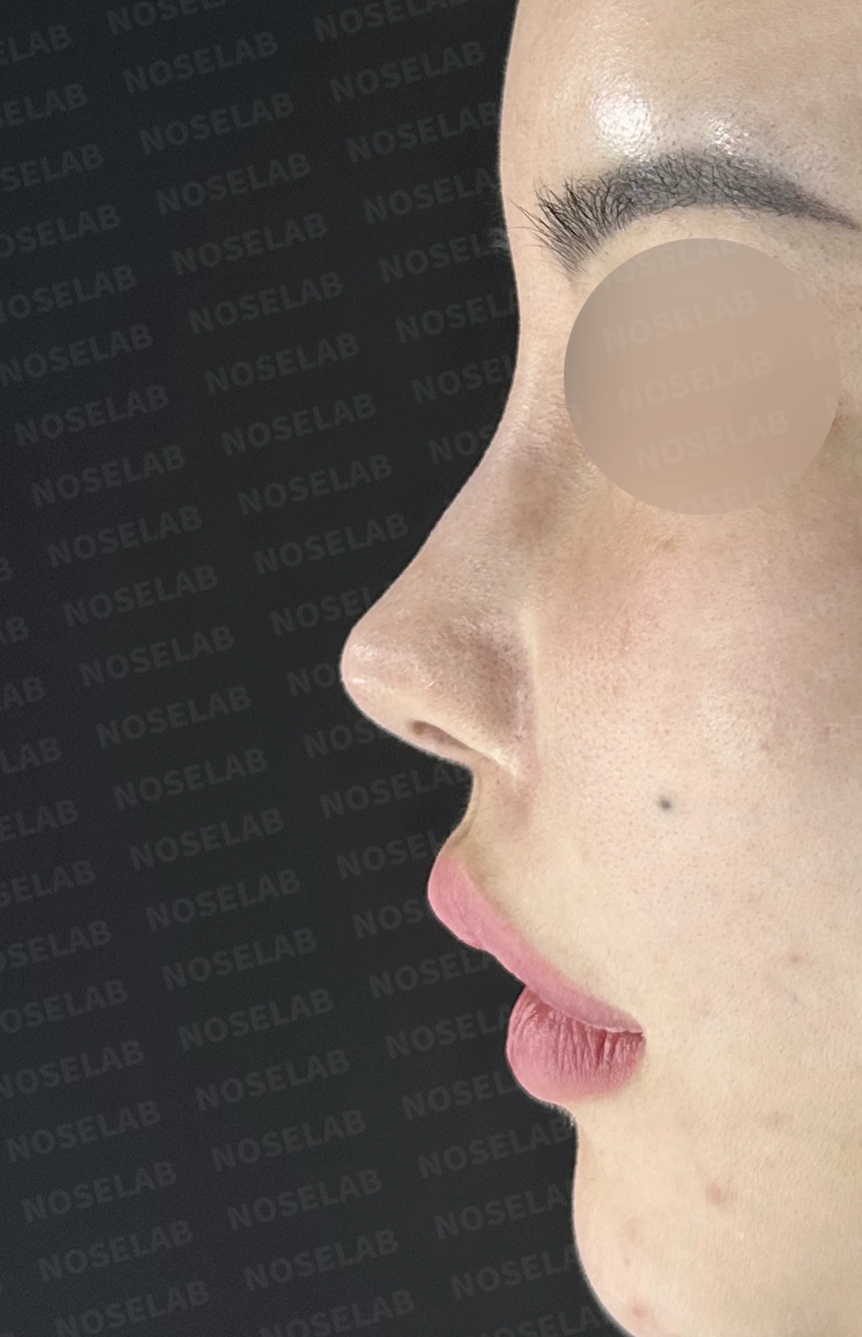
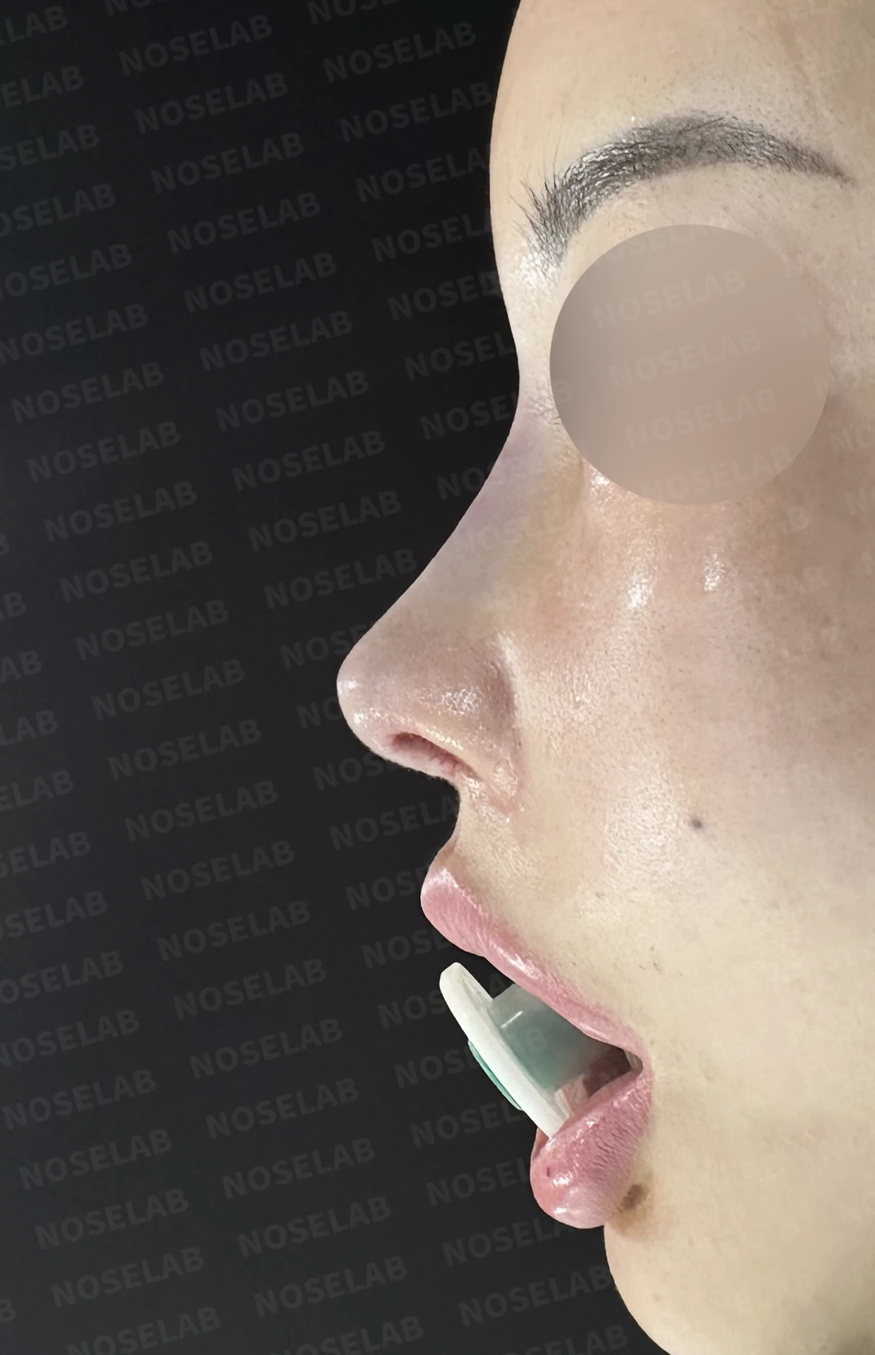
Before surgery / Day 0 immediately after surgery Photos taken immediately after surgery show swelling and may differ from the final result.
Front view changes:
The crooked nose was straightened
Improved nostril asymmetry
Overall stable and natural contour
Before surgery / Day 0 immediately after surgery Photos taken immediately after surgery show swelling and may differ from the final result.
Profile view changes:
Nasal bridge height secured within a safe margin
Formation of a natural bridge line
A soft tip shape that is not overly sharp
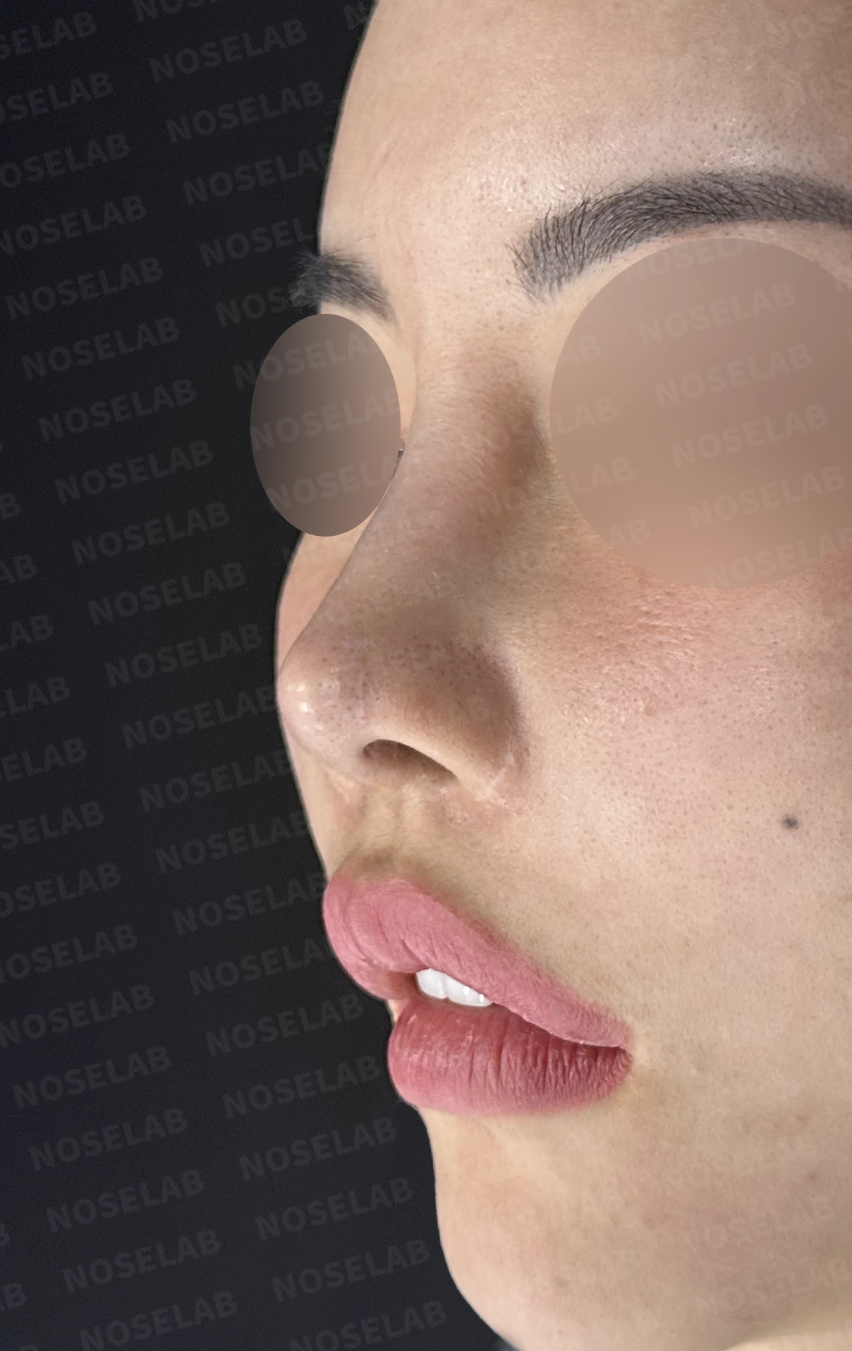
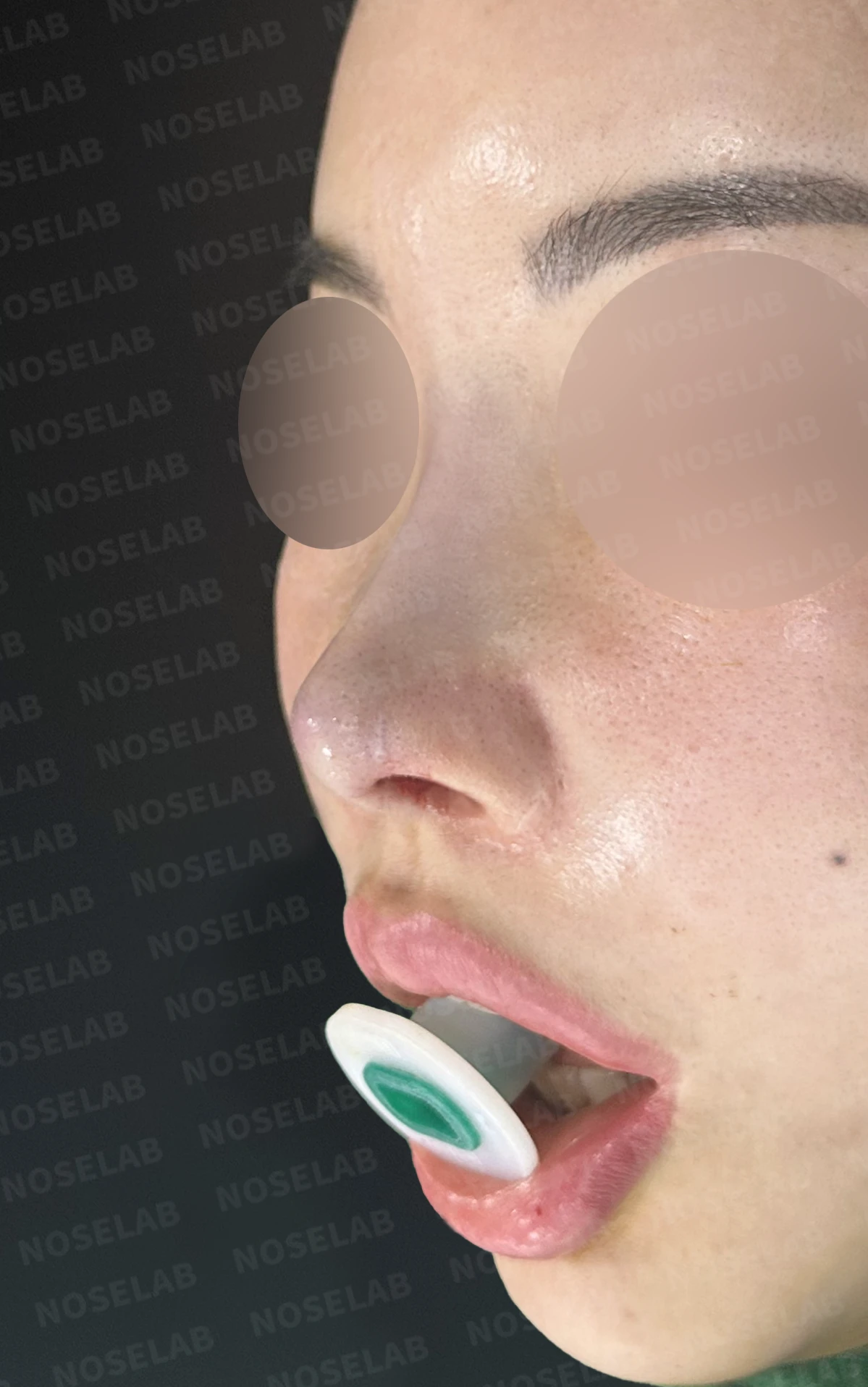
Before surgery / Day 0 immediately after surgery Photos taken immediately after surgery show swelling and may differ from the final result.
Structural changes:
Secured a stable framework through complete reconstruction of the septum and lower lateral cartilages
Maintains long-term structural stability
Established a healthy tissue foundation
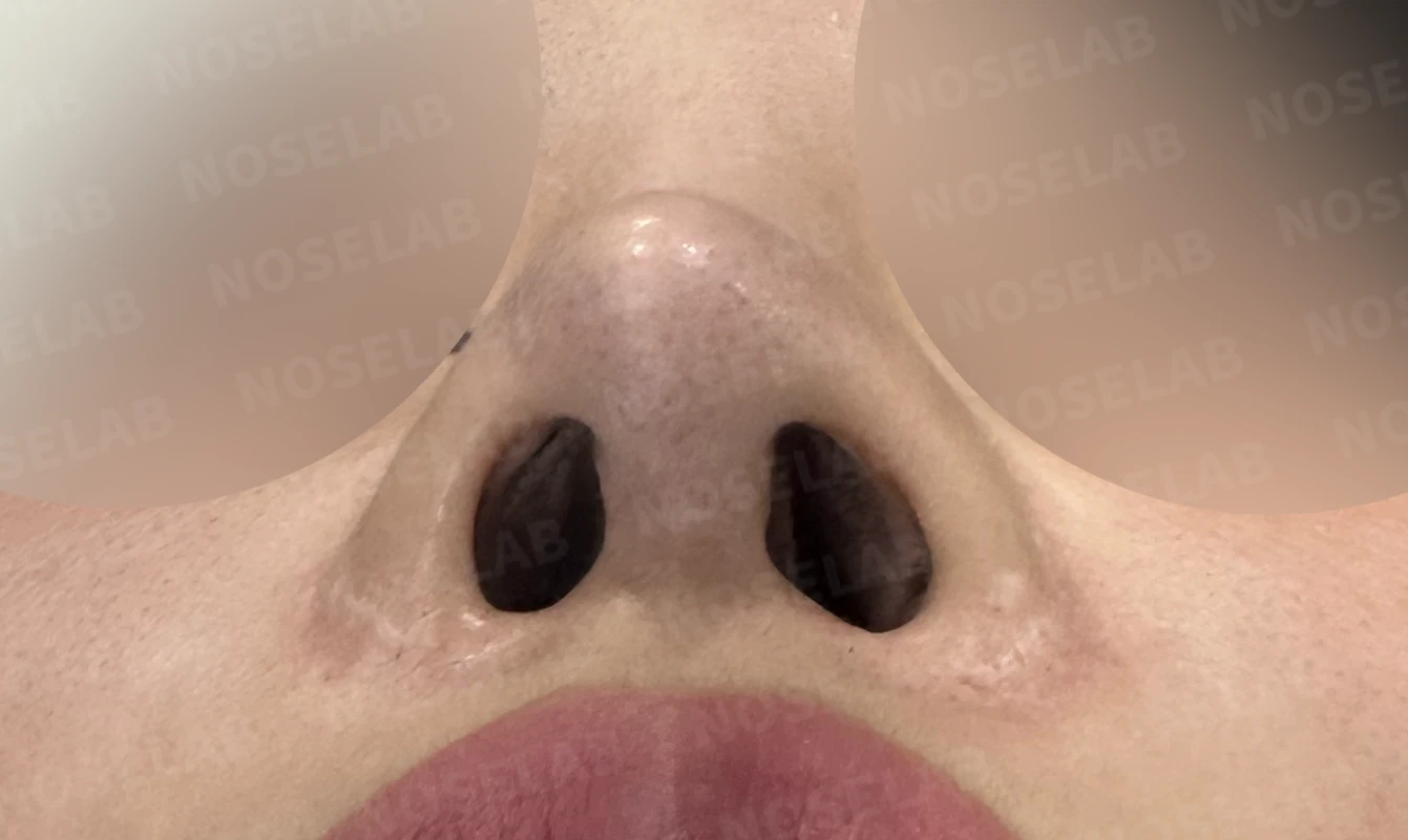
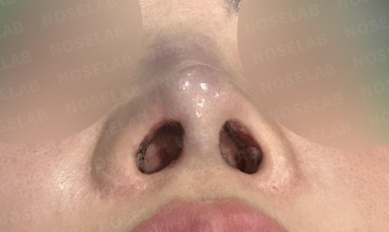
Before surgery / Day 0 immediately after surgery Photos taken immediately after surgery show swelling and may differ from the final result.
💡 Specialist opinion
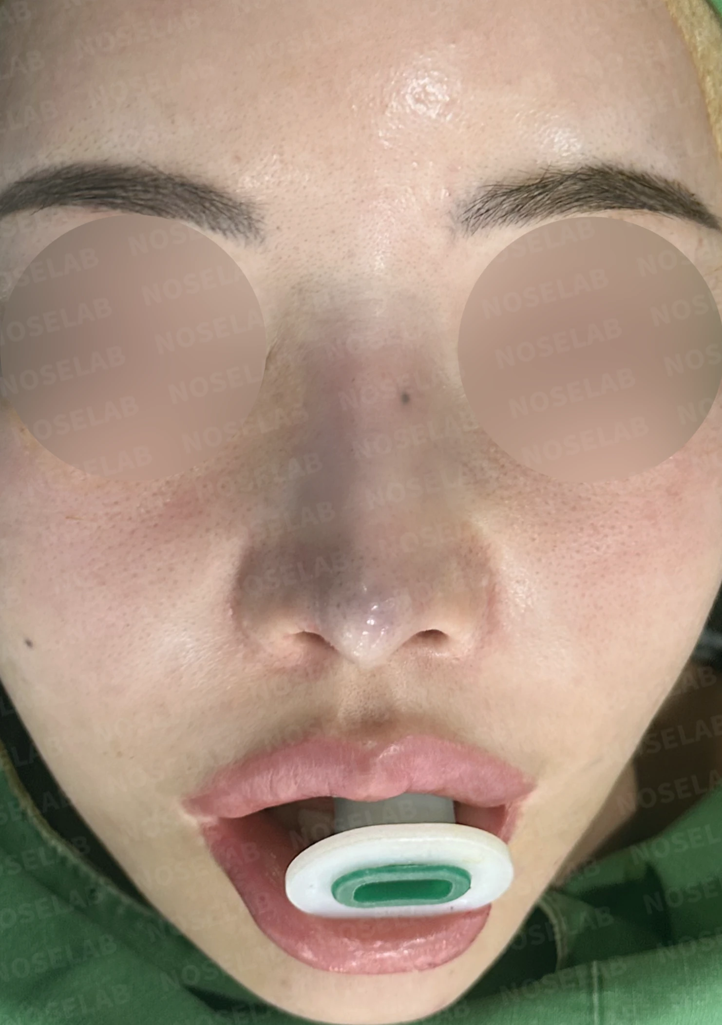
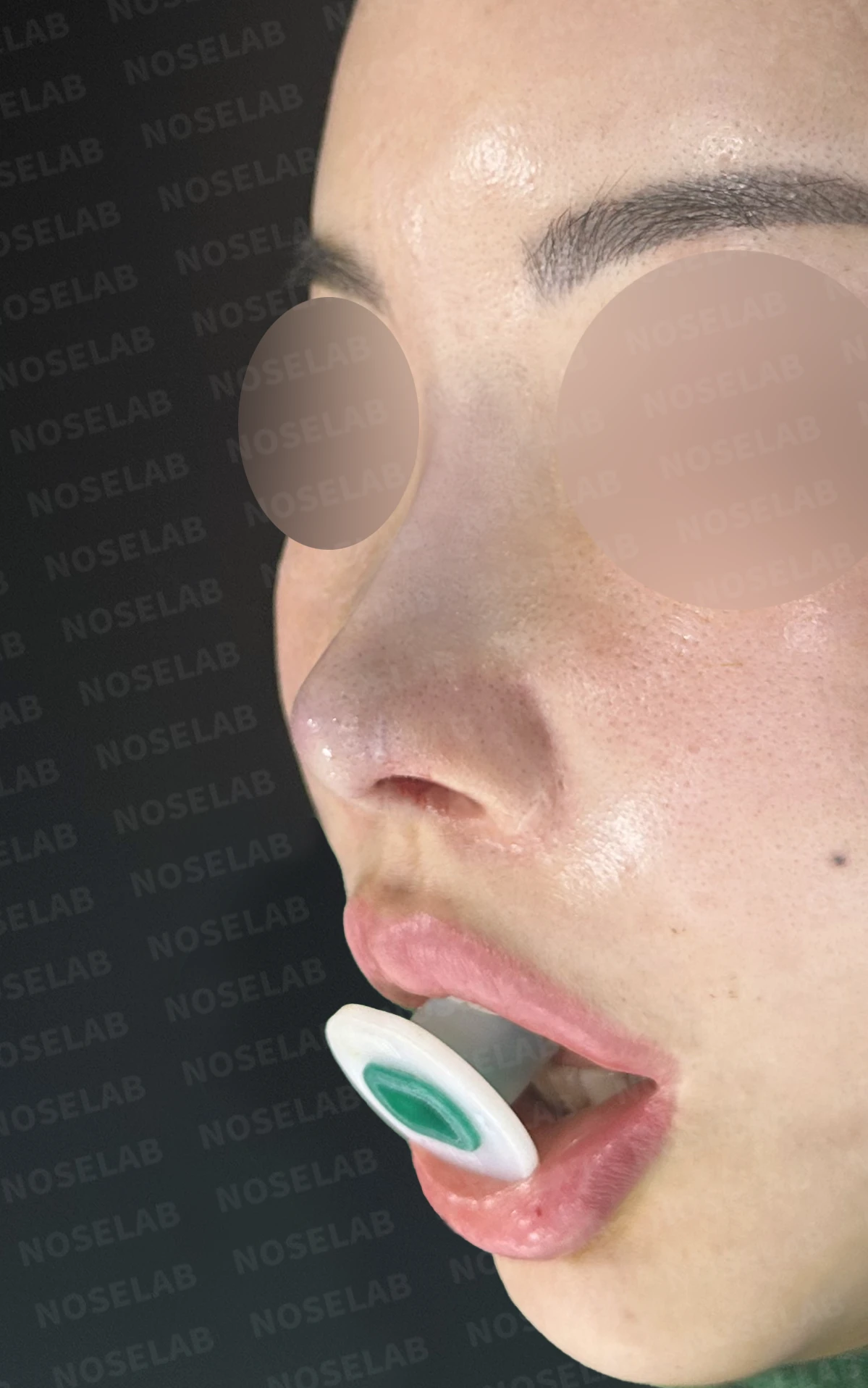
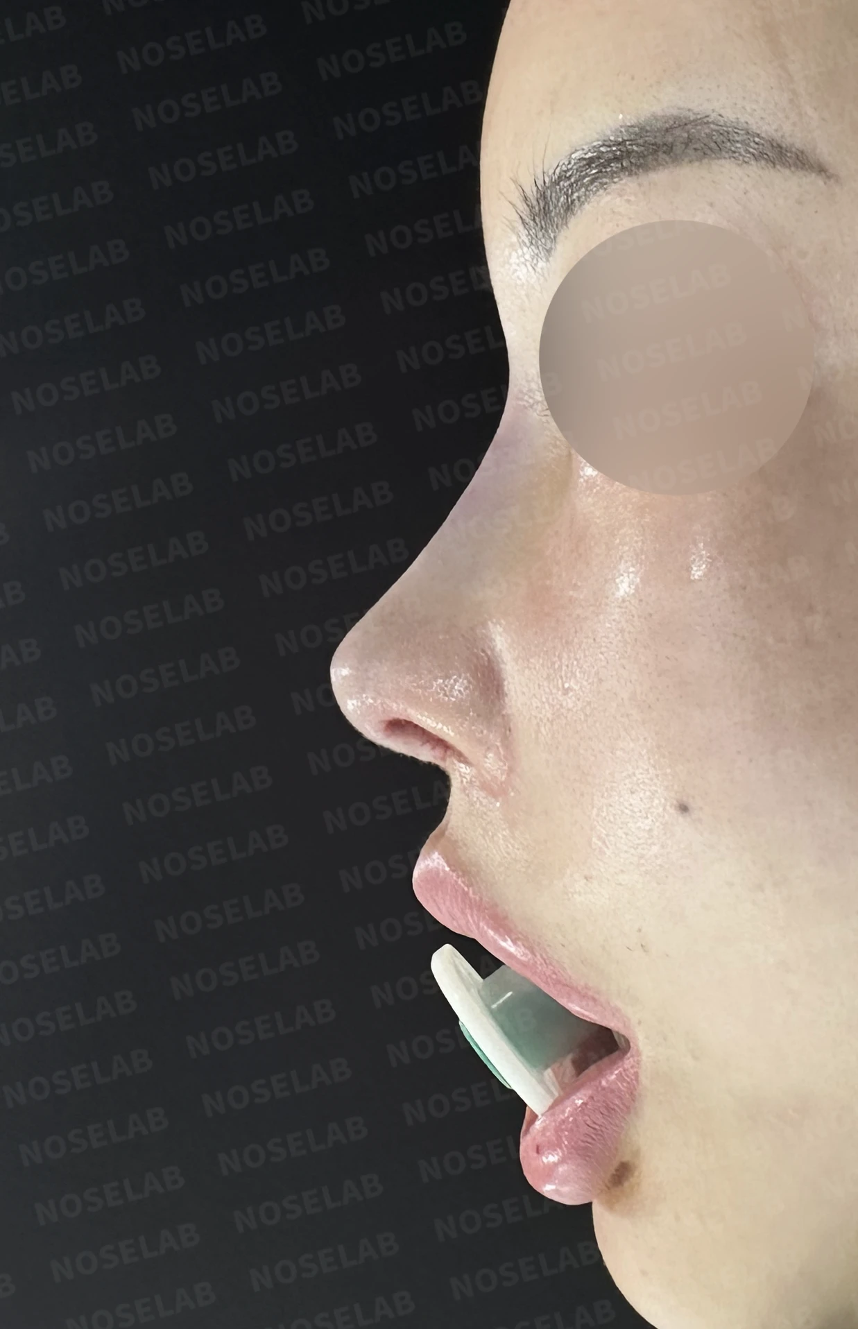
Day 0 immediately after surgery: front, oblique, and profile viewsPhotos taken immediately after surgery show swelling and may differ from the final result.
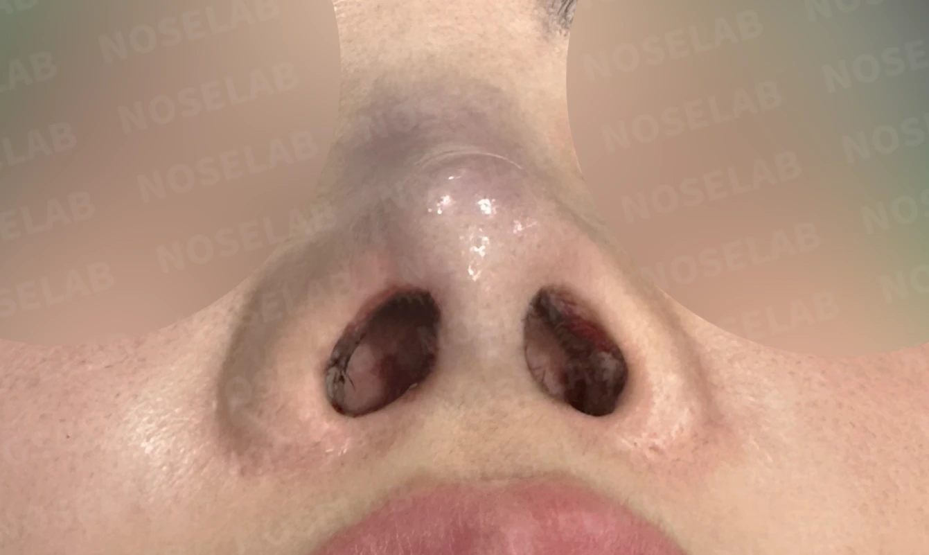
Day 0 immediately after surgery: nostril viewPhotos taken immediately after surgery show swelling and may differ from the final result.
In patients who have undergone multiple rhinoplasties, the normal anatomical structures—the septal cartilage and lower lateral cartilages—are often damaged with little to none remaining. In this case, as expected, barely any septal or lower lateral cartilage remained, so both were completely reconstructed using autologous rib cartilage.
Particularly in this scenario, because abnormal findings were observed around the implant on CT, identifying the exact cause during surgery was essential. Serous fluid was noted and sent for bacterial culture. Diagnosing it as a chronic foreign body reaction rather than purulent infection allowed us to safely proceed with surgery following thorough antibiotic irrigation and complete capsule removal.
Furthermore, because part of the nasal bone was eroded following removal of the foreign material, the bridge height was markedly reduced. However, because the patient's native tissues were severely damaged and weakened, we did not aggressively raise the bridge. We established a safe, aesthetically sound structural framework; if additional height is desired, it can be slightly increased after 6 months once the surgical result has fully stabilized.
In revision rhinoplasty, it is important to accurately assess the current condition of the patient's tissues and pursue the intended result while prioritizing safety. Rather than an overly aggressive procedure, a stepwise approach can lead to better long-term results.
I will continue to prioritize patient safety and dedicate myself to achieving the intended results. I hope this serves as helpful information for those facing similar concerns. This has been Dr. Peter C. Kang, Medical Director. Thank you.
#closedrhinoplasty #closedrevisionrhinoplasty #autologousribrhinoplasty #septalreconstruction #alarreconstruction #foreignbodyremoval #revisionrhinoplastyspecialist #multiplerevisionrhinoplasties #nostrilasymmetry

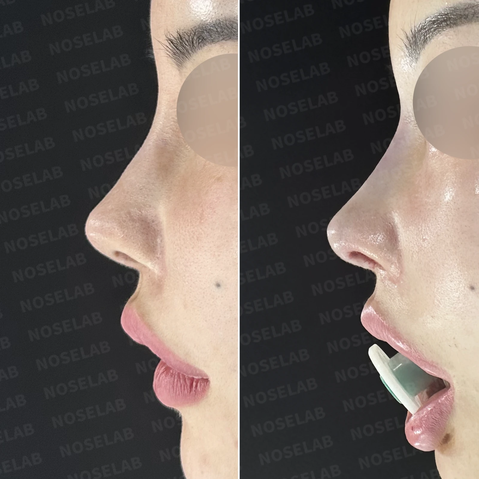
Limitations and risks
When autologous rib cartilage is used, scarring and pain may remain at the donor site on the chest, and pneumothorax (pleural injury) can occur rarely. Over time, the grafted cartilage may warp or undergo partial resorption.
When using donor rib cartilage, long-term follow-up is necessary because the grafted cartilage may resorb or warp.
When revising after implant removal, contracture may progress again after removal, the nasal height and length may decrease, and staged surgery may be necessary depending on the skin condition.
When foreign materials such as injected fillers are removed and reconstructed, contracture may recur, nasal height and length may decrease, and staged surgery may be necessary depending on the skin condition.
When the septum is corrected at the same time, nasal obstruction may persist or recur, and in rare cases, septal perforation or changes in smell may occur.
Common potential risks include infection, bleeding, asymmetry, scarring, skin necrosis, and insufficient improvement; results vary depending on individual skin condition and previous surgical history.