English日本語简体中文
Skip to main content
Dr. Peter C. Kang's Rhinoplasty Stories
PrologueBlogMy thoughtsNasal obstructionSurgical journalSurgical photos
About the surgeon

Thin nasal skin cases: distinguishing implant show-through from reinforcing materials

Records of thin nasal skin often appear alongside visible cartilage beneath the skin or a history of multiple surgeries. One original record explained that previously crushed and inserted cartilage became clumped together, causing the skin to thin. Another record addressed thin nasal tip skin separately within a plan to correct a long nose and a dorsal hump.

This page distinguishes the finding of thin skin from the actual materials used for reinforcement. Some cases note dermal grafts, while others describe removing autologous dermis from the existing nasal bridge and reusing it to reinforce the nasal tip skin. Compare how the restructuring of cartilage and the covering or reinforcement of skin are recorded by following the original texts.

View before-and-after cases ↓Comprehensive rhinoplasty guide

Searching for surgical cases matching your criteria.
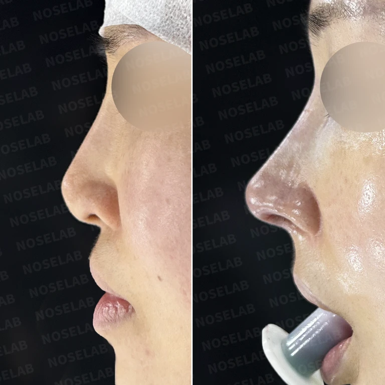
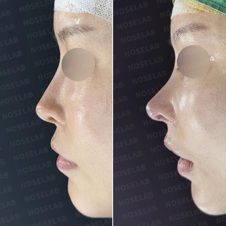
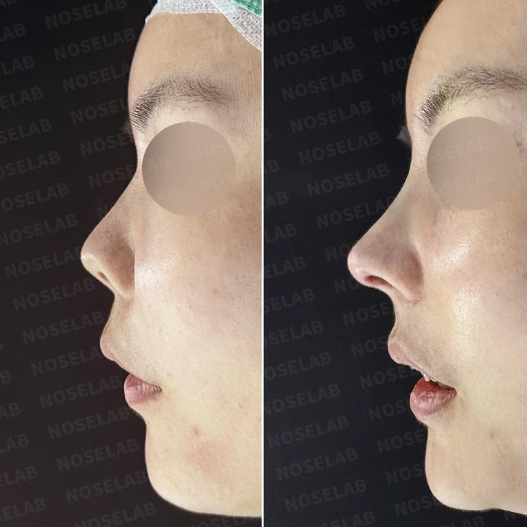
Why an L-shaped silicone implant gradually causes the nasal tip to become upturned: removal and reconstruction — Before-and-after comparison
Before-and-after comparison

Why an L-shaped silicone implant gradually causes the nasal tip to become upturned: removal and reconstruction

A case where an L-shaped implant placed about 10 years ago caused skin thinning and an upturned nasal tip. The L-shaped silicone implant was removed, and nasal tip reconstruction with autologous rib cartilage, septal correction, and dermal tissue reinforcement were performed.

Preoperative condition
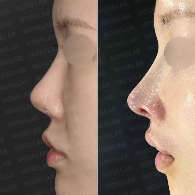
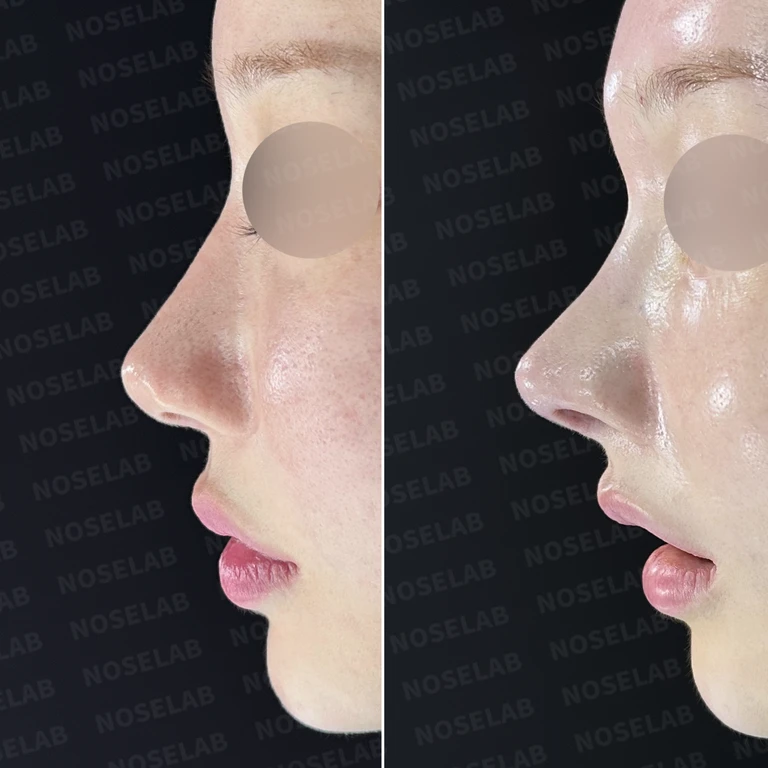
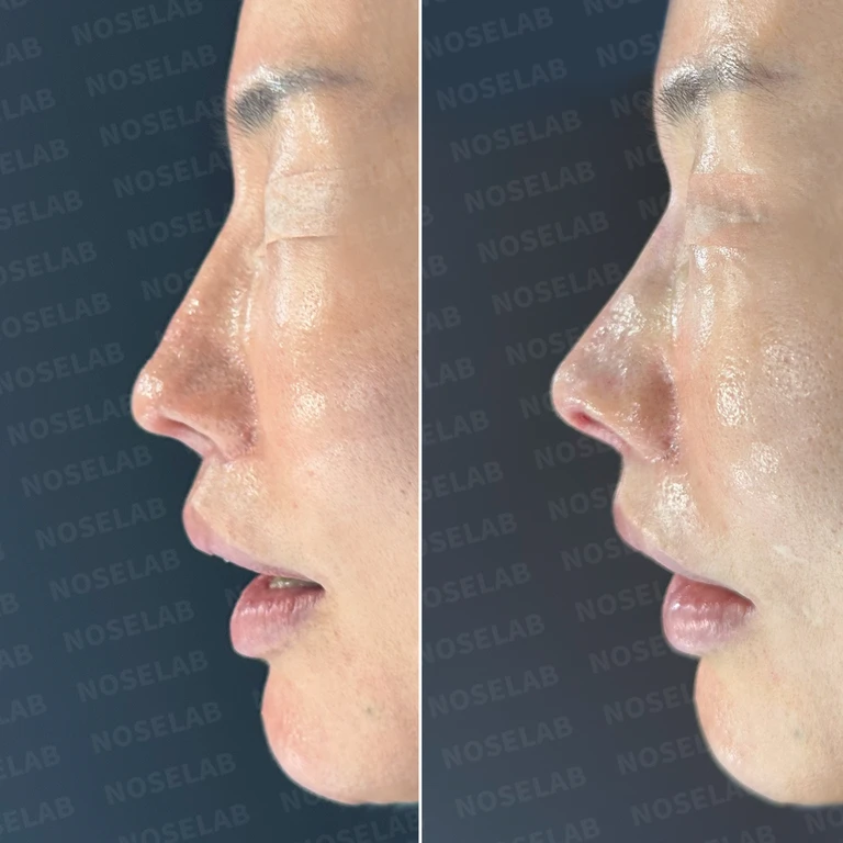
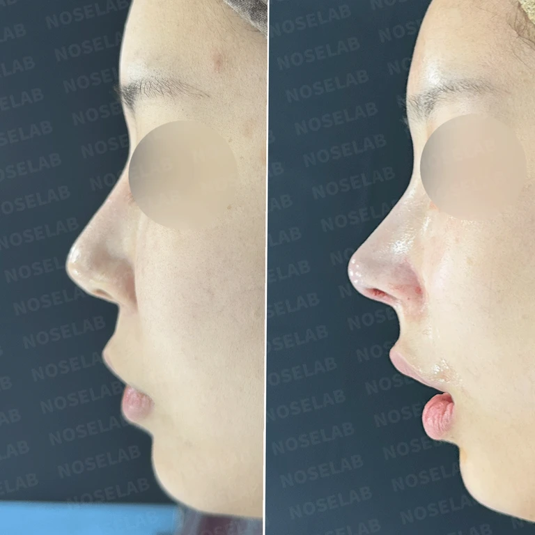
Why an implant is palpable and visible through the nasal tip: rib cartilage reconstruction and dermis reinforcement — Before-and-after comparison
Before-and-after comparison

Why an implant is palpable and visible through the nasal tip: rib cartilage reconstruction and dermis reinforcement

A case where a silicone implant became palpable, the nasal tip skin thinned, and asymmetry developed after a previous rhinoplasty. The implant was removed, the nasal tip and septum were reconstructed using sliced autologous rib cartilage, and artificial dermis was used for reinforcement.

Preoperative condition

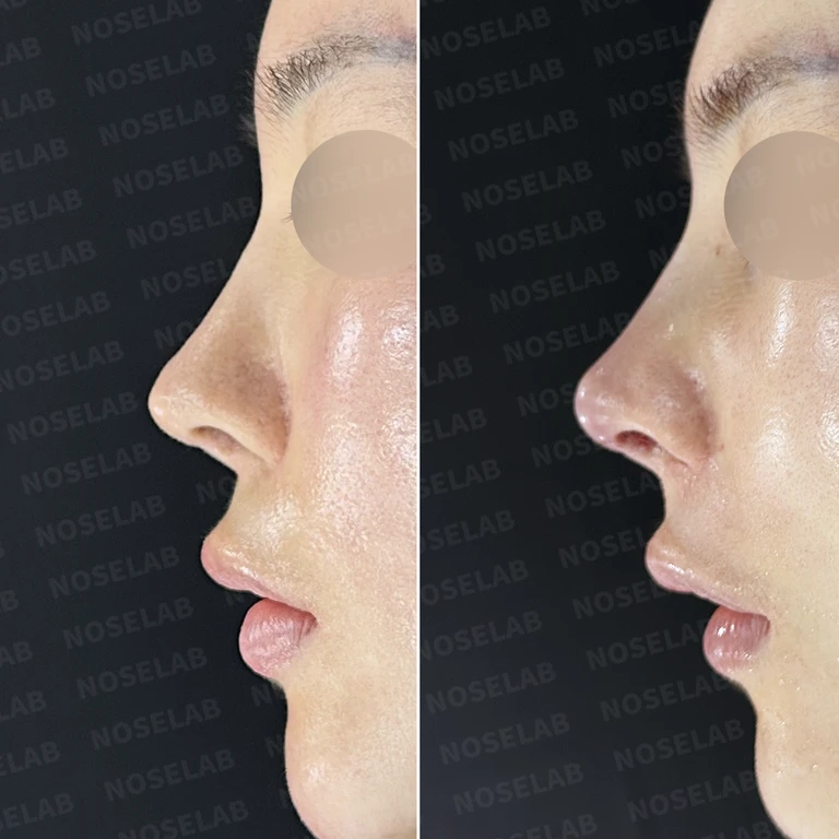
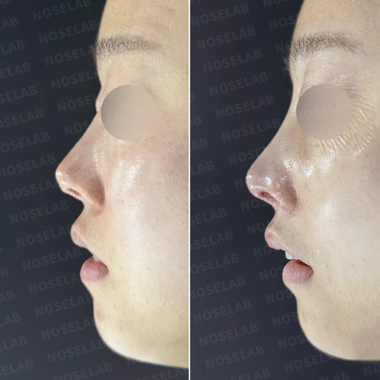
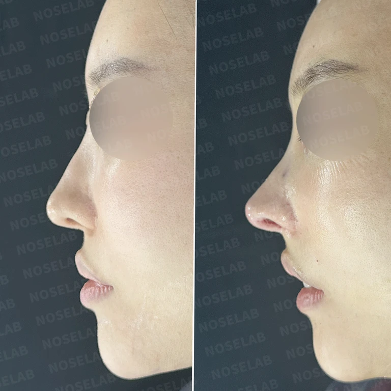
Why mesh remaining in the septum causes an upturned nose, and reconstruction after removal — Before-and-after comparison
Before-and-after comparison

Why mesh remaining in the septum causes an upturned nose, and reconstruction after removal

This is a case where mesh left in the septum after previous surgeries resulted in an upturned nose and excessive nostril show. All foreign material was removed, followed by septal reconstruction using autologous rib cartilage, replacement of the silicone implant, and reinforcement of the skin with acellular dermal matrix.

Preoperative condition

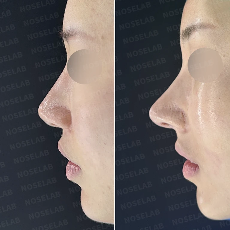
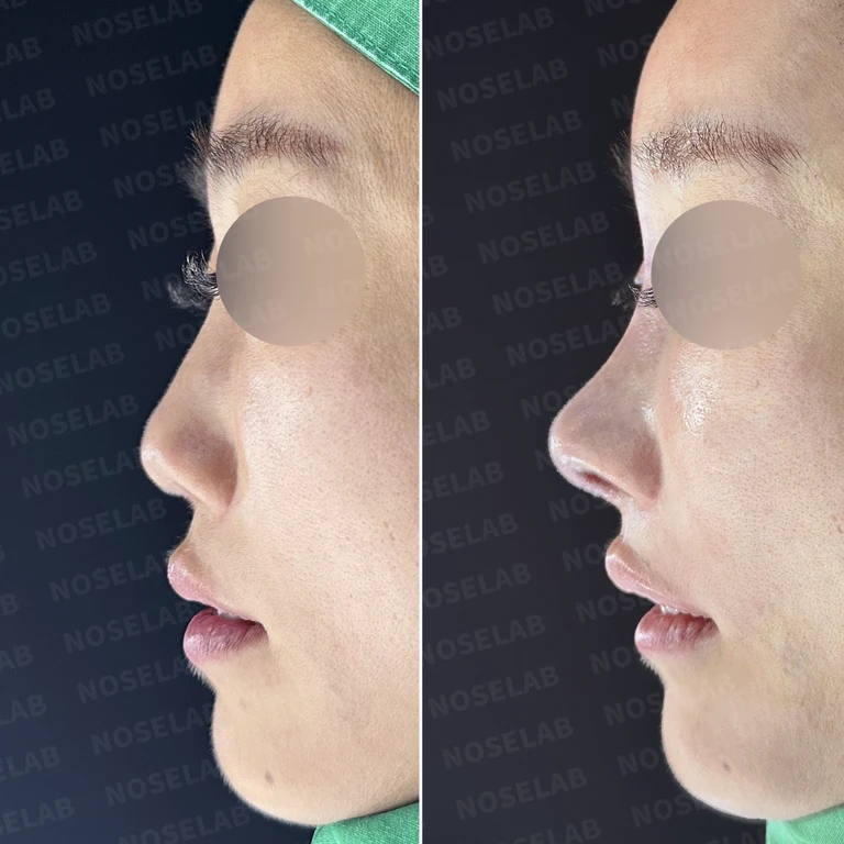
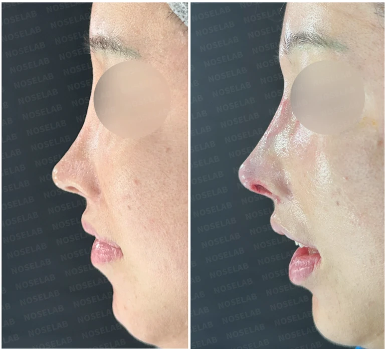
Why implants show through and nostrils look M-shaped, and how to reconstruct them — Before-and-after comparison
Before-and-after comparison

Why implants show through and nostrils look M-shaped, and how to reconstruct them

Implant show-through at the nasal tip and thinned skin were reinforced with donor dermis, and foreign substances were removed. The nasal tip was reconstructed using autologous rib cartilage, along with dragon nose correction and lateral osteotomy.

Preoperative condition

Why nasal tip skin thins after multiple surgeries: autologous dermis reinforcement and reconstruction methods — Before-and-after comparison
Before-and-after comparison

Why nasal tip skin thins after multiple surgeries: autologous dermis reinforcement and reconstruction methods

We removed previous foreign materials and implants, reconstructed the nasal tip using autologous rib cartilage, reinforced the thinned skin with autologous dermis, recarved a silicone implant, and performed fat grafting between the eyebrows.

Preoperative condition

Why nasal obstruction persists after multiple surgeries: How to reconstruct the septum and nasal valve — Before-and-after comparison
Before-and-after comparison

Why nasal obstruction persists after multiple surgeries: How to reconstruct the septum and nasal valve

For a patient whose septum had collapsed after repeated surgeries, causing internal nasal valve stenosis and inferior turbinate adhesions, we performed septal reconstruction with autologous rib cartilage, osteotomy reversal, and adhesiolysis.

Preoperative condition

Why nasal tip cartilage shows through the skin and how dermis grafting reinforces it — Before-and-after comparison
Before-and-after comparison

Why nasal tip cartilage shows through the skin and how dermis grafting reinforces it

For a nose showing cartilage show-through through thinned skin, a widened nasal bridge, and nasal obstruction 7 years after surgery, autologous dermis reinforcement, autologous rib cartilage tip plasty, lateral osteotomy, and septoplasty were performed.

Preoperative condition
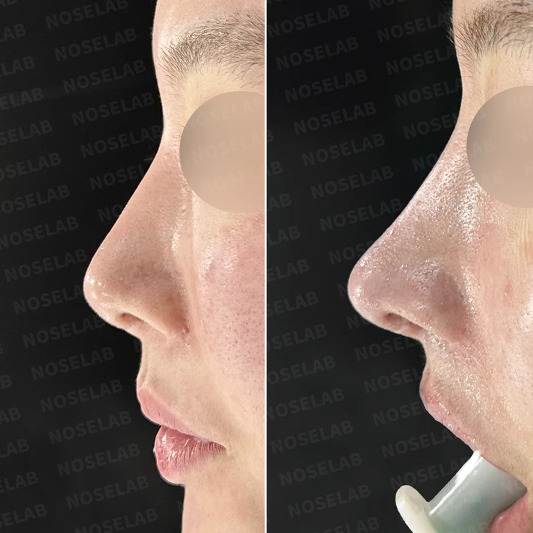
A nose with almost no septal cartilage left: why the nasal tip cannot be elevated — Before-and-after comparison
Before-and-after comparison

A nose with almost no septal cartilage left: why the nasal tip cannot be elevated

A case where the septal support was largely lost from previous surgeries, resulting in nostril asymmetry. The central support was reconstructed with autologous rib cartilage, and the nasal tip was elevated with dermis and cartilage grafts.

Preoperative condition
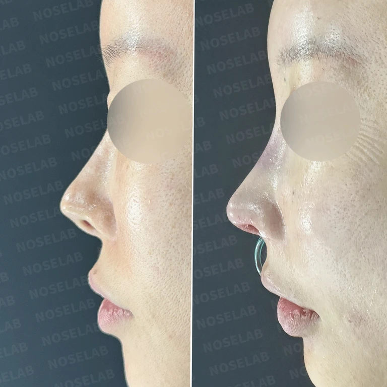
How rib cartilage reconstruction is done when the septum is damaged after multiple surgeries — Before-and-after comparison
Before-and-after comparison

How rib cartilage reconstruction is done when the septum is damaged after multiple surgeries

A case where a septal defect and filler-induced deformity were identified after five prior surgeries. Following filler removal, the septum was reconstructed with autologous rib cartilage, and the nasal tip was reinforced using existing deep temporal fascia.

Preoperative condition
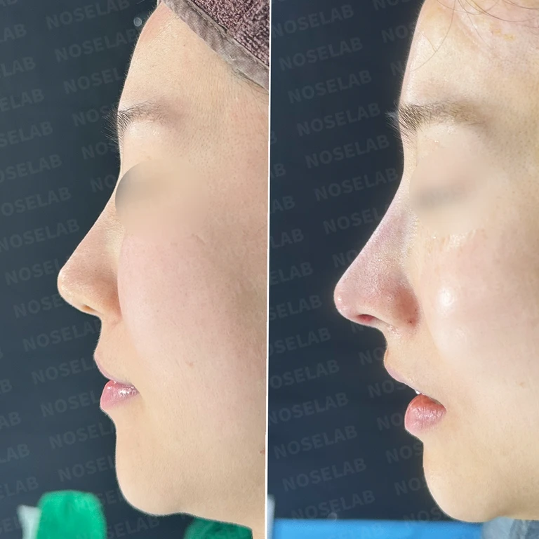
Why nasal tip skin thins and cartilage shows through, and how skin reinforcement is done — Before-and-after comparison
Before-and-after comparison

Why nasal tip skin thins and cartilage shows through, and how skin reinforcement is done

A case of skin thinning, cartilage show-through, and an upturned nose following contracted nose surgery. The skin was reinforced with dermis and autologous rib perichondrium, along with septal extension.

Preoperative condition

Foreign materials from illegal procedures in the nasal bridge: how to perform removal and total septal reconstruction — Before-and-after comparison
Before-and-after comparison

Foreign materials from illegal procedures in the nasal bridge: how to perform removal and total septal reconstruction

We removed foreign materials from illegal injections in the nasal bridge, completely reconstructed the cartilage-depleted septum using autologous rib cartilage, and performed dermal grafting on the thinned skin.

Preoperative condition

Why an L-shaped silicone implant can nearly perforate the columellar mucosa: removal and reconstruction with autologous rib cartilage — Before-and-after comparison
Before-and-after comparison

Why an L-shaped silicone implant can nearly perforate the columellar mucosa: removal and reconstruction with autologous rib cartilage

We removed an L-shaped silicone implant that was at imminent risk of perforating the left columellar mucosa, then reconstructed the contracted nose using autologous rib cartilage for septal extension and autologous dermis for tip reinforcement.

Preoperative condition

Why cartilage shows through after multiple rhinoplasties and how to restore the mucosa and structure — Before-and-after comparison
Before-and-after comparison

Why cartilage shows through after multiple rhinoplasties and how to restore the mucosa and structure

A case where autologous rib cartilage was used to reinforce soft tissue, reconstruct nasal structure, and perform functional surgery to improve mucosal thinning, lateral cartilage show-through, and nostril asymmetry resulting from repeated surgeries.

Preoperative condition · Postoperative record

Septal perforation and nasal tip cartilage show-through after repeated rhinoplasties: how reconstruction is performed — Before-and-after comparison
Before-and-after comparison

Septal perforation and nasal tip cartilage show-through after repeated rhinoplasties: how reconstruction is performed

A case where autologous rib cartilage was used for full septal reconstruction alongside dermis grafting to address repeated surgical history, septal perforation, and thinned nasal tip skin.

Preoperative condition

Why donor dermis absorbs after multiple revision surgeries and how to reconstruct the septum — Before-and-after comparison
Before-and-after comparison

Why donor dermis absorbs after multiple revision surgeries and how to reconstruct the septum

For a patient who experienced partial absorption of donor dermis on the nasal bridge and septal damage after multiple revision surgeries, the donor dermis was removed, the septum was reconstructed with autologous rib cartilage, and the skin was reinforced with retroauricular dermis.

Preoperative condition

Changes following nasal bridge skin necrosis after rhinoplasty and how reconstruction is performed — Before-and-after comparison
Before-and-after comparison

Changes following nasal bridge skin necrosis after rhinoplasty and how reconstruction is performed

In a case with signs of nasal bridge skin necrosis and alar asymmetry after multiple rhinoplasties, the nasal bridge and nostril shape were reconstructed using autologous rib cartilage grafting, alar repositioning, and growth factor application.

Preoperative condition

Why nasal tip cartilage shows through the skin and turns upward: reinforcement and reconstruction methods — Before-and-after comparison
Before-and-after comparison

Why nasal tip cartilage shows through the skin and turns upward: reinforcement and reconstruction methods

For a patient presenting with visible nasal tip cartilage and an upturned nose following previous surgery with a silicone implant and ear cartilage, we performed silicone implant removal, septal extension using autologous rib cartilage, fat grafting, and skin reinforcement with donor dermis.

Preoperative condition

Why nasal bridge and tip show-through happens after old silicone rhinoplasty: dermal reinforcement and rib cartilage reconstruction — Before-and-after comparison
Before-and-after comparison

Why nasal bridge and tip show-through happens after old silicone rhinoplasty: dermal reinforcement and rib cartilage reconstruction

To resolve show-through of an L-shaped silicone implant and cartilage beneath thinned skin, we removed the previous silicone implant, reinforced the skin with dermis, adjusted the tip height, reconstructed the nasal tip with autologous rib cartilage, and replaced the implant with a new one.

Preoperative condition

No search results found

Please try searching with different keywords.

Scroll to keep reading

1 / 2Next case

Common questions before reading the cases

Was the same type of dermis used in all thin nasal skin cases?

No. One original record notes a plan to reinforce the skin with artificial dermis, while another explains that autologous dermis removed from the existing nasal bridge was reused to reinforce the thinned skin of the nasal tip. Rather than assuming the material is identical simply from the word dermis, you can check whether it was autologous or artificial, and whether it was newly applied or reused.

Are skin reinforcement and nasal tip cartilage reconstruction the same thing?

They are explained separately in the original texts. One patient record states that problematic cartilage was adjusted and the structure was rebuilt with autologous rib cartilage, after which a dermal graft was placed over the thinned skin. This was a case where addressing the internal structure and reinforcing the skin occurred together. Rather than looking only at the before-and-after photos of show-through, examine the distinct procedures that were performed.

Related topics to explore

Implant show-through and palpabilitySkin and soft tissue reinforcementAutologous dermisArtificial and allogeneic dermis

Surgical records referenced in this guide

  • Septal perforation and nasal tip cartilage show-through after repeated rhinoplasties: how reconstruction is performed
  • Revision rhinoplasty with autologous rib cartilage and columella correction for a long nose and dorsal hump
  • Is it possible to correct a philtrum reduction and remove autologous dermis?
Original article

Read more records

Planning a visit to Seoul?Prepare your consultation with the NOSELAB team.

International patient guideWhatsApp ↗
Dr. Peter C. Kang's Rhinoplasty Stories
Rhinoplasty guideCases by topicDr. Peter C. KangNaver Blog ↗
View original photo
Enlarged photo
WhatsAppChat with NOSELAB↗