Hello.
I am Dr. Peter C. Kang, medical director.
I wore a short-sleeved shirt today. Even though it is still April, summer seems to have arrived quickly. Please take care not to catch a cold during this change of seasons.
Today, I would like to discuss a somewhat complex surgery. It is not an exceedingly difficult procedure, but it is an unfortunate situation. I will discuss a case where an unsuccessful philtrum reduction or alar reduction directly affected the shape of the nose.
Personally, I find these cases very upsetting, as it feels as though such procedures take away opportunities to achieve a better result.
Preoperative design consultation
This patient had undergone 4 previous surgeries before visiting our clinic. Autologous dermis from the buttock had been placed on the nasal bridge, and multiple procedures including a philtrum reduction and nasal bone osteotomy had been performed. The patient visited our clinic seeking correction for a nose that appeared crooked, a low, pinched nasal tip, and noticeable asymmetry.
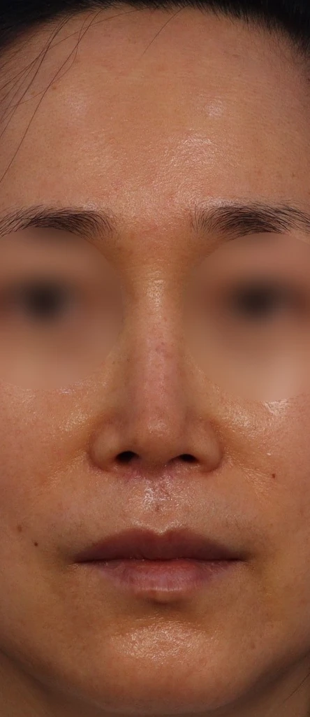
Before surgery: front view
In the preoperative front view, asymmetry of the alae is noticeable, and the difference in nostril size appears severe. Additionally, the alae appear pinched, and the nasal tip appears deviated to the right.
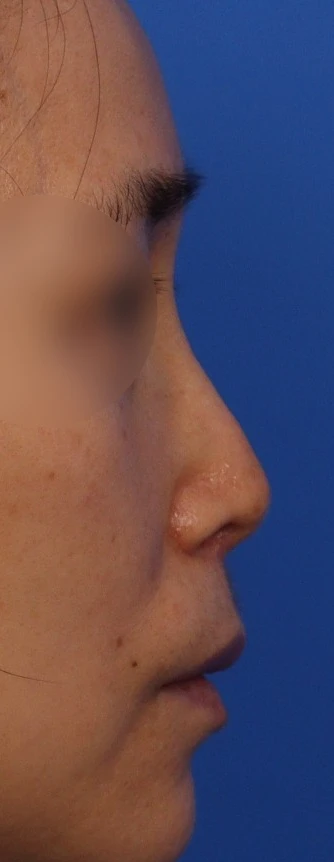
Before surgery: profile view
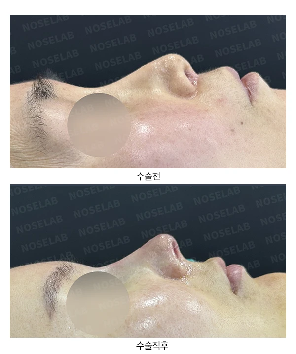
In the profile view, the nasal bridge is high relative to the nasal tip, the tip is short and drooping, and the junction between the tip and bridge appears depressed.
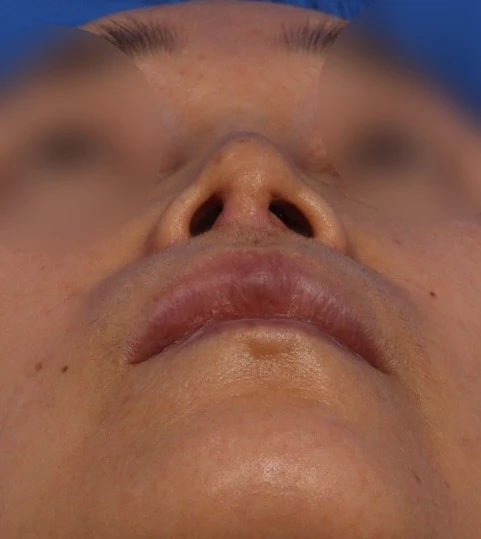
Before surgery: basal (nostril) view
The nostril asymmetry appears severe, and the right nasal sill looks much shorter than the left.
Surgical plan
On the nasal CT scan taken at the initial visit, both nasal bones had collapsed inward due to excessive osteotomy, significantly narrowing the nasal vault. In the front view, the sides of the bridge implant appeared compressed, creating a stark chalk-like look. Therefore, a lateral osteotomy restoration was planned.
The plan was to remove the autologous dermis and reuse it on the nasal tip.
The plan was to reposition the tip cartilage and reinforce the lower lateral cartilage using autologous rib cartilage to correct the pinched appearance.
Because a significant difference in the width of the nasal sill was expected due to the overly aggressive philtrum reduction, a rotation flap was planned to expand the deficient skin below the right columella.
Surgical findings
After removing the autologous dermis and checking the condition of the nasal bones, most of the roof of the nasal bone was missing, leaving little bone behind. The remaining nasal bones on both sides were moved outward, the gap between the bones was filled with autologous rib cartilage, and a silicone implant was placed broadly over them. The autologous dermis on the bridge, along with the ear cartilage previously placed in front, was completely removed intact and reused to reinforce the thinned skin of the nasal tip.
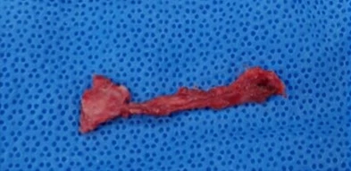
Photograph of the removed ear cartilage (left) and autologous dermis (right).
The most challenging part of today's surgery was correcting the nostril asymmetry caused by the previous philtrum reduction. To shorten the philtrum, the columella must be separated from the philtrum, the philtrum excised appropriately, and the tissues resutured precisely at the midline. In this case, as seen in the photo, the columella was anchored onto the right side of the philtrum base, leaving the right nasal sill excessively short compared to the left. To correct this, a rotation flap that transferred skin from inside the nostril outward was performed to widen the nasal sill.
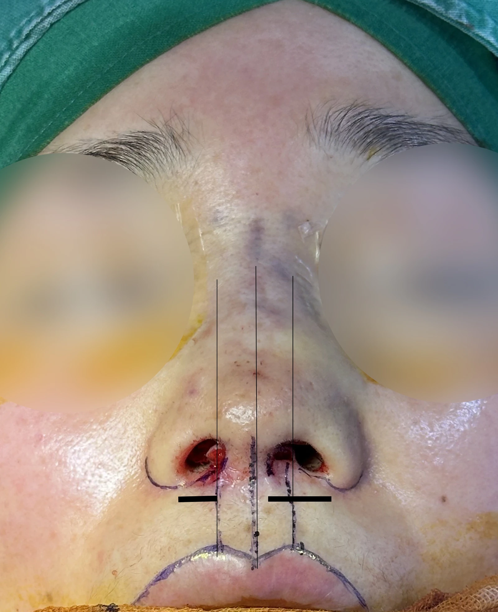
A case of improper suturing during a previous philtrum reduction.
Before surgery vs. immediately after surgery
Let us compare the preoperative photos with the photos immediately after surgery (Day 0).Immediate postoperative photos show swelling and may differ from the final result.
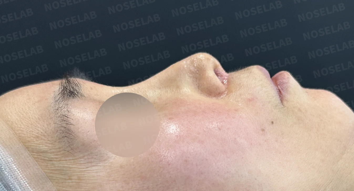
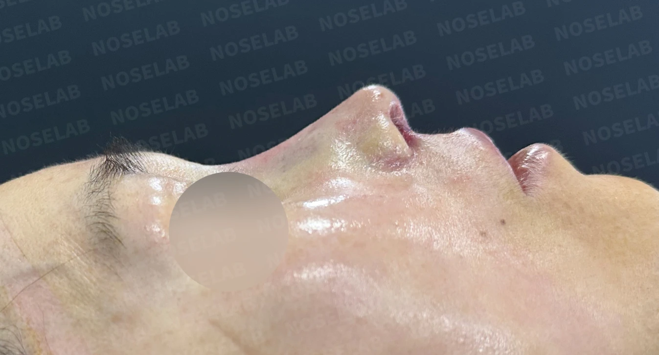
Before surgery / Day 0 immediately after surgery Photos taken immediately after surgery show swelling and may differ from the final result.
In the profile view immediately after surgery (day 0), the previously drooping nasal tip is well supported, and the nasolabial angle appears natural. Immediate postoperative photos show swelling and may differ from the final result.
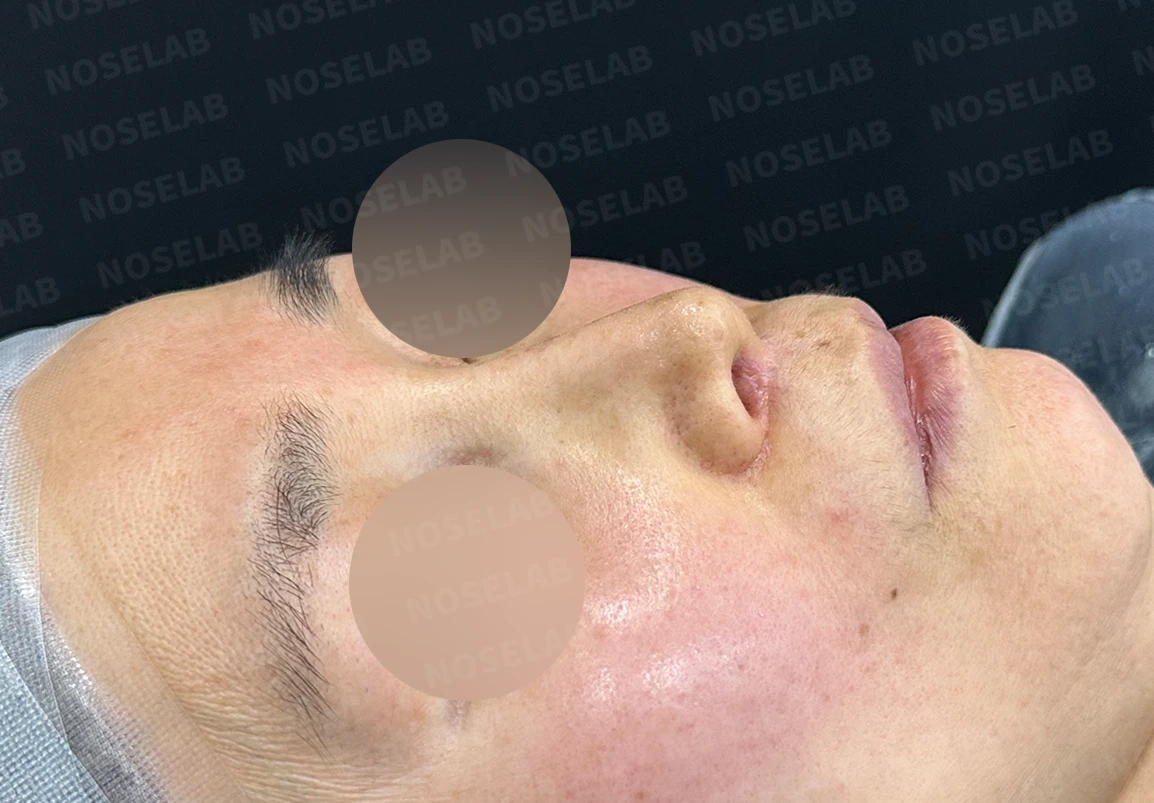
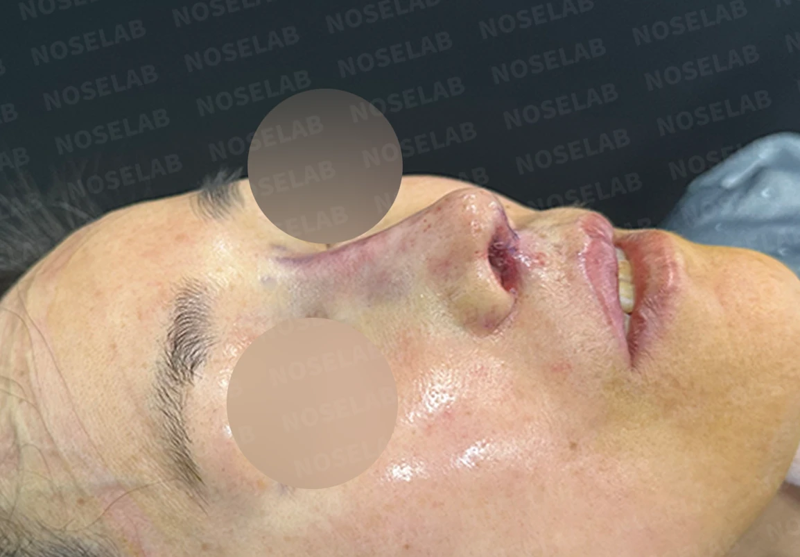
Before surgery / Day 0 immediately after surgery Photos taken immediately after surgery show swelling and may differ from the final result.
Looking at the 45-degree oblique view photo, you can see that the radix has lowered and the nasal tip has become more defined.
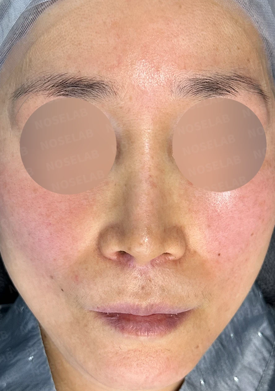
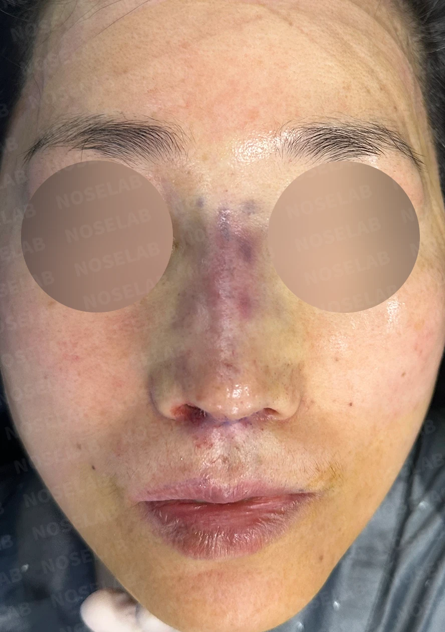
Before surgery / Day 0 immediately after surgery Photos taken immediately after surgery show swelling and may differ from the final result.
In the front view, bruising from removing the autologous dermis is visible along the dorsum, but because it is not deep bruising, it will mostly resolve within 3 weeks. The severe difference in the nasal sill and the crooked appearance have significantly improved.
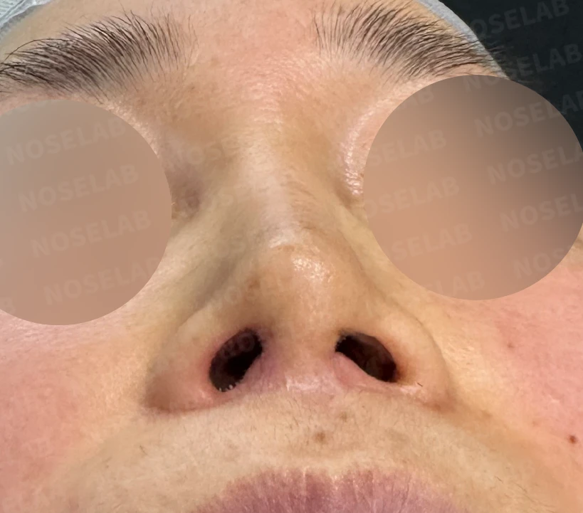
Before surgery / Day 0 immediately after surgery Photos taken immediately after surgery show swelling and may differ from the final result.
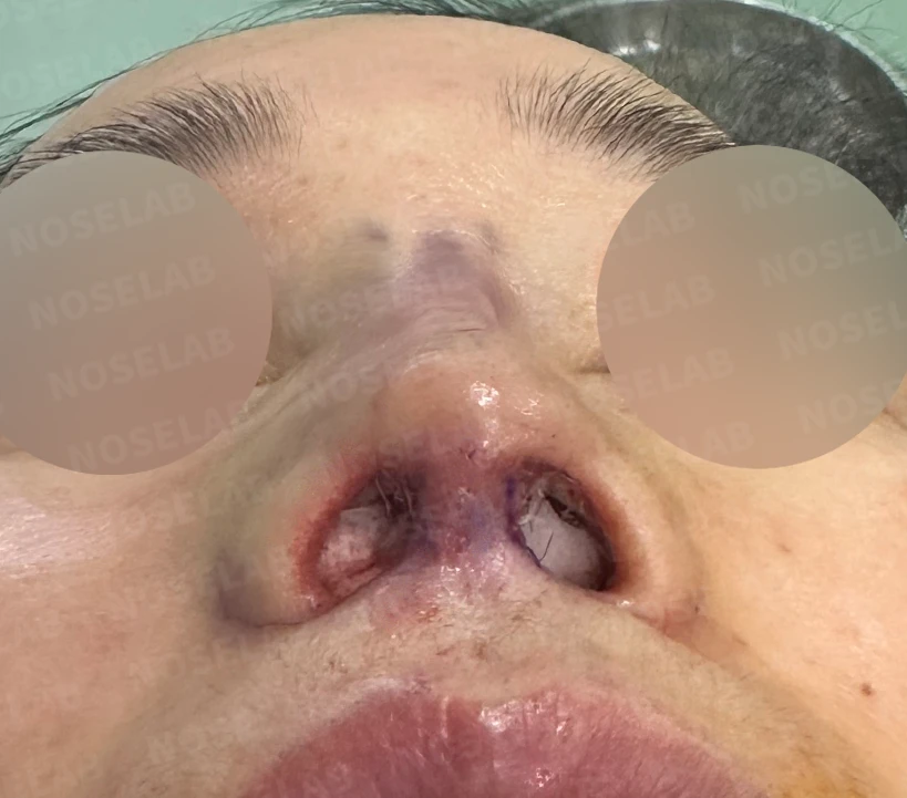
Looking at the nostrils from below, the severely narrowed right nostril has been restored to some extent, though it may decrease by about 10% during the healing process. However, because part of the internal nostril skin was brought forward to widen the space, the widened nostril is not expected to narrow significantly.
Photos immediately after surgery
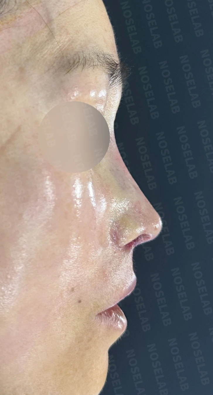
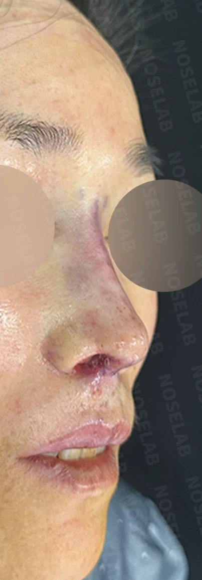
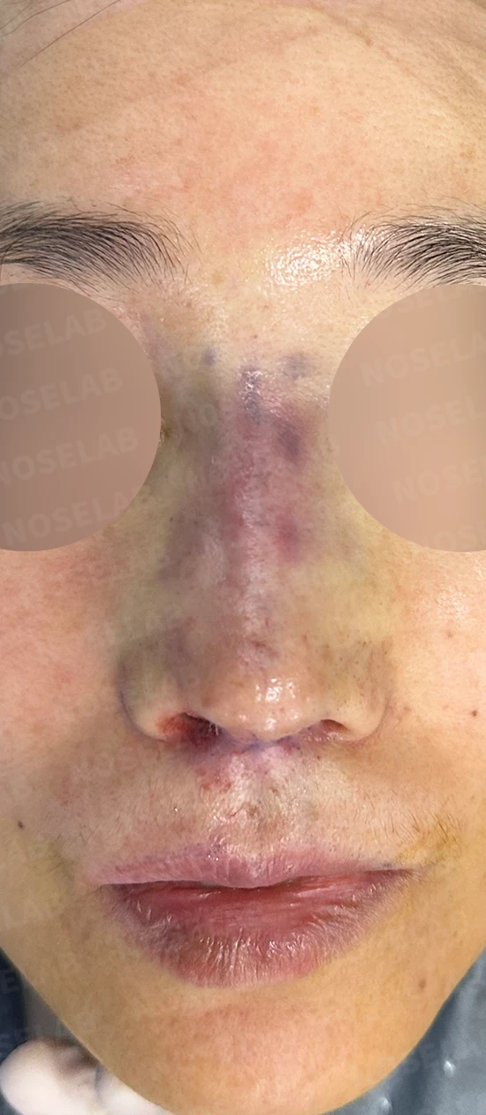
Photos immediately after surgery (Day 0): 90-degree profile, 45-degree oblique, and front view.Immediate postoperative photos show swelling and may differ from the final result.
Basal (nostril) view immediately after surgery (Day 0)Immediate postoperative photos show swelling and may differ from the final result.
My thoughts on philtrum reduction and rhinoplasty
First, my thoughts regarding philtrum reduction and rhinoplasty are entirely my personal views, so I hope there is no misunderstanding. Having performed rhinoplasty for a long time, there are 2 procedures that I personally disfavor. Those procedures are alar reduction and philtrum reduction. Because I perform surgeries using the closed approach, I may be more sensitive to nostrils becoming smaller, but I do not favor these procedures because they often prevent me from achieving a better nasal shape for the patient.
Furthermore, from an anatomical standpoint, these areas are prone to suboptimal scarring. No matter how meticulously they are sutured, these tissues are constantly pulled when talking, eating, smiling, crying, and making facial expressions, making it difficult for scars to heal cleanly. In addition, everyone has some degree of asymmetry, so separating and reattaching the alae and columella inevitably worsens that asymmetry.
As in this case, the previous surgeon likely did not intentionally anchor the columella to the right side of the philtrum; however, it ended up misaligned by more than 2 mm to the right, causing the deviated tip and nostril asymmetry.
Therefore, I believe one should decide on alar reduction or philtrum reduction very carefully, and if undergoing surgery, it is advisable to choose a technique with minimal incisions.
Today, we reviewed rhinoplasty in patients who have undergone philtrum reduction. I hope this provides helpful insight for those dealing with similar concerns.
This was Dr. Peter C. Kang, medical director.

Limitations and risks
When autologous rib cartilage is used, scarring and pain may remain at the donor site on the chest, and pneumothorax (pleural injury) can occur rarely. Over time, the grafted cartilage may warp or undergo partial resorption.
Common potential risks include infection, bleeding, asymmetry, scarring, skin necrosis, and insufficient improvement; results vary depending on individual skin condition and previous surgical history.