Hello.
I am Dr. Peter C. Kang, medical director.
Today, I would like to talk about the procedure known as nostril lowering (alar rim lowering).
Preoperative design
These are photos taken before surgery.
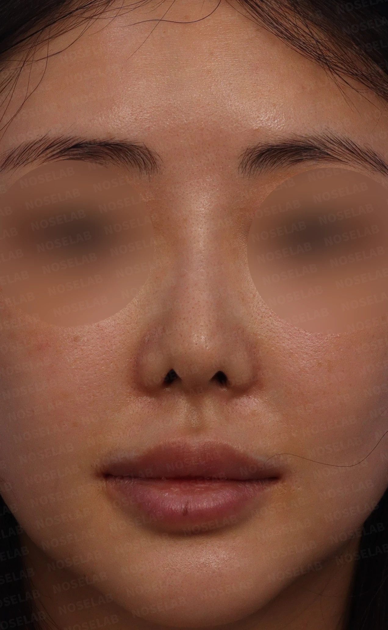
Before surgery: front view
Looking at the front view, the starting point of the nasal bridge, situated in the center of the glabella, looks unnatural,
there is excessive nostril show along with nostril asymmetry,
and the nostrils look excessively pinched and peaked, appearing unnatural.
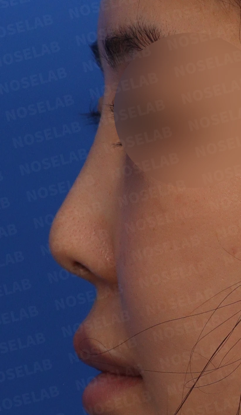
Before surgery: profile view
In the profile view, the nasal tip appears drooping,
and the radix starting point between the eyebrows appears notably high.
A protruding mouth can also be observed.
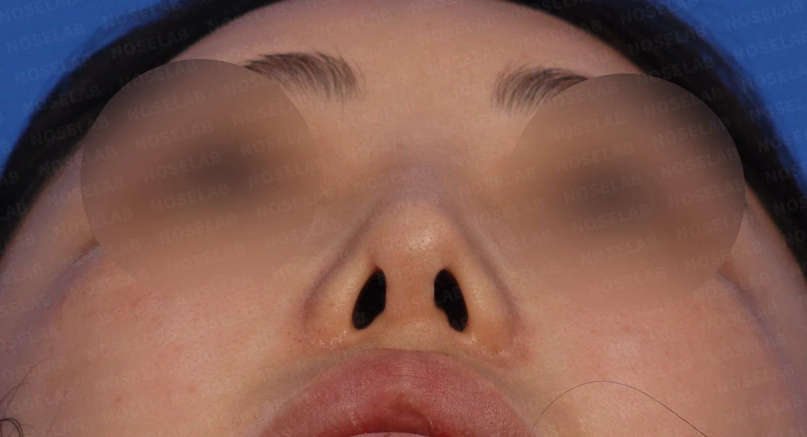
Before surgery: basal (nostril) view
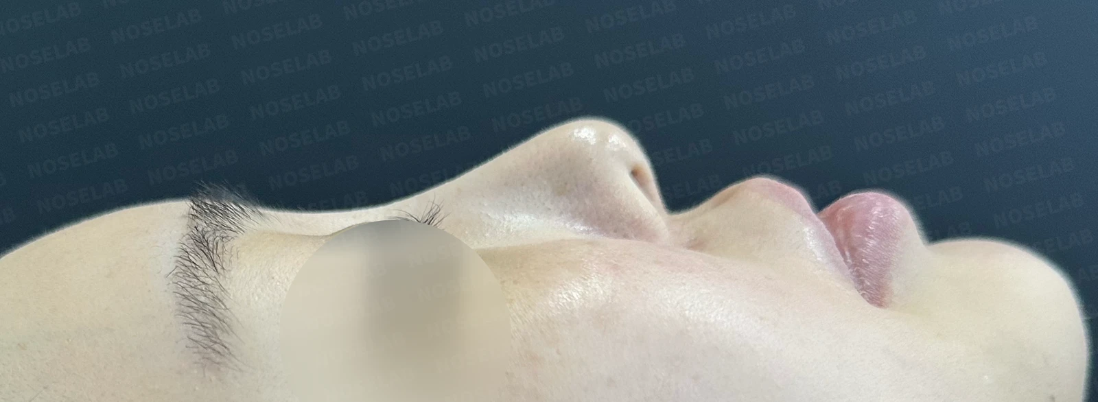
The basal view showing the nostrils demonstrates nostril asymmetry, and the columellar area above the right nostril appears significantly depressed.
Preoperative nasal obstruction examination
These are the nasal computed tomography (CT) scans and nasal endoscopy photos taken at our clinic.
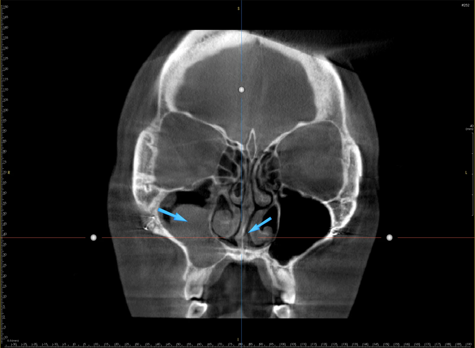
Coronal view
On the coronal view, findings of chronic sinusitis in the right maxillary sinus and a deviated septum can be observed.
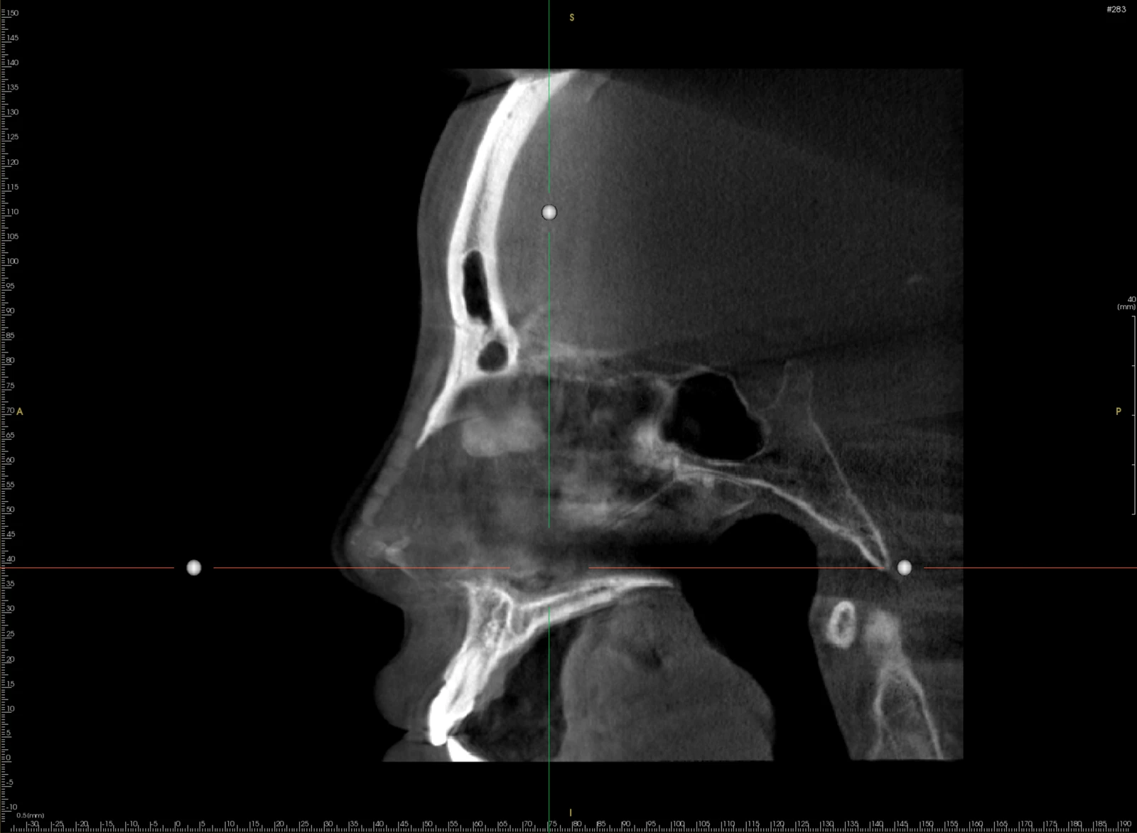
Sagittal view
On the sagittal view, a silicone implant extending from the nasal bridge to the nasal tip is visible,
and the cartilage grafted in the septal area shows partial progression of calcification.
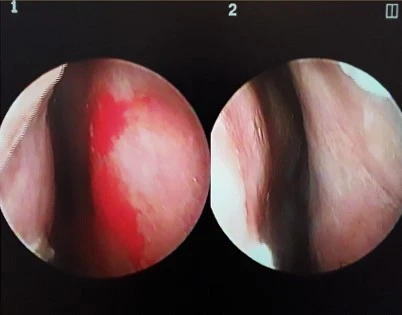
These are nasal endoscopy images (1 right, 2 left).
Endoscopic findings show that the nasal mucosa on both sides appears pale,
with hypertrophied inferior turbinates on both sides,
and septal deviation to the right can be observed.
Surgical plan
Upturned nasal tip -> Planned to lower the nasal tip as much as possible by performing septal extension
Short nose -> Planned to lengthen and raise the nasal tip in a downward, elongating direction
Thin skin -> Planned to reuse the autologous gluteal dermis placed during the previous surgery
Lowering the starting point -> Planned to lower the nasal bridge radix starting point and insert a new silicone implant
Surgical procedure
Because too much septal cartilage had been harvested and used at the previous clinic, the remaining septum was very small and weak; therefore, it was reinforced using donor rib cartilage to provide structural support.
To relieve nasal obstruction, septoplasty was performed to correct the deviated septum, bilateral spreader grafts were placed, and radiofrequency submucosal resection of the inferior turbinates was carried out.
To correct the peaked appearance of the right nostril on the front view, ear cartilage was grafted to the anterior alar rim to perform nostril lowering, while on the left side, the lower lateral cartilage was repositioned to achieve nostril lowering.
The gluteal dermis that covered the dorsum down to the nasal tip was cleanly removed and then reused at the nasal tip to prevent cartilage show-through.
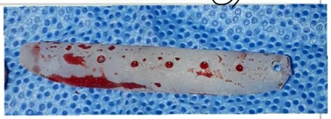
This is the removed silicone implant.
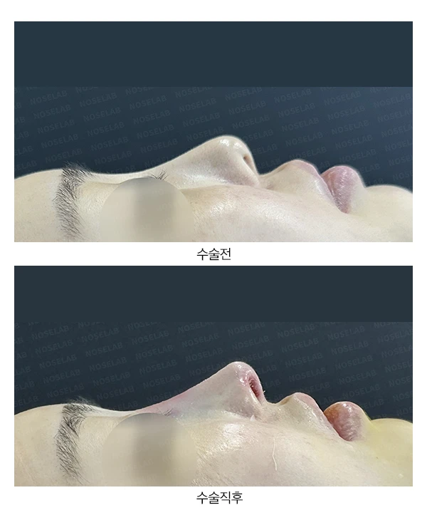
Before surgery vs. immediately after surgery
Let us compare the preoperative photos with the photos immediately after surgery (Day 0).Immediate postoperative photos show swelling and may differ from the final result.
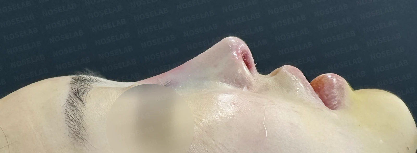
Before surgery / Day 0 immediately after surgery Photos taken immediately after surgery show swelling and may differ from the final result.
This is the profile view. The radix starting point between the eyebrows was lowered naturally, and the blunt, heavy look of the nasal tip improved significantly. The columella was lowered slightly to create a natural nasolabial angle.
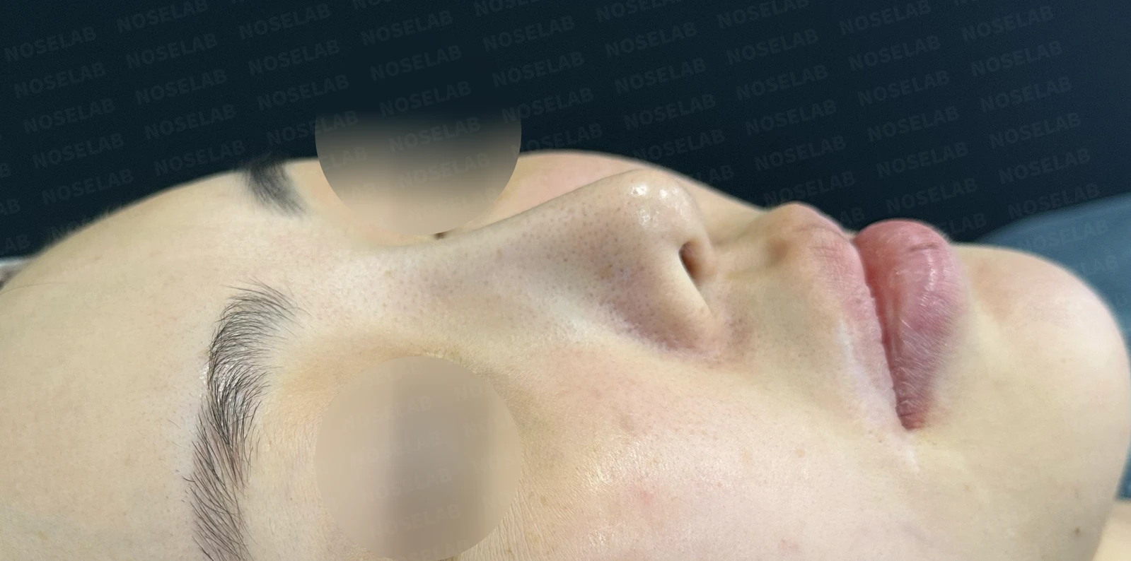
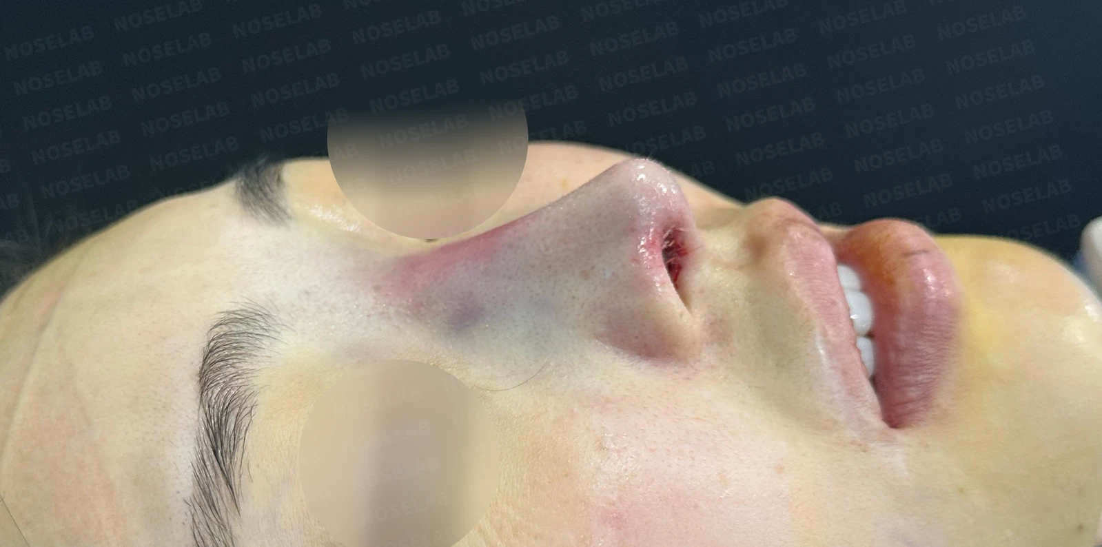
Before surgery / Day 0 immediately after surgery Photos taken immediately after surgery show swelling and may differ from the final result.
The nose, which previously appeared pulled excessively upward toward the forehead, now looks naturally lowered.
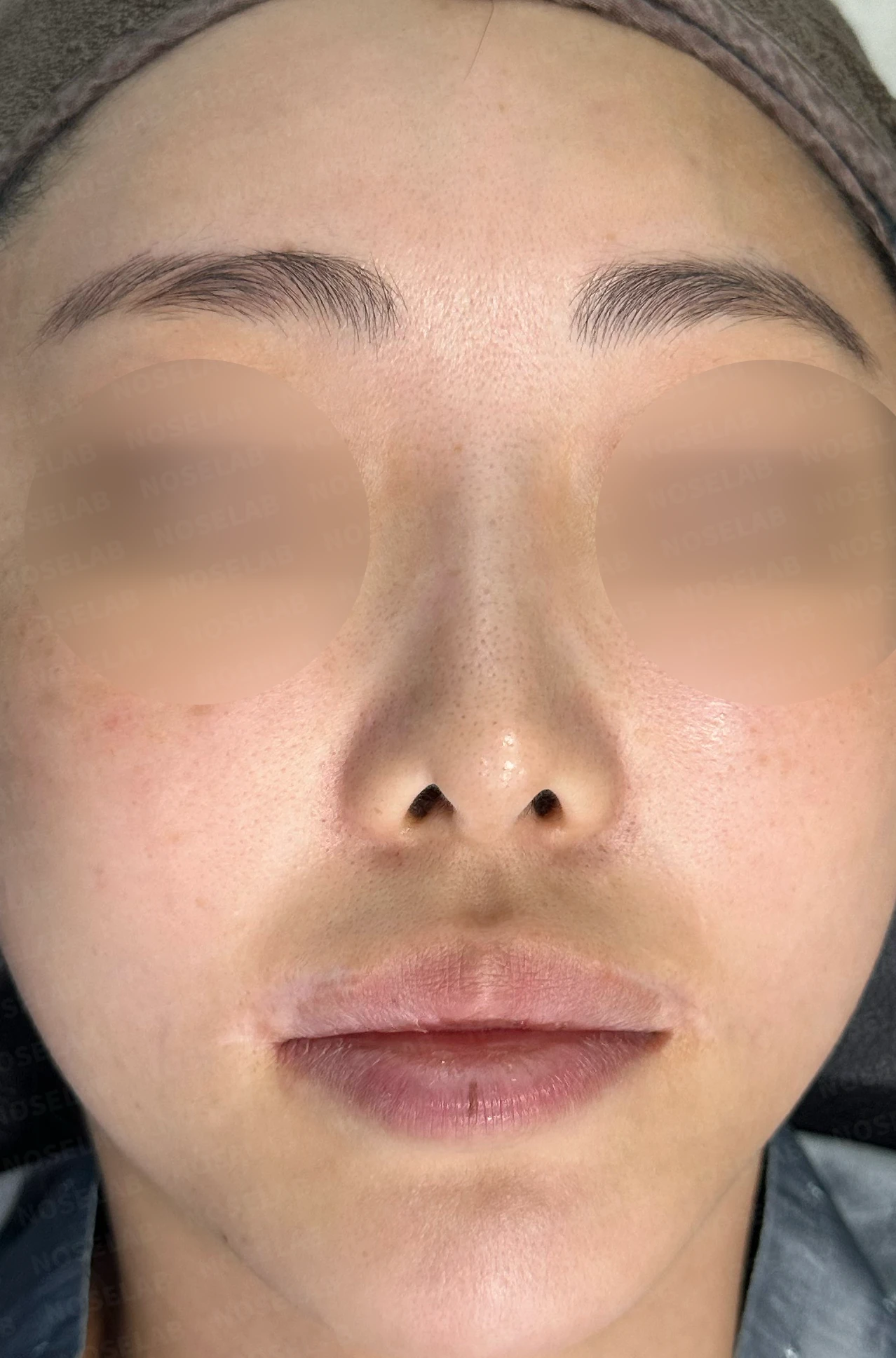
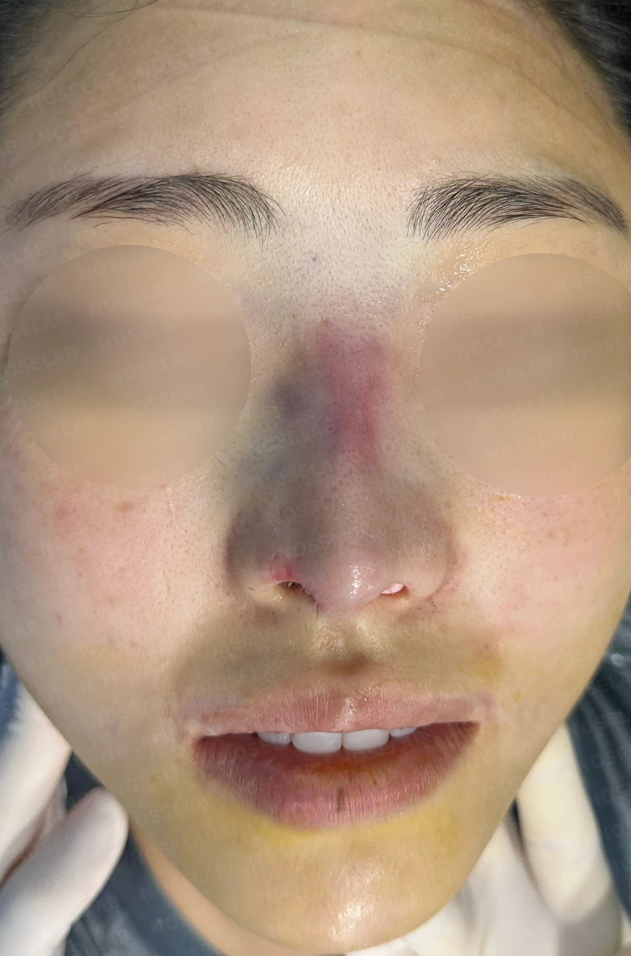
Before surgery / Day 0 immediately after surgery Photos taken immediately after surgery show swelling and may differ from the final result.
The deviation visible on the front view has improved, and the pinched, sharply pulled-up appearance of the right nostril has also resolved substantially.
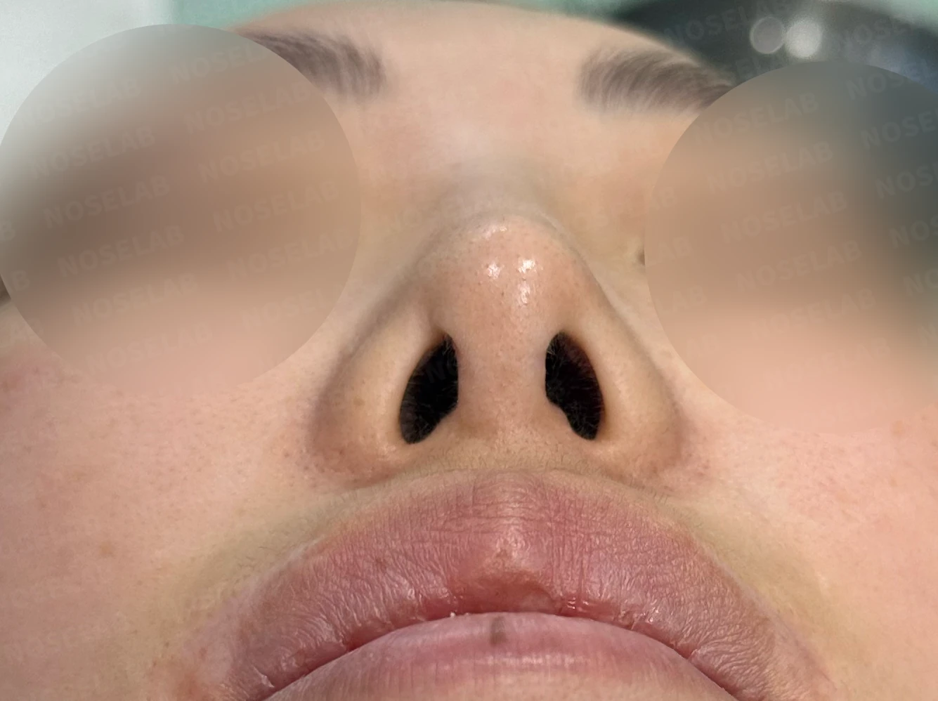
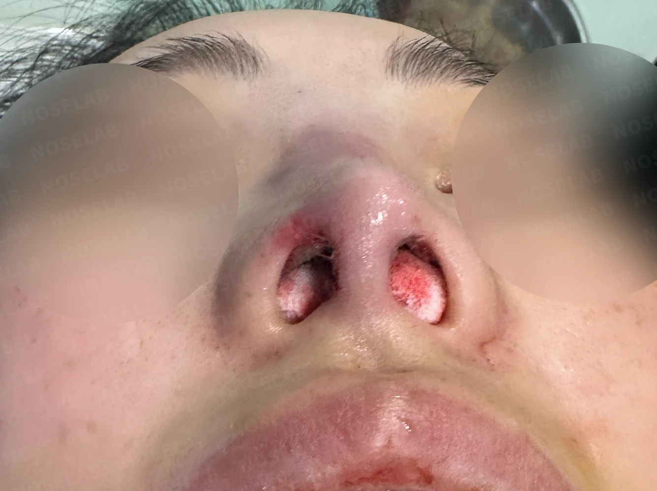
Before surgery / Day 0 immediately after surgery Photos taken immediately after surgery show swelling and may differ from the final result.
The depression above the right nostril has been well restored, and the nostril asymmetry now appears much more balanced and natural.
Photos immediately after surgery
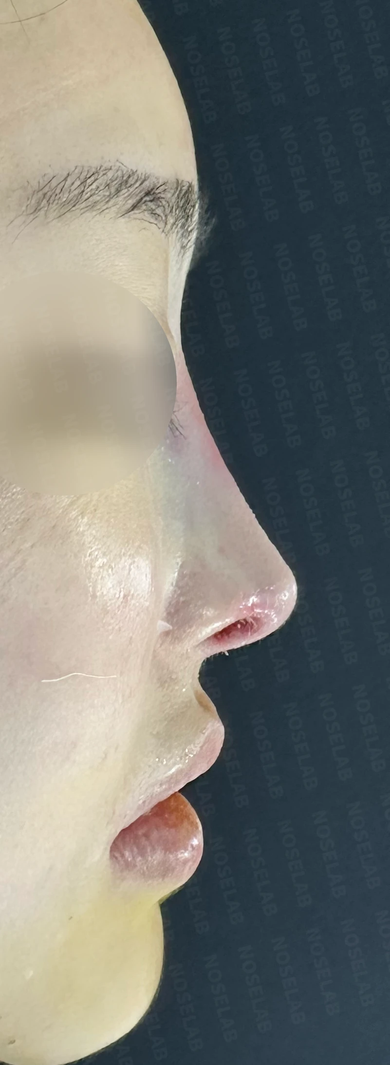
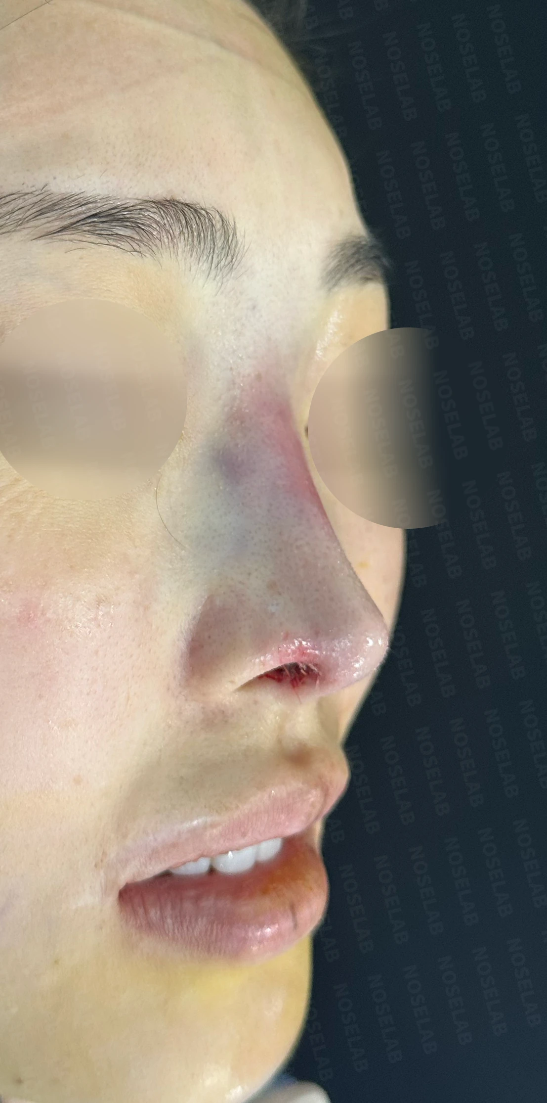
Photos immediately after surgery (Day 0): 90-degree profile, 45-degree oblique, and front view.Immediate postoperative photos show swelling and may differ from the final result.
Basal (nostril) view immediately after surgery (Day 0).Immediate postoperative photos show swelling and may differ from the final result.
My thoughts on nostril lowering surgery
When should nostril lowering surgery be considered? Most patients assume it is a surgery that must be done whenever nostril show is prominent from the front. However, my view is somewhat different. It certainly has the effect of reducing visible nostril show to some degree.
However, in many cases where excessive nostril show is seen from the front, it is not due to the shape of the alar rim itself, but rather because the nasal tip is genuinely upturned—in other words, the nasolabial angle is too wide. In such situations, rather than performing alar rim elongation, the primary surgery should be directed toward lowering the nasal tip and reducing the nasolabial angle.
Therefore, I view nostril lowering (alar rim extension) as one of the surgical options that can be utilized primarily when there is frontal nostril asymmetry that needs correction.
When congenital differences in alar rim shape cause one nostril to show more than the other, that asymmetry cannot be corrected without directly addressing that area. In such cases, nostril lowering surgery is performed through cartilage grafting, cartilage repositioning, or skin grafting.
There are several surgical techniques for nostril lowering, which I will explain in detail another time.
Today, we discussed nostril lowering surgery. I hope this provides helpful insight for those considering this procedure.
This was Dr. Peter C. Kang. Thank you.

Limitations and risks
When using donor rib cartilage, long-term follow-up is necessary because the grafted cartilage may resorb or warp.
When the septum is corrected at the same time, nasal obstruction may persist or recur, and in rare cases, septal perforation or changes in smell may occur.
Common potential risks include infection, bleeding, asymmetry, scarring, skin necrosis, and insufficient improvement; results vary depending on individual skin condition and previous surgical history.