Hello, this is Dr. Peter C. Kang, medical director.
Today, I would like to introduce a complex revision rhinoplasty case of a patient who was concerned about an upturned nose and cartilage showing through so thinly that it seemed ready to break through the nasal tip skin.
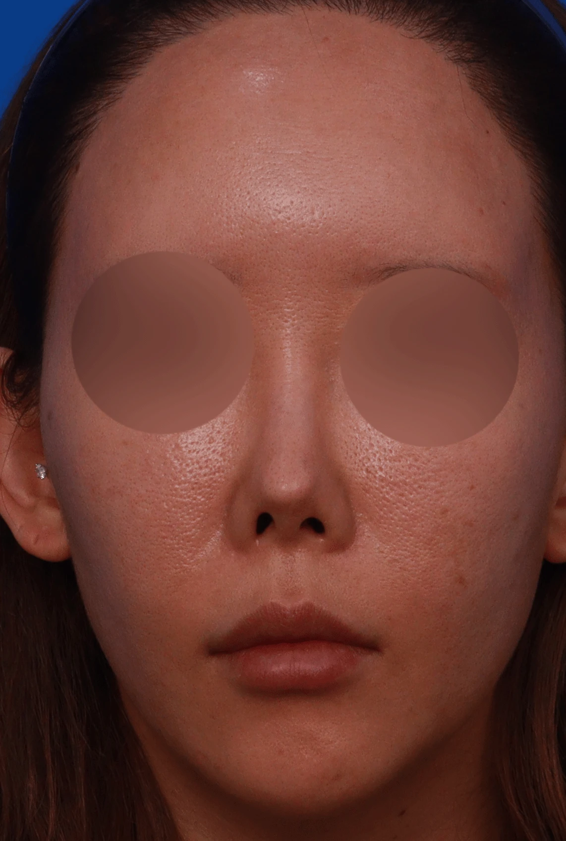
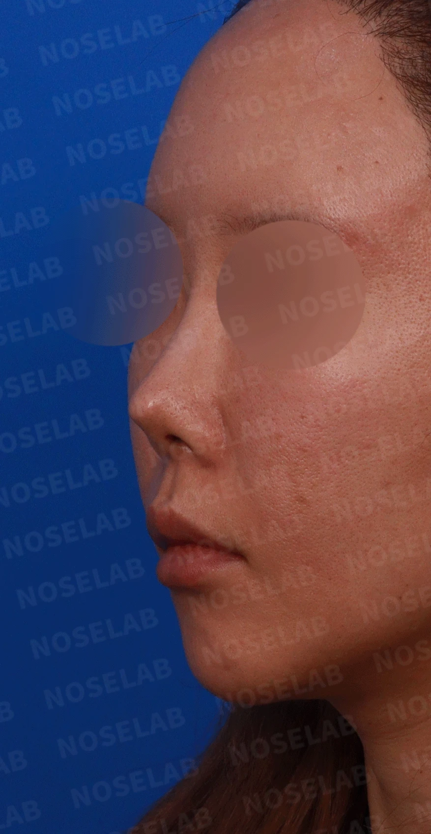
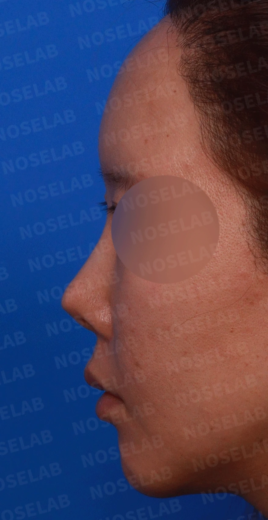
Design consultation front, left profile, and oblique views
📍 Surgical history
15 years ago in Bucheon: surgery with an I-shaped silicone implant and ear cartilage (septal cartilage use unclear)
🔎 Initial consultation findings
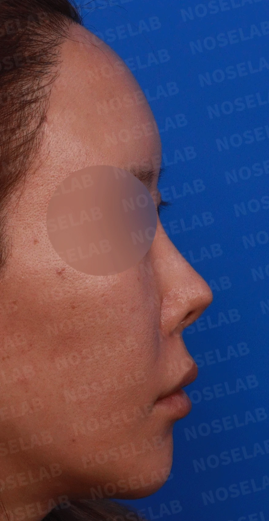
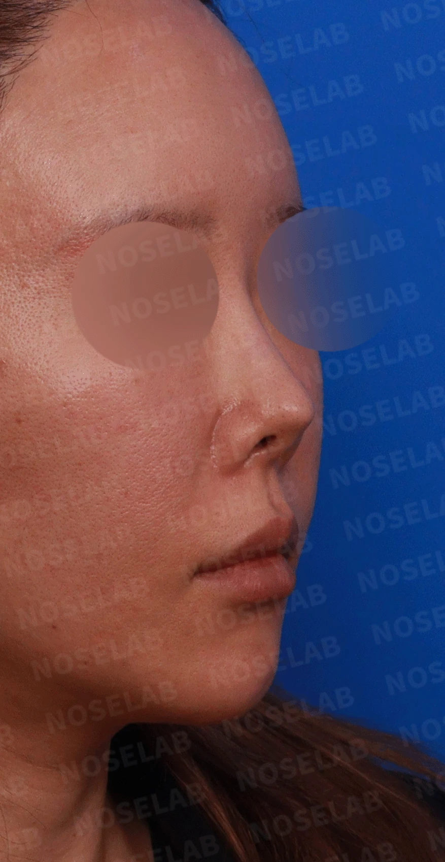
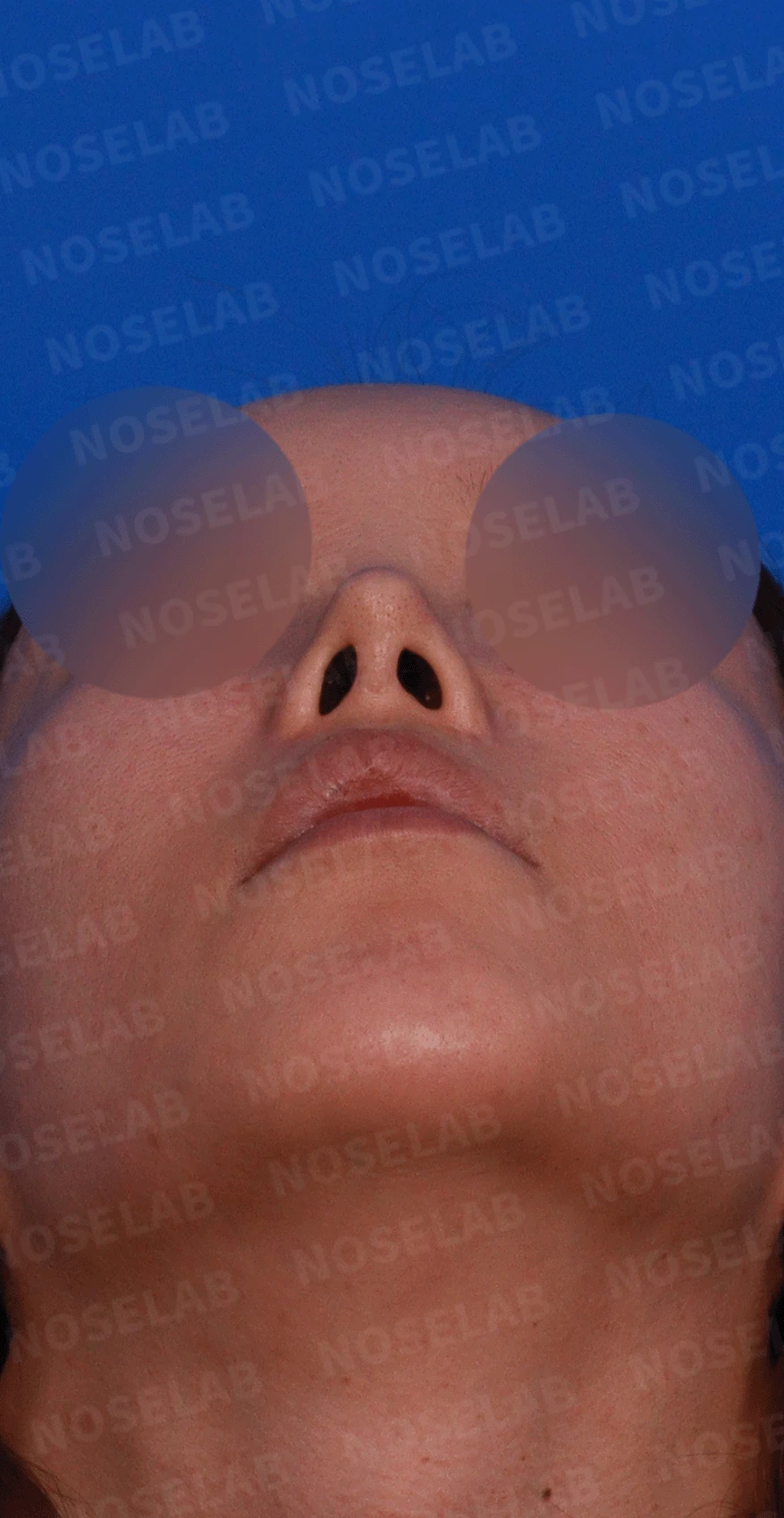
Design consultation right profile, oblique, and nostril views
Primary concerns
Nasal tip cartilage visible through extremely thinned skin, as if about to protrude
Pointed and severely upturned nasal tip
Front view
Upturned nasal shape
Excessive nostril show
Nostril asymmetry
High radix
Profile view
Pointed nasal tip
Retracted columella with poor visibility
Wide nasolabial angle
Nostrils
Nostril asymmetry
Columella tilted to the left
✨ Patient's requests
Resolving the visibility of the nasal tip cartilage
Correction of an upturned nose
Correction of nostril asymmetry
Achieving a natural nasal line
💉 Surgical procedure details
Structural improvement
Septal extension using autologous cartilage
Nasal tip cartilage repositioning and skin reinforcement using donor dermis
Removal of existing silicone implant
Correction of the nasolabial angle
Lower lateral cartilage repositioning to improve nostril symmetry
Deviated septum and rhinitis surgery to improve nasal function
Improvement in shape
Correction of upturned nose
Improving the pointed nasal tip
Adjustment of columellar position and angle
Lowered glabella radix
Skin reinforcement surgery
Peritip fat grafting for thinned nasal tip skin with cartilage show-through
Improving nasal tip skin thickness and texture using donor dermis
✅ Surgical results
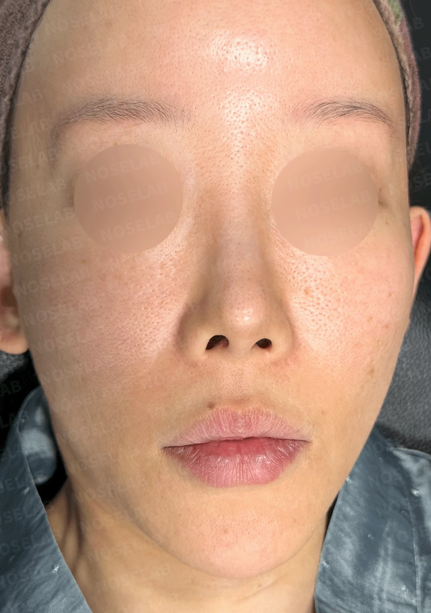
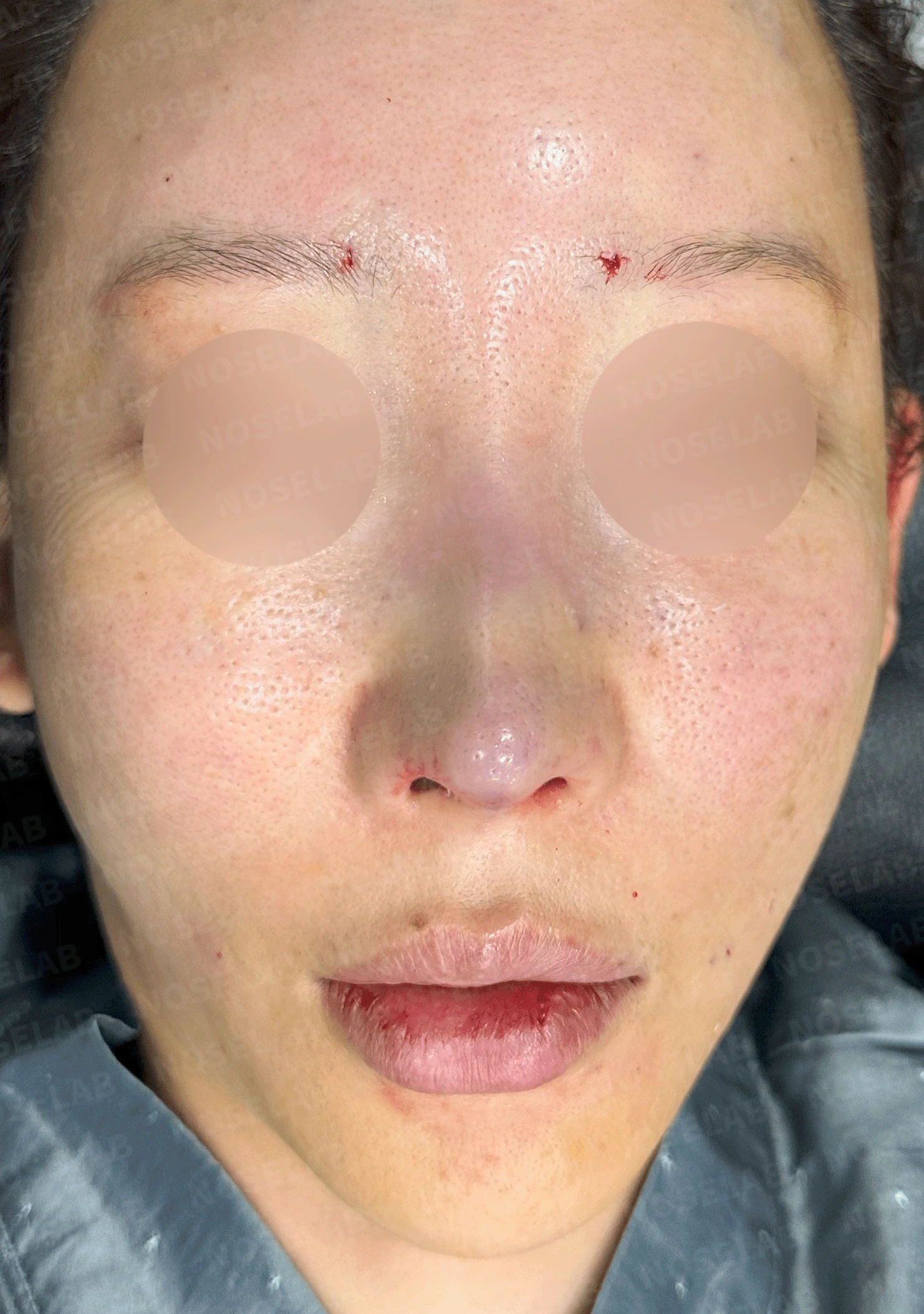
Before surgery / Day 0 immediately after surgery Photos taken immediately after surgery show swelling and may differ from the final result.
Front view
Improvement in upturned nasal shape
Significant reduction in nasal tip cartilage visibility
Reduction of nostril show
Enhanced nostril symmetry
More natural glabella
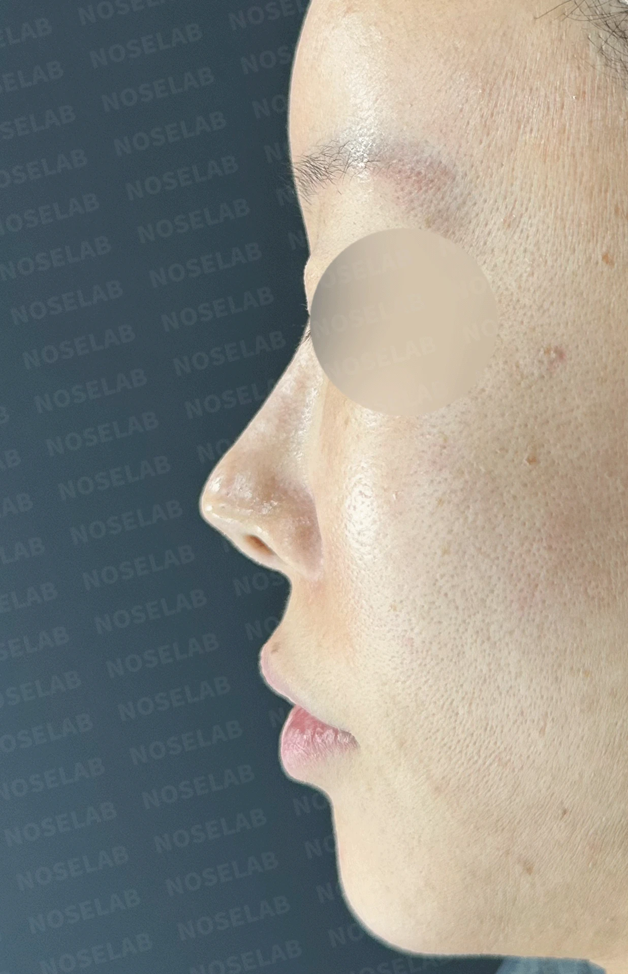
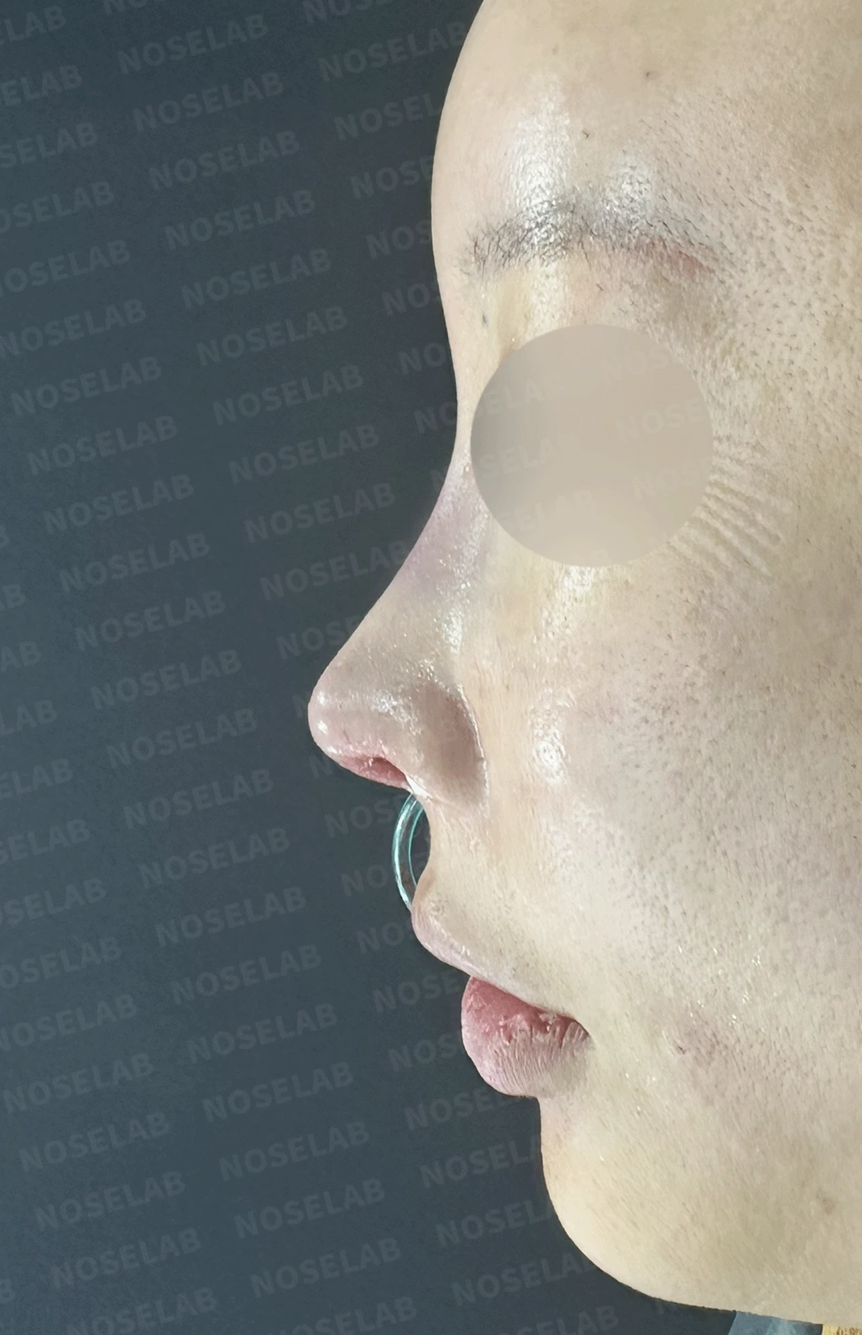
Before surgery / Day 0 immediately after surgery Photos taken immediately after surgery show swelling and may differ from the final result.
Profile view
Natural nasal tip shape
Correction of the upturned nasal tip
Adequate columellar show
Harmonious nasolabial angle
Balanced nasal line
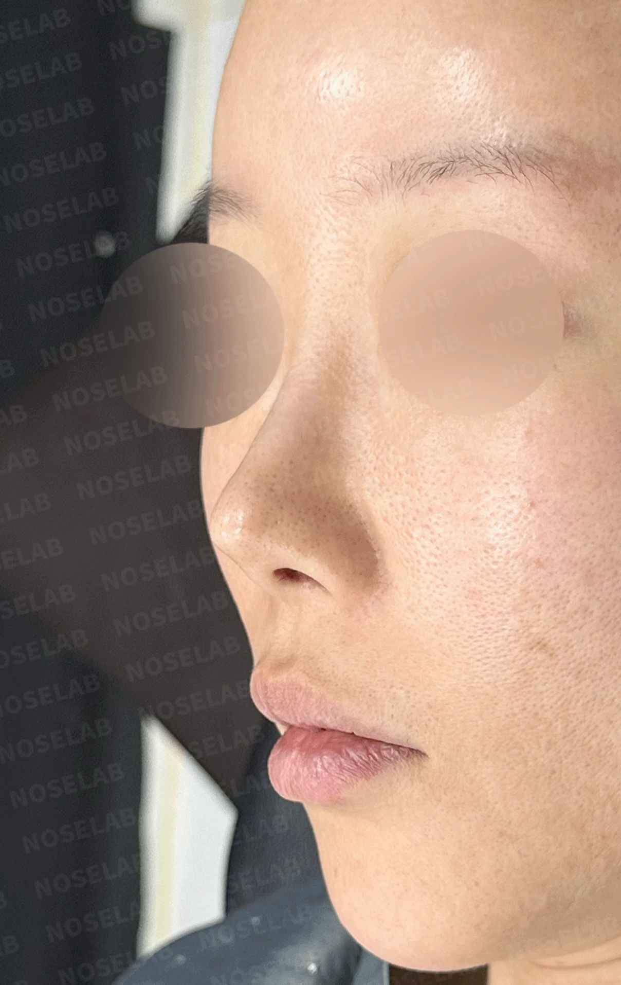
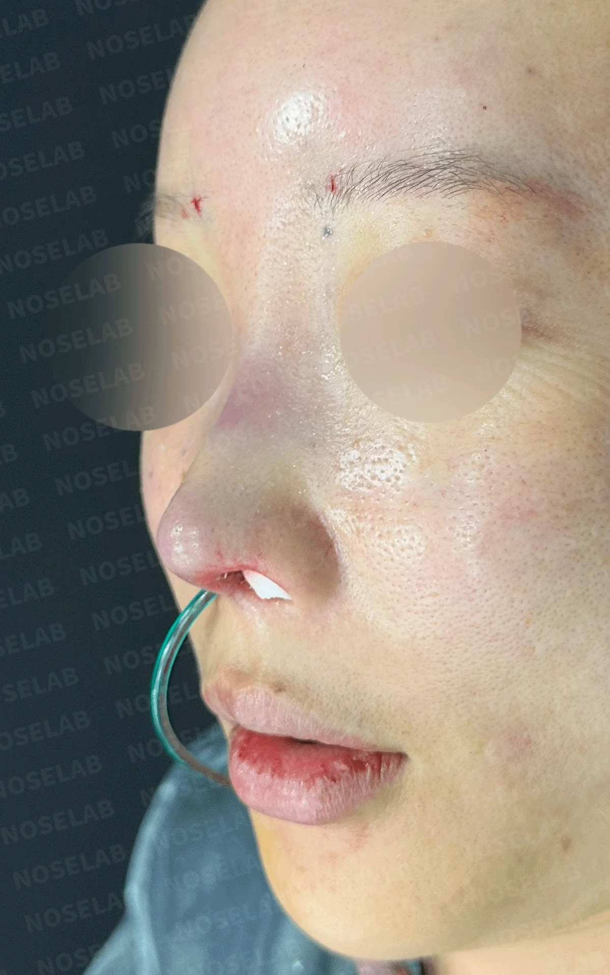
Before surgery / Day 0 immediately after surgery Photos taken immediately after surgery show swelling and may differ from the final result.
Nostrils
Symmetric nostril shape
Straightened columella
Stable nostril architecture
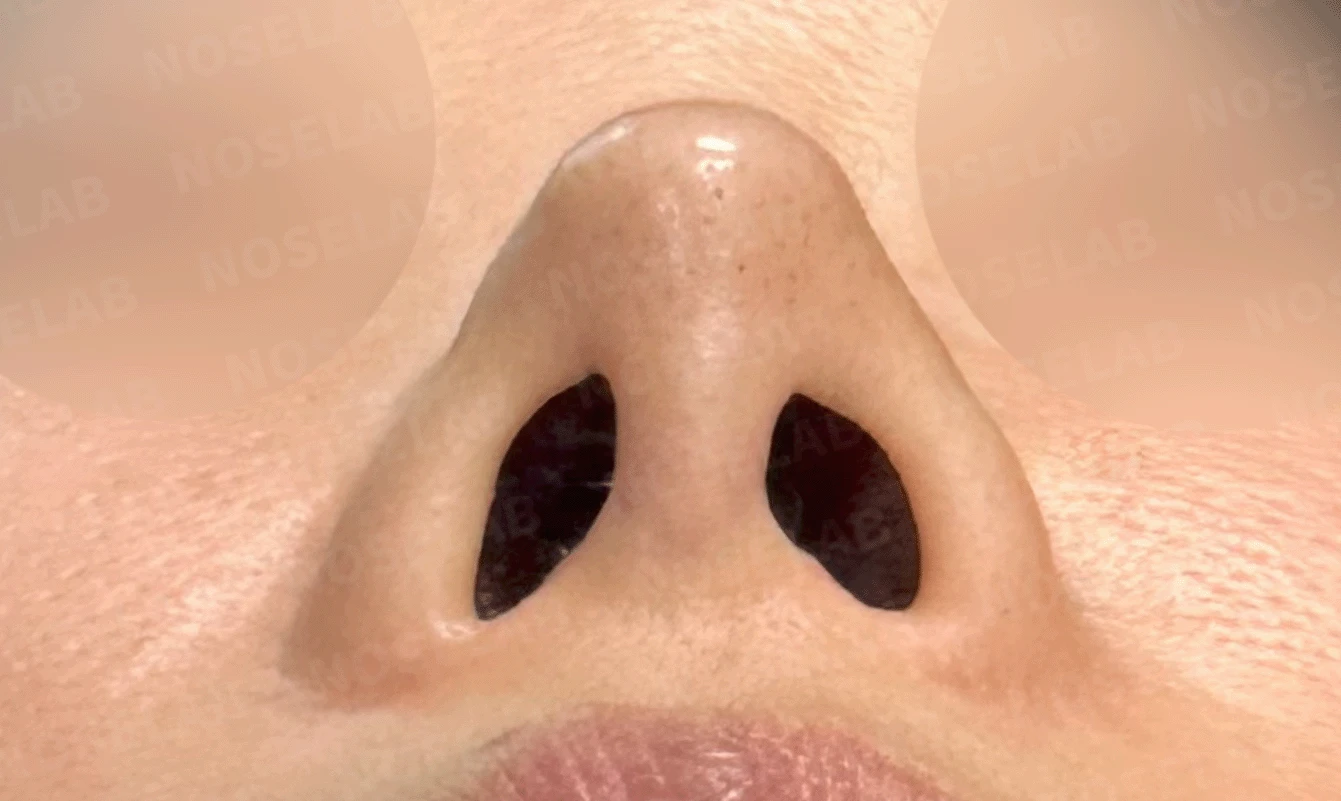
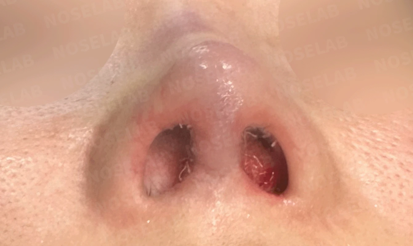
Before surgery / Day 0 immediately after surgery Photos taken immediately after surgery show swelling and may differ from the final result.
👨⚕️ Specialist's assessment
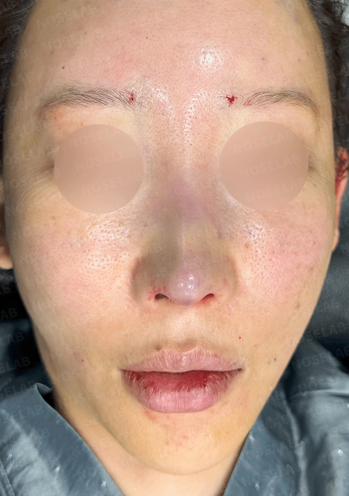
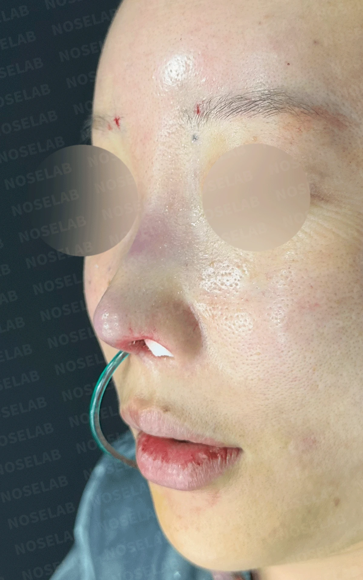
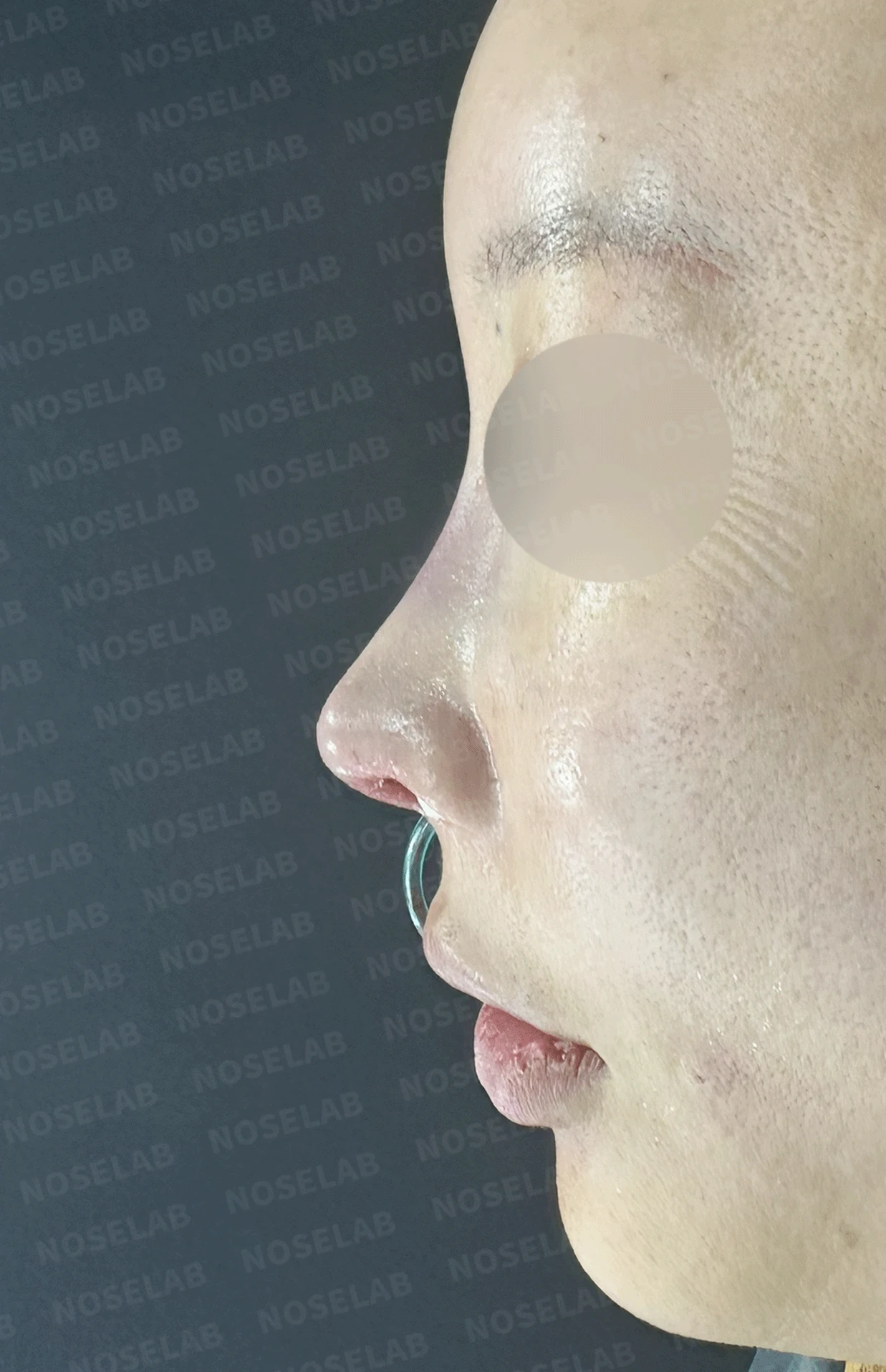
Day 0 immediately after surgery: front, profile, and oblique viewsImmediate postoperative photos show swelling and may differ from the final outcome.
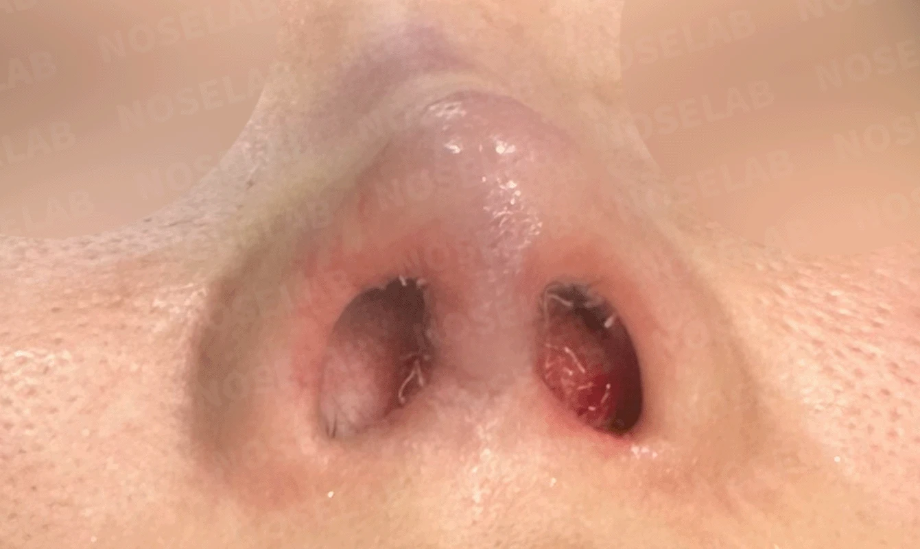
Nostril view on day 0 immediately after surgery Photos taken immediately after surgery show swelling and may differ from the final result.
The patient had undergone surgery 15 years prior using an I-shaped silicone implant and ear cartilage. The main concerns were cartilage showing through the nasal tip so thinly that it looked as though it might break through the skin, along with a severely upturned nasal tip. In addition, nostril asymmetry and a columella tilted toward the left required correction.
The highest priority was placed on reinforcing the nasal tip skin. Visibility of the cartilage beneath the nasal tip occurs when the overlying skin has thinned excessively, and this was addressed through skin reinforcement using fat grafting and donor dermis. By restoring appropriate thickness and elasticity to the thin skin, we significantly reduced the cartilage visibility and achieved a natural tip shape.
The upturned nose was corrected using autologous cartilage for septal extension and cartilage repositioning at the nasal tip. By adjusting the orientation and angle of the tip cartilages, the upturned tip was lowered to a natural position, and the sharp tip contour was softened. During this process, the position and angle of the columella were also adjusted to straighten the columella, which had been tilted to the left.
The previous silicone implant was removed, and autologous rib cartilage was used to reposition the tip cartilages and secure a stable support structure, while the nasolabial angle was adjusted to create a harmonious profile line. In addition, nostril asymmetry was addressed by meticulously repositioning the cartilages and correcting the deviated columella, achieving a balanced appearance on the frontal view as well.
This was a meaningful case where we focused on the primary issues of thinned tip skin and an upturned nose while comprehensively addressing various secondary problems, including nostril asymmetry and columellar deviation. In particular, the use of fat grafting and donor dermis to reinforce the nasal tip skin served as the key technique that effectively resolved the patient's unique concerns.
This has been Dr. Peter C. Kang, medical director.
Thank you.
#closedrhinoplasty #cartilageshowthrough #upturnednosecorrection #fatgraft #donordermis #customrhinoplasty

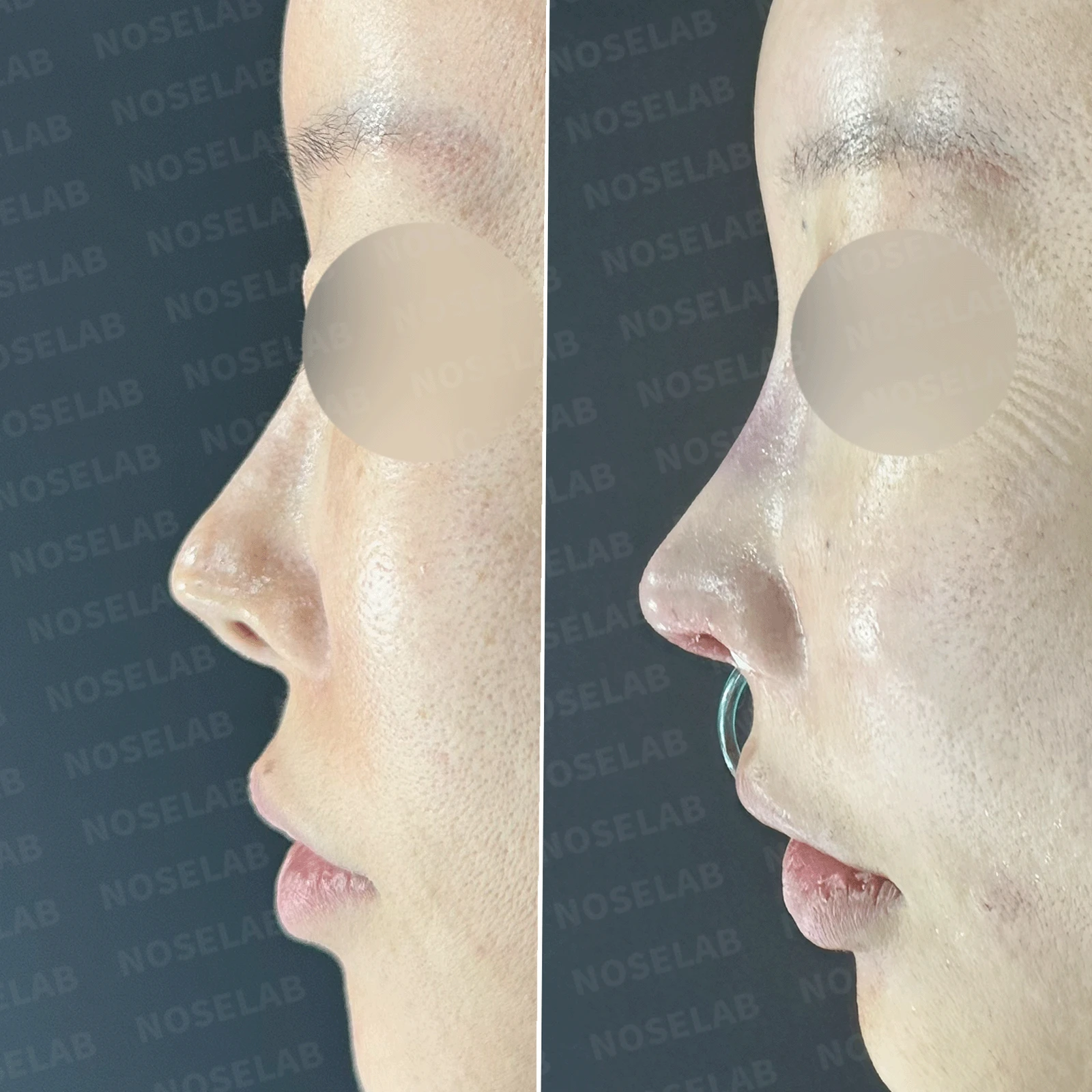
Limitations and risks
When autologous rib cartilage is used, scarring and pain may remain at the donor site on the chest, and pneumothorax (pleural injury) can occur rarely. Over time, the grafted cartilage may warp or undergo partial resorption.
When revising after implant removal, contracture may progress again after removal, the nasal height and length may decrease, and staged surgery may be necessary depending on the skin condition.
When the septum is corrected at the same time, nasal obstruction may persist or recur, and in rare cases, septal perforation or changes in smell may occur.
Common potential risks include infection, bleeding, asymmetry, scarring, skin necrosis, and insufficient improvement; results vary depending on individual skin condition and previous surgical history.