Hello, I am Dr. Peter C. Kang.
Today, I would like to introduce the case of a patient who visited our clinic concerned about cartilage show-through at the nasal tip and a widened nose following previous surgery.
🔍 Surgical history
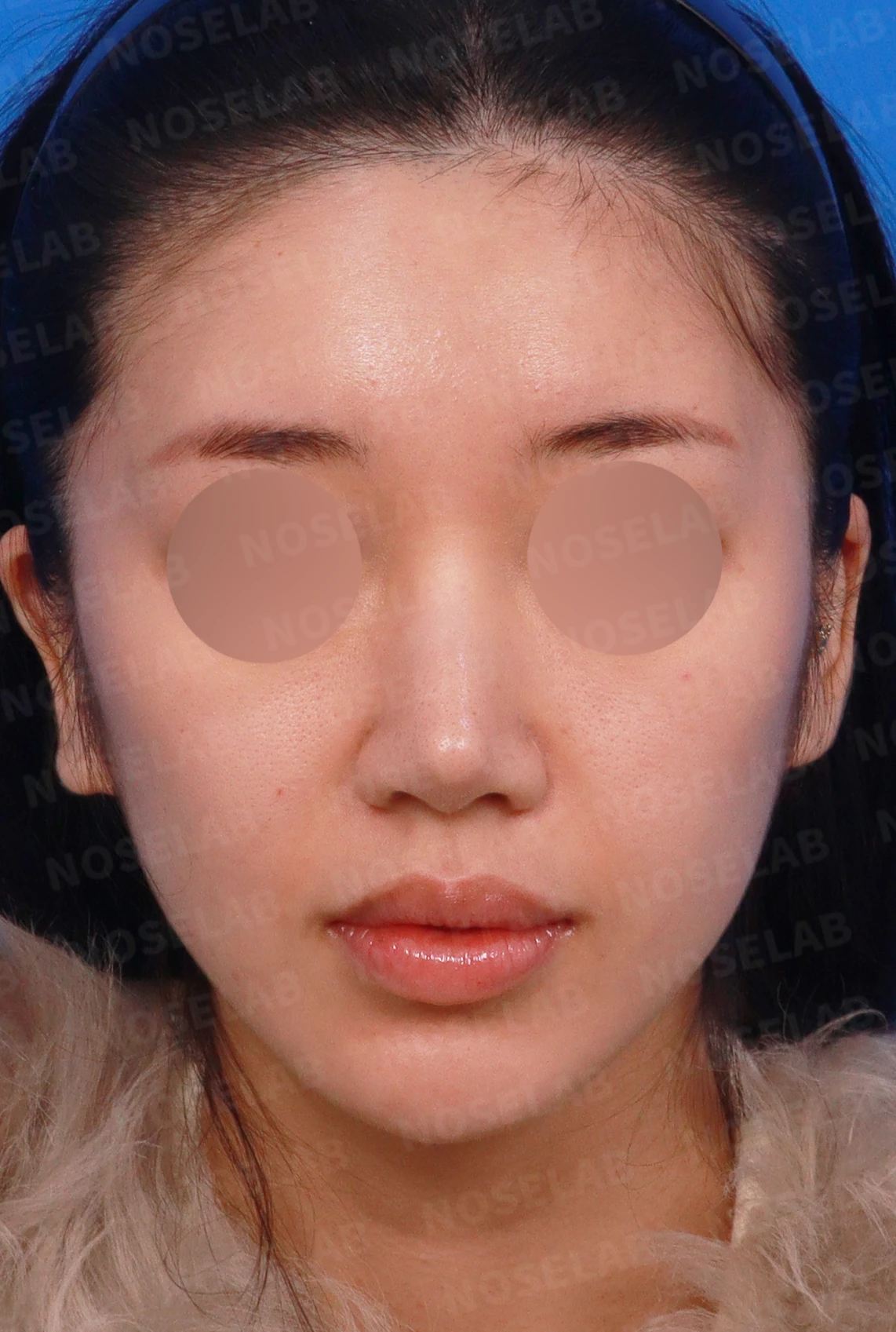
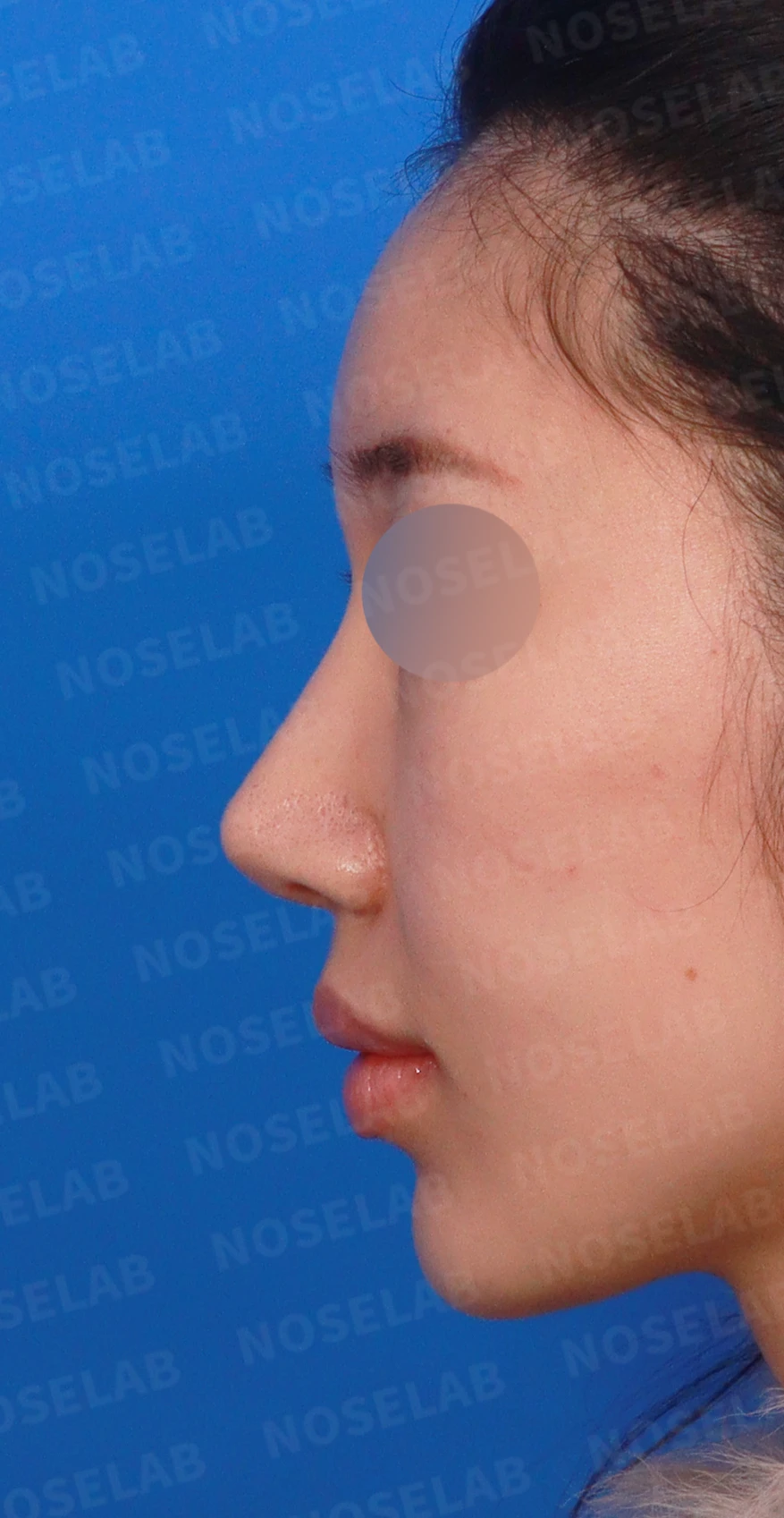
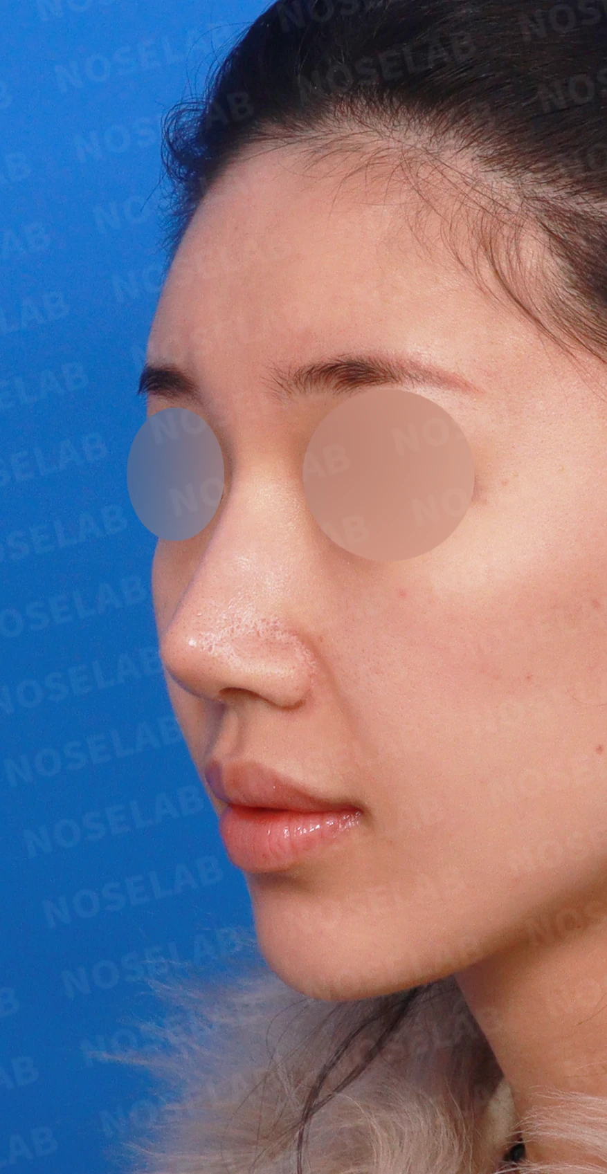
Front, left profile, and oblique views during design consultation.
7 years ago: silicone implant, ear cartilage, septum, alar reduction
Although overall rhinoplasty had been performed
Over time, the nasal tip cartilage began to show through the skin
The nose also began to appear wide and crooked
💉 Patient requests
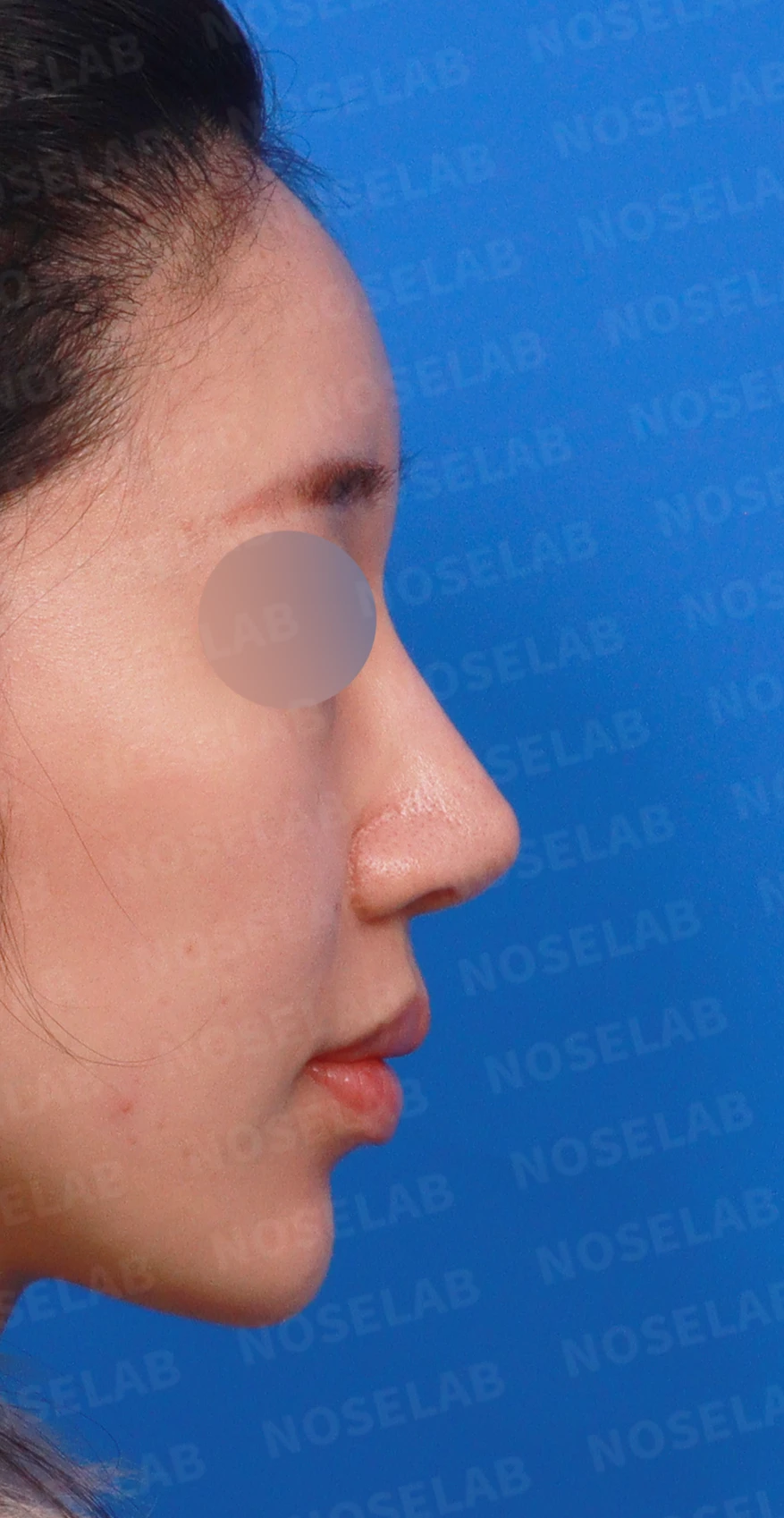
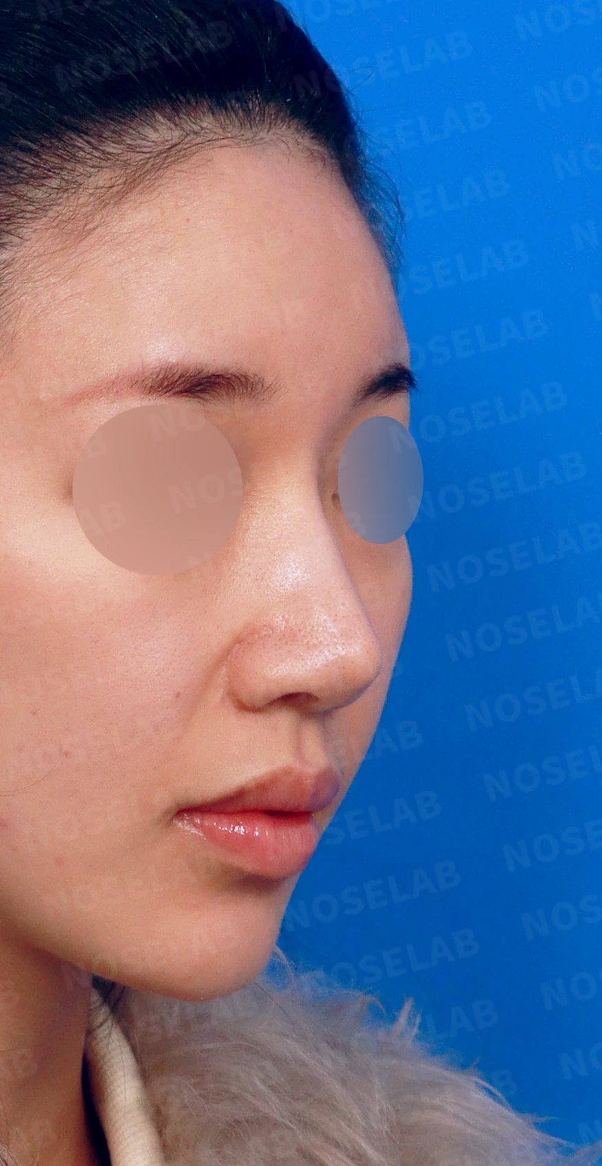
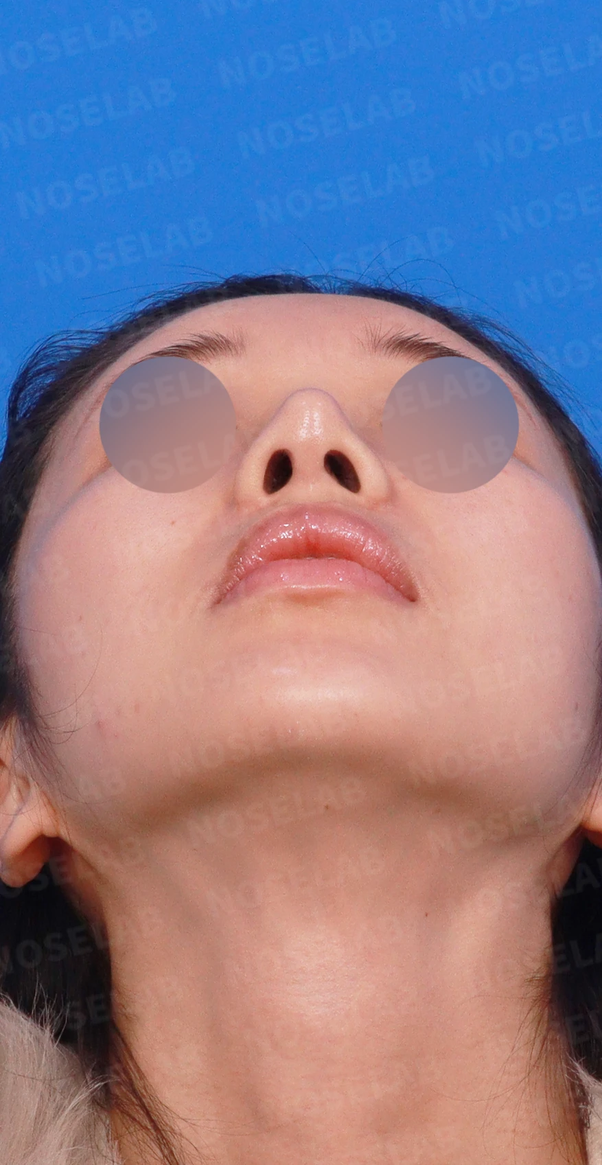
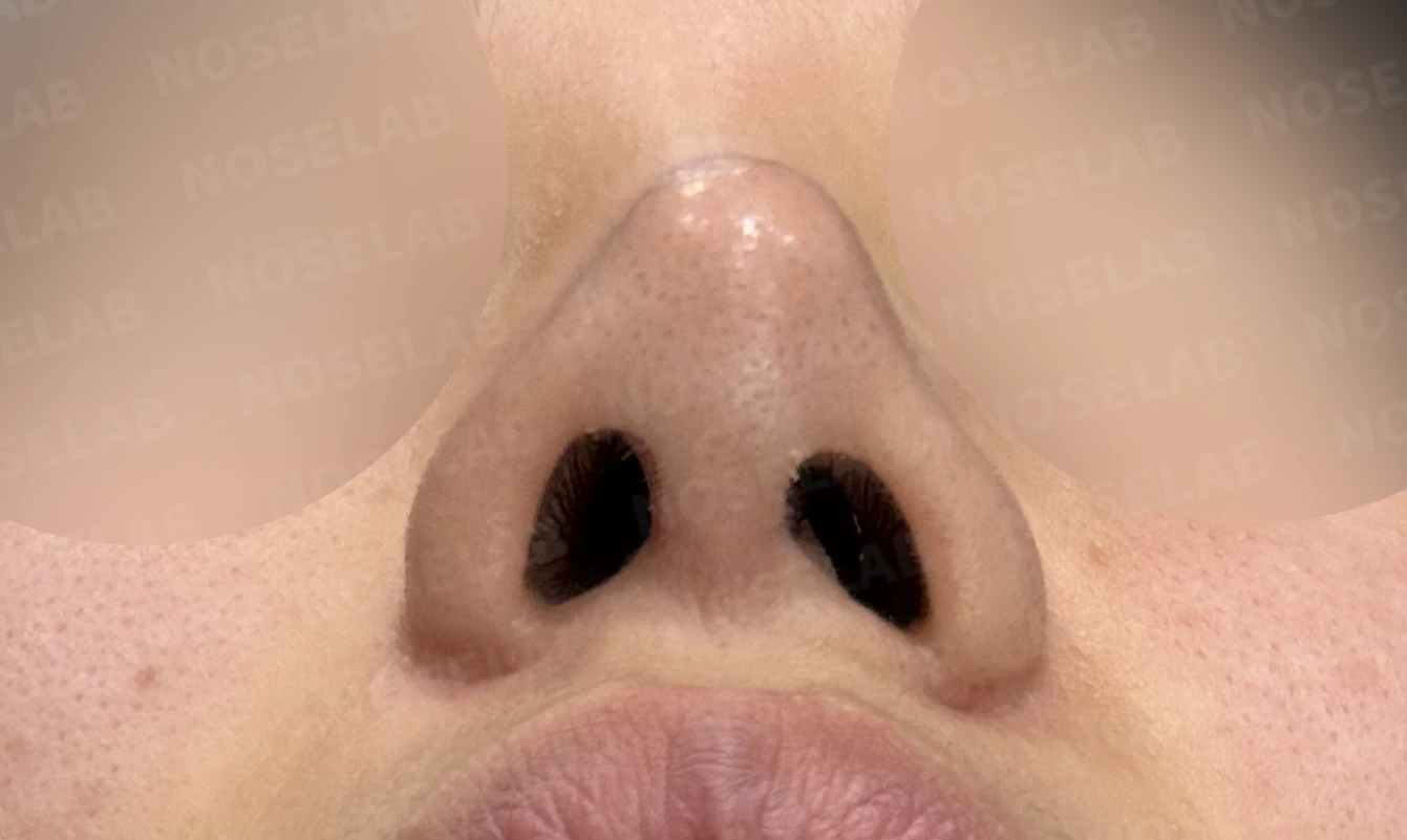
Right profile, oblique, and nostril views during design consultation.
The patient had concerns about the following issues after a previous surgery:
Nasal tip cartilage show-through: cartilage visible through the nasal tip skin
Distinct contour: wanting a clearly defined nasal contour from the front view
Slim, defined tip: a refined, sharper nasal tip
Straight line: a bridge line closer to straight than a straight-to-slightly-curved line
Correcting a wide nose: correcting widened nasal bones
Correcting a crooked nose: securing symmetry for a slightly deviated nose
Improving nasal obstruction: breathing difficulties caused by a deviated septum and hypertrophic rhinitis
🔧 Surgical plan and procedure
In this patient's case, cartilage show-through from the previous surgery was the primary aesthetic concern. In addition, nasal obstruction caused by a deviated septum and hypertrophic rhinitis needed to be addressed.
1. Skin reinforcement using autologous dermis
Autologous dermis grafting over the area of nasal tip cartilage show-through
Reinforcing skin thickness to prevent cartilage from showing through
Maintaining natural skin texture
Establishing stable, long-term skin thickness
2. Widening the nasal tip cartilage width
Broadening and repositioning the previously used cartilage
Structurally preventing recurrent cartilage show-through
Securing both tip volume and structural stability
Creating a defined yet natural nasal tip shape
3. Nasal tip plasty using autologous rib cartilage
Precise sculpting for a slim, defined tip
Forming a sharp, refined nasal tip line
Achieving a bridge line close to straight
Emphasizing a distinct contour on the front view
4. Lateral nasal osteotomy
Accurately correcting deviated nasal bones
Narrowing widened nasal bones for a slimmer impression
Achieving bilateral symmetry
Balanced proportions on the front view
5. Septoplasty
Correction of deviated septum
Restoring internal nasal cavity symmetry
Improvement of breathing function
6. High-frequency radiofrequency turbinoplasty
Reducing inferior turbinates enlarged by hypertrophic rhinitis
Securing internal nasal airway space
Addressing the root cause of nasal obstruction
Rapid recovery through a minimally invasive approach
7. Nasolabial angle correction
Improving the angle between the nose and the upper lip
A refined silhouette in the profile line
Harmony in overall facial proportions
🌟 Surgical results
1. Resolution of nasal tip cartilage show-through
Skin thickness reinforced with autologous dermis grafting
Cartilage completely covered for a natural nasal tip
Widened cartilage width prevents recurrence
Smooth skin texture maintained
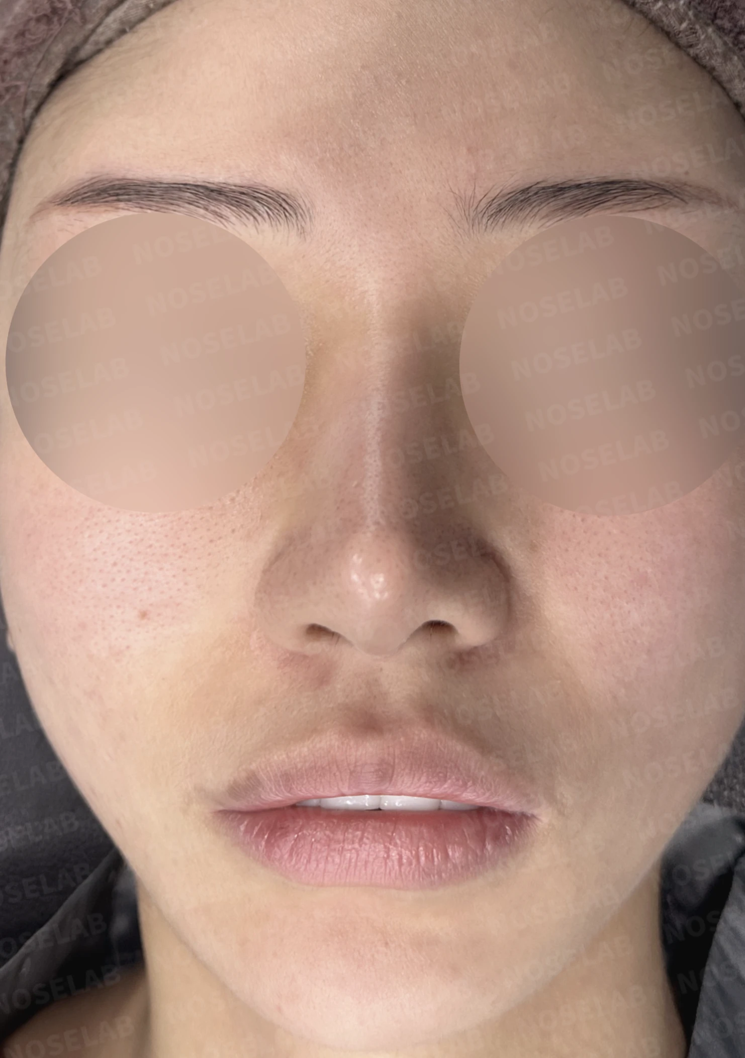
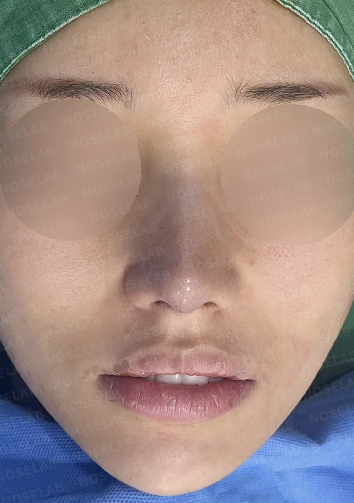
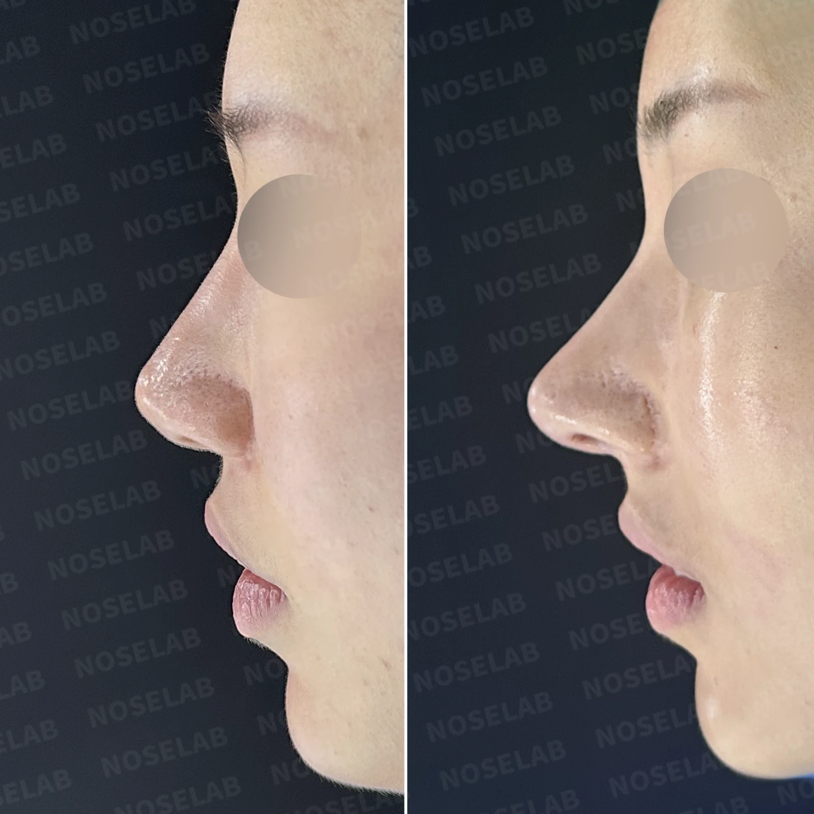
Before surgery / Day 0 immediately after surgery Photos taken immediately after surgery show swelling and may differ from the final result.
2. Aesthetic improvement
Defined nasal contour on the front view
Slim, defined nasal tip with a sharp aesthetic
Refined bridge line close to straight
Better symmetry achieved by correcting deviated nasal bones
A previously wide nose transformed into a slim, refined appearance
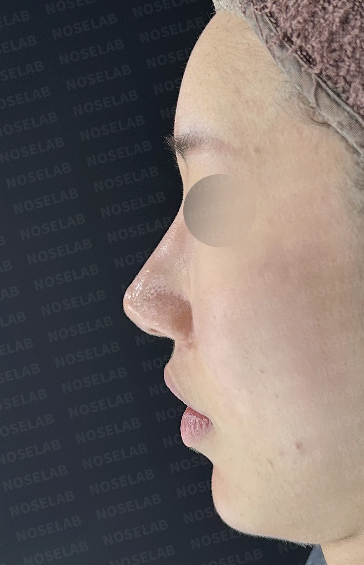
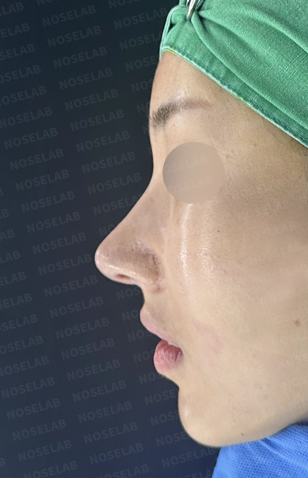
Before surgery / Day 0 immediately after surgery Photos taken immediately after surgery show swelling and may differ from the final result.
3. Functional improvement
Structural stability achieved through deviated septum correction
Securing internal nasal airway space with turbinoplasty
Smooth breathing through both sides
Chronic nasal obstruction significantly improved
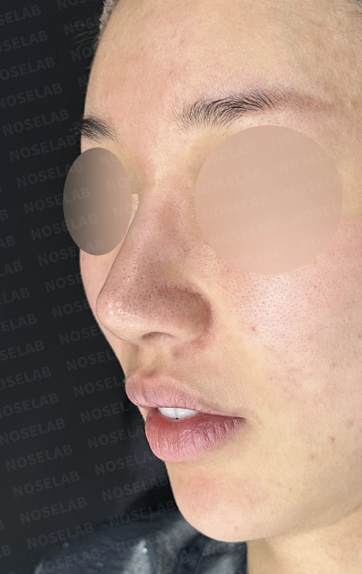
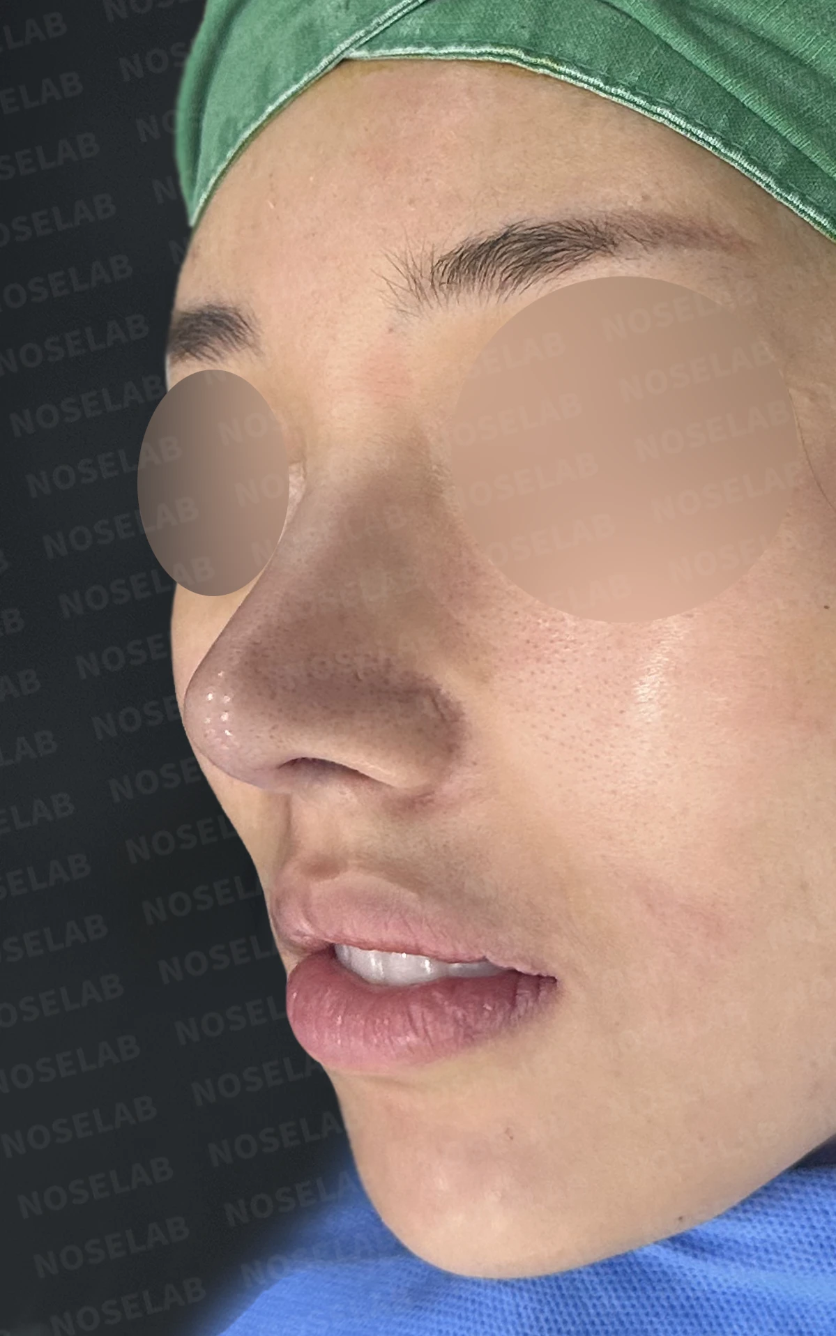
Before surgery / Day 0 immediately after surgery Photos taken immediately after surgery show swelling and may differ from the final result.
4. Overall harmony
Refining the profile line through nasolabial angle correction
Balanced proportions so the midface does not appear elongated
Proportions in harmony with the whole face
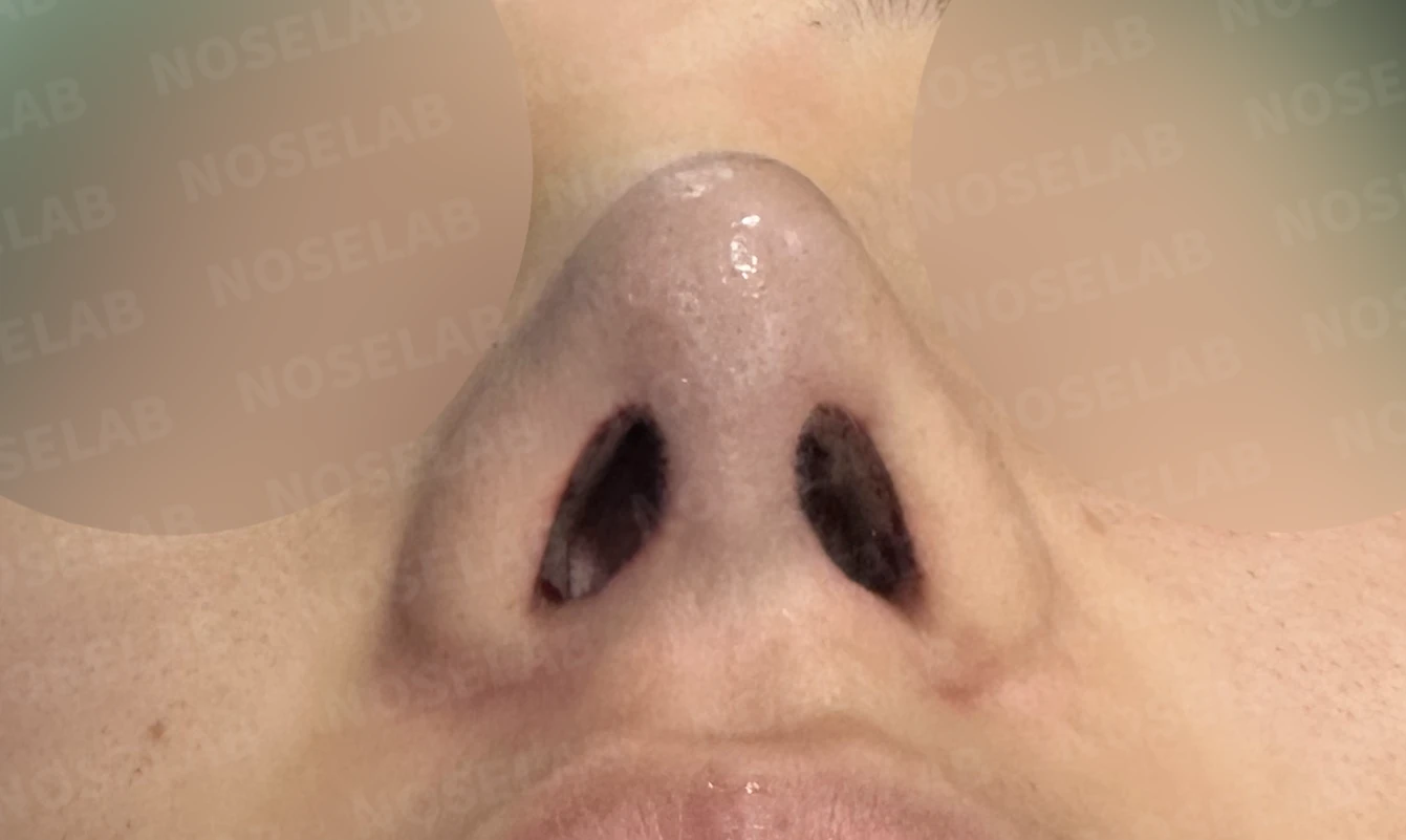
Before surgery / Day 0 immediately after surgery Photos taken immediately after surgery show swelling and may differ from the final result.
💡 Specialist opinion
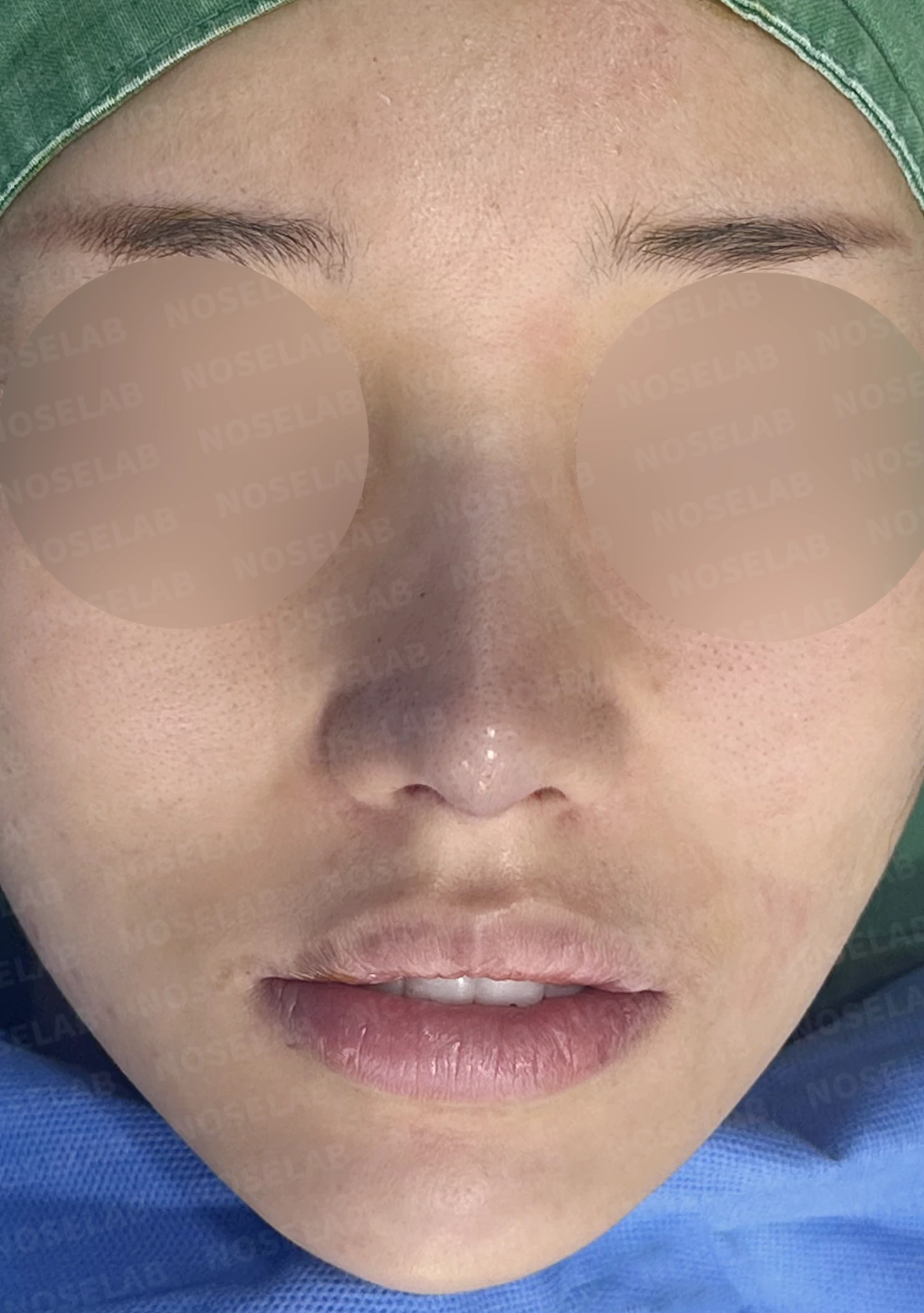
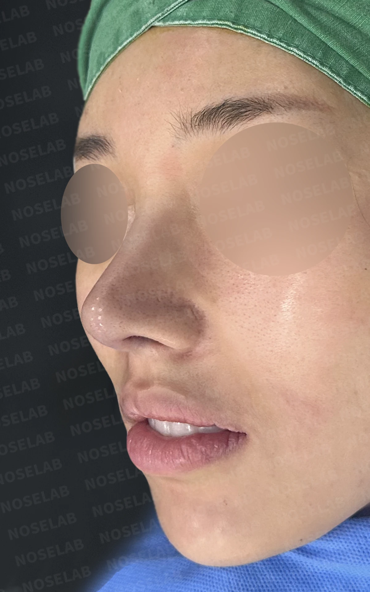
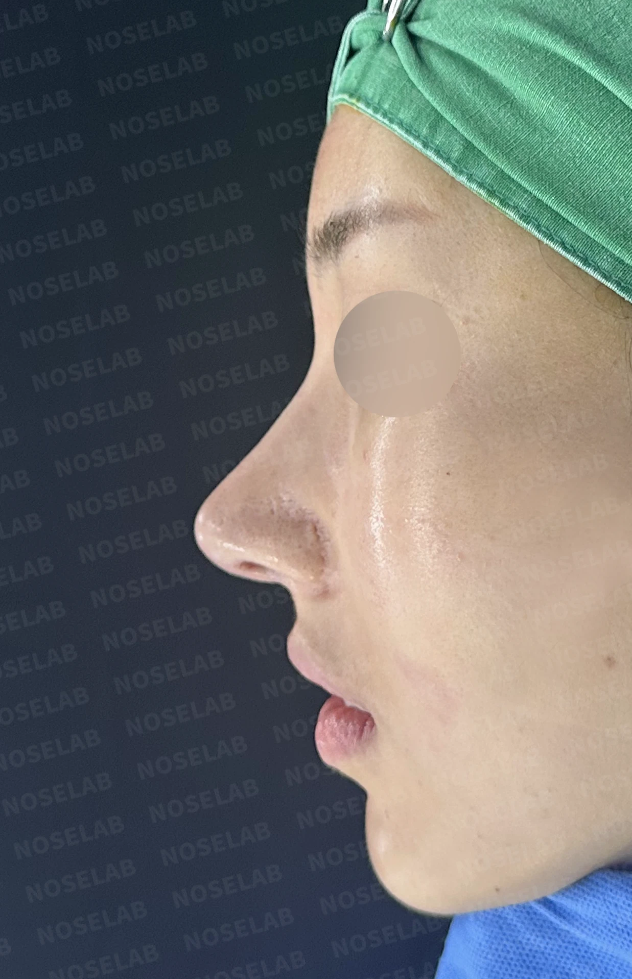
Front, oblique, and profile views on day 0 immediately after surgery. Photos taken immediately after surgery show swelling and may differ from the final result.
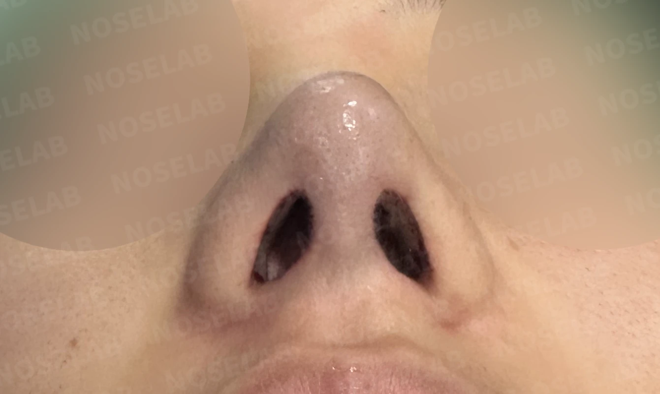
Nostril view on day 0 immediately after surgery. Photos taken immediately after surgery show swelling and may differ from the final result.
Cartilage show-through at the nasal tip occurs when the skin is thin or when the cartilage framework is overly sharp. In this patient's case, the cartilage used in the previous surgery was relatively sharp, and because the skin was thin, the cartilage gradually became more visible over time.
Resolving this problem requires a two-step approach. First, skin thickness must be reinforced. Because autologous dermis is harvested from the patient's own body, there is no risk of rejection, and it remains stable over time, making it an ideal material for reinforcing nasal tip skin.
Second, the cartilage structure itself must be modified. Rather than merely thickening the skin, the cartilage width was widened and repositioned so that sharp edges no longer irritate the skin. This not only resolves the current issue but also prevents future cartilage show-through.
At the same time, autologous rib cartilage was sculpted to create the slim, defined nasal tip desired by the patient. Achieving a sharp definition without allowing cartilage to show through requires a delicate balance; performing both skin reinforcement and cartilage repositioning enabled us to satisfy both goals.
Additionally, lateral nasal osteotomy corrected the widened and deviated nasal bones, creating a clear and slim contour from the front view. Nasal obstruction caused by a deviated septum and hypertrophic rhinitis was also addressed, yielding a result that was satisfying both functionally and aesthetically.
Revision rhinoplasty should not simply correct the problem area; it is essential to understand and treat the underlying cause. NOSELAB will continue to pursue systematic analysis and delicate surgical techniques to achieve the intended results for our patients.
This was Dr. Peter C. Kang. Thank you.
#closedrhinoplasty #closedrevisionrhinoplasty #revisionrhinoplasty #cartilageshowthrough #autologousdermisgraft #skinreinforcement #definedtip #widenosecorrection #crookednosecorrection #lateralosteotomy #deviatedseptum #hypertrophicrhinitis #nasalobstructionrelief #nasolabialanglecorrection

Limitations and risks
When autologous rib cartilage is used, scarring and pain may remain at the donor site on the chest, and pneumothorax (pleural injury) can occur rarely. Over time, the grafted cartilage may warp or undergo partial resorption.
When the septum is corrected at the same time, nasal obstruction may persist or recur, and in rare cases, septal perforation or changes in smell may occur.
Common potential risks include infection, bleeding, asymmetry, scarring, skin necrosis, and insufficient improvement; results vary depending on individual skin condition and previous surgical history.