Hello, I am Dr. Peter C. Kang.
Today, I would like to share the case of a patient who underwent autologous dermis grafting to improve tip sharpness and cartilage show-through.
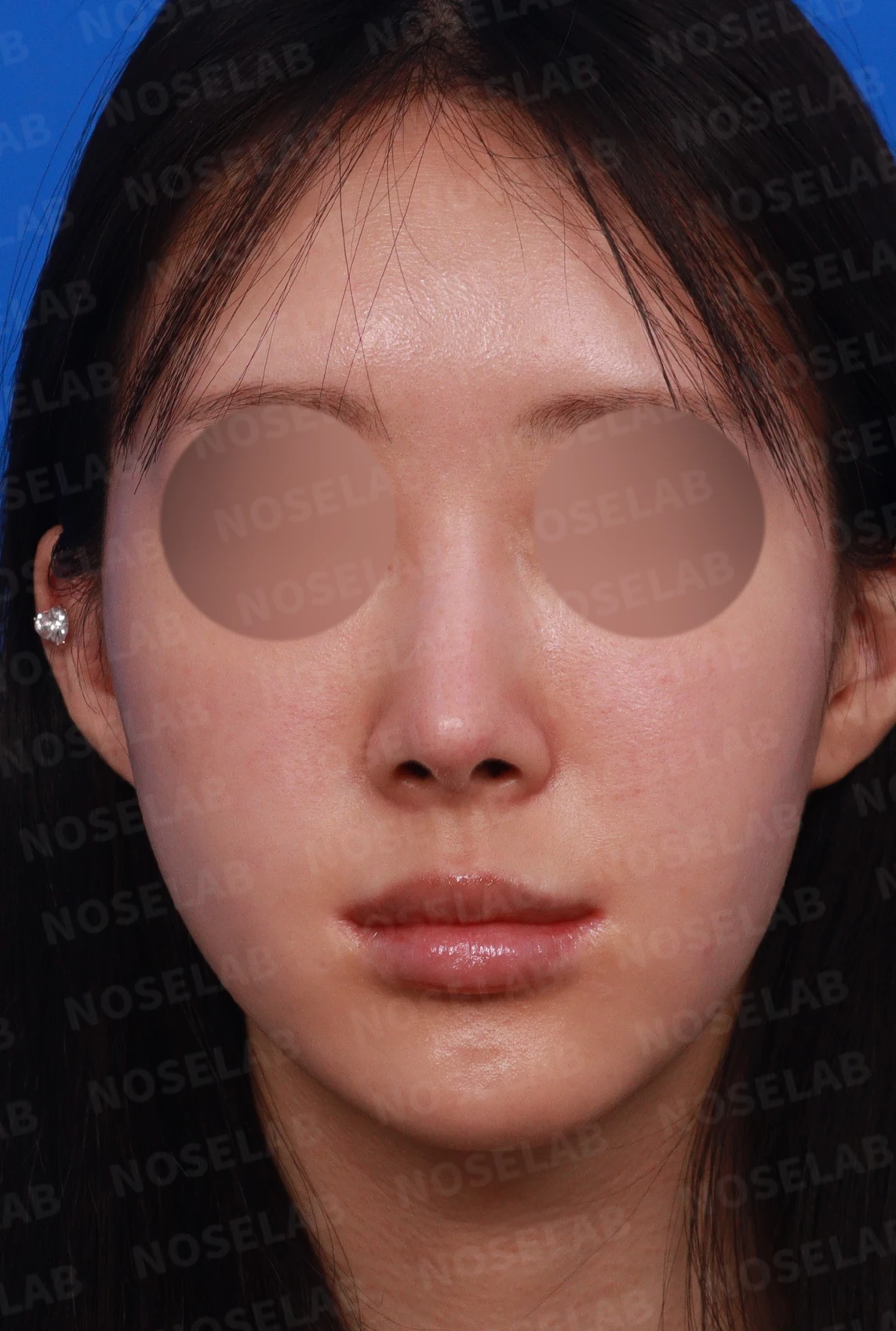
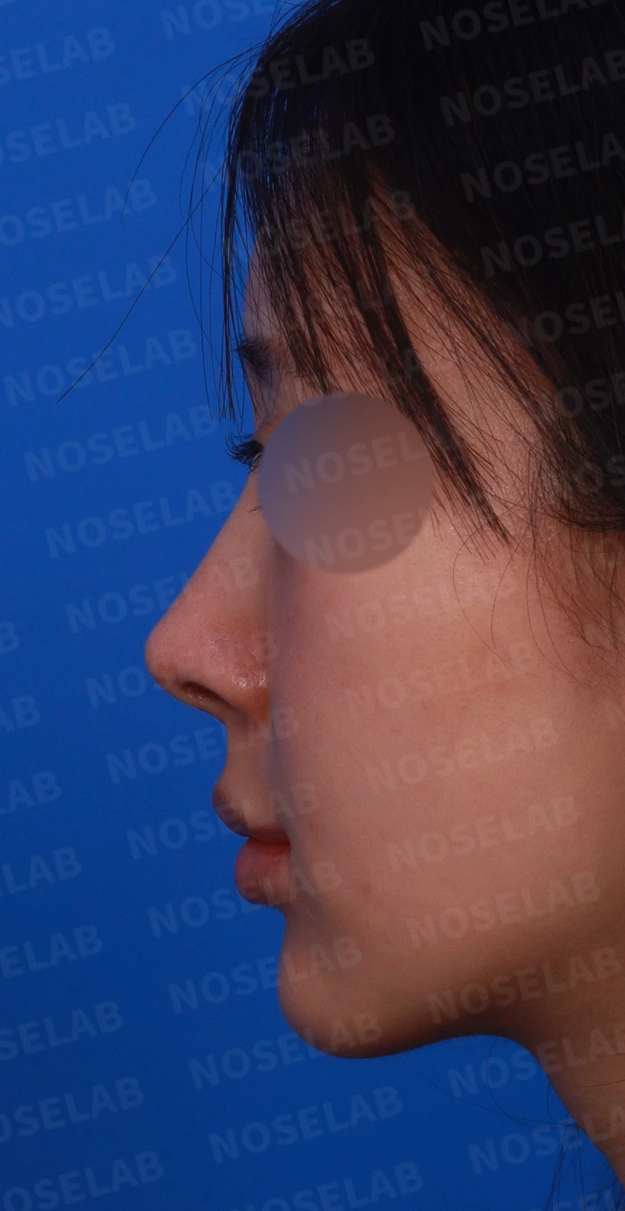
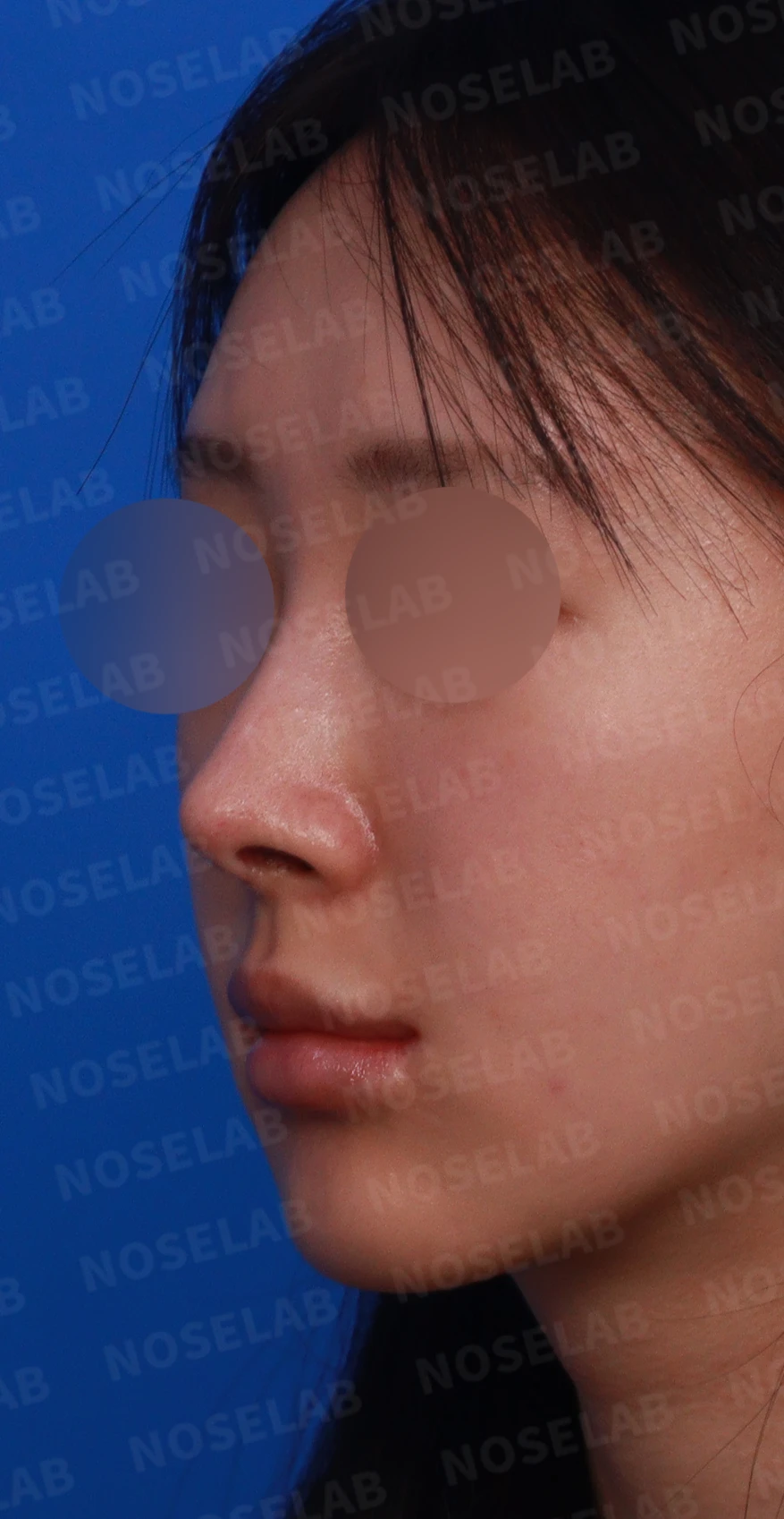
Front, left profile, and oblique views during design consultation
📍 Surgical history
7–8 years ago: Nose filler procedure, subsequently dissolved
3 years ago: Septum + ear cartilage (nasal tip)
March 2024: Silicone implant + autologous rib cartilage
August 2024: Alloderm + nasal tip cartilage trimming
🔎 Initial consultation findings
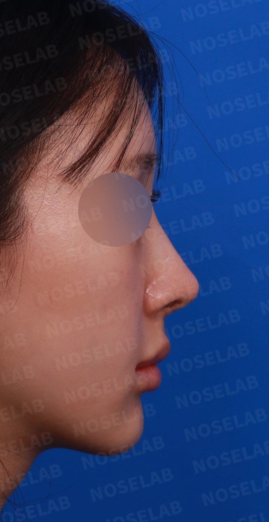
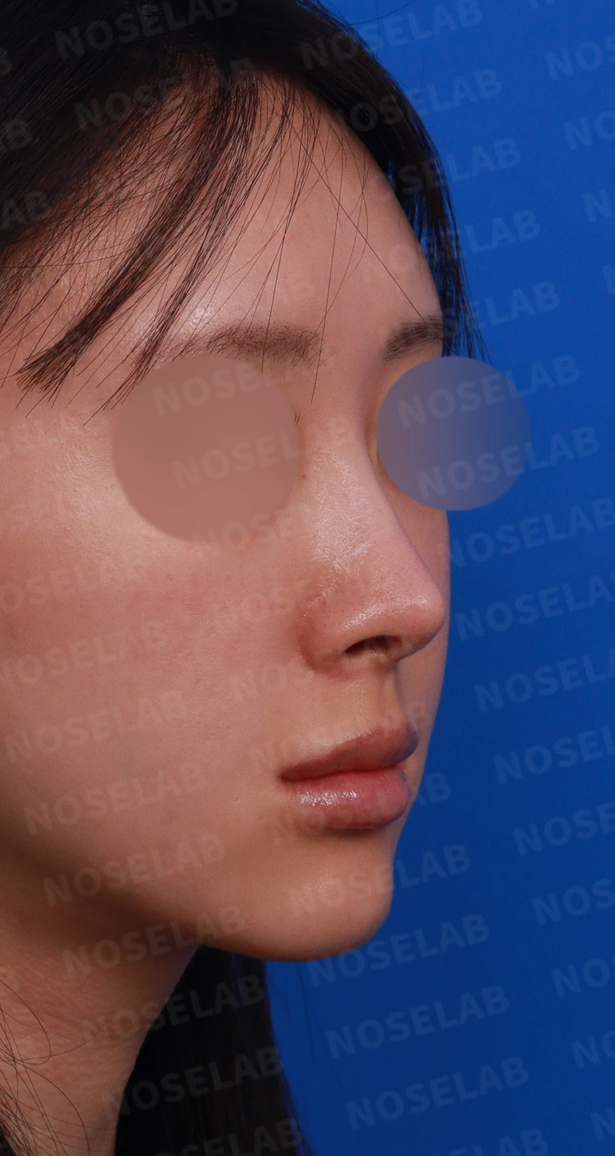
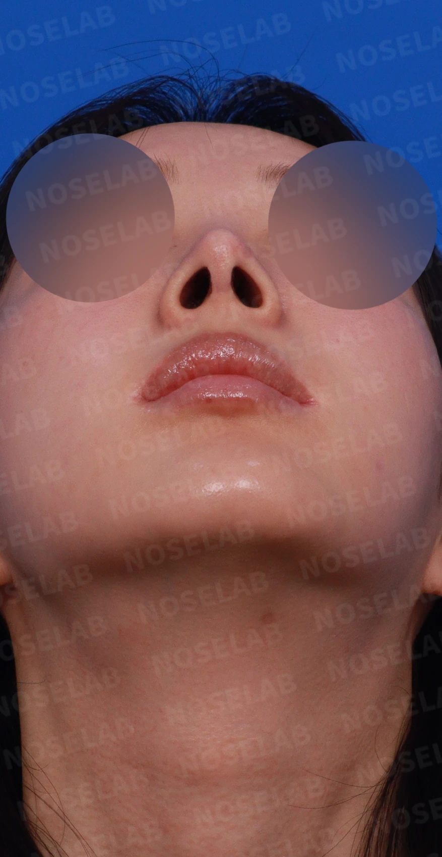
Right profile, oblique, and nostril views during design consultation
Thinned nasal tip skin
Cartilage show-through
Pointed nasal tip
Long nasal shape
Deviated nose
Narrow nasal bridge and nasal tip width
Large nostrils
Thin columella
Rhinitis symptoms
Nasal obstruction
✨ Patient's requests
Improving the pointed nasal tip
Decreased nostril show
Correction of hanging columella
Resolution of nasal tip cartilage show-through
💉 Surgical procedure details
1. Structural improvement
Correction of deviated nose with lateral osteotomy
Retroauricular autologous dermis harvest
Reinforcement of thinned tip skin
2. Shape improvement
Raising the columella
Natural nasolabial angle correction
Overall nasal profile
3. Functional improvement
Treatment of rhinitis
Relief of nasal obstruction
Overall structural stability secured
✅ Surgical results
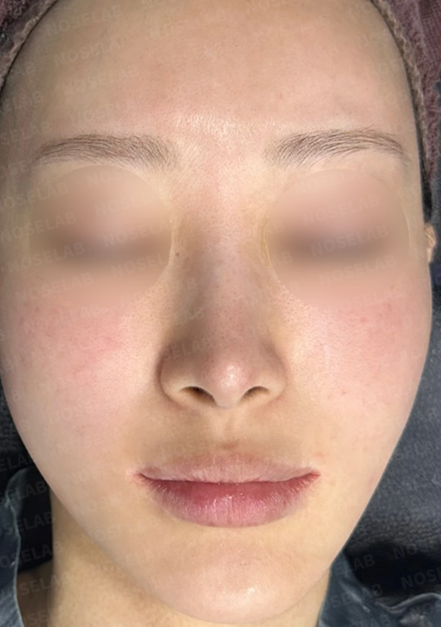
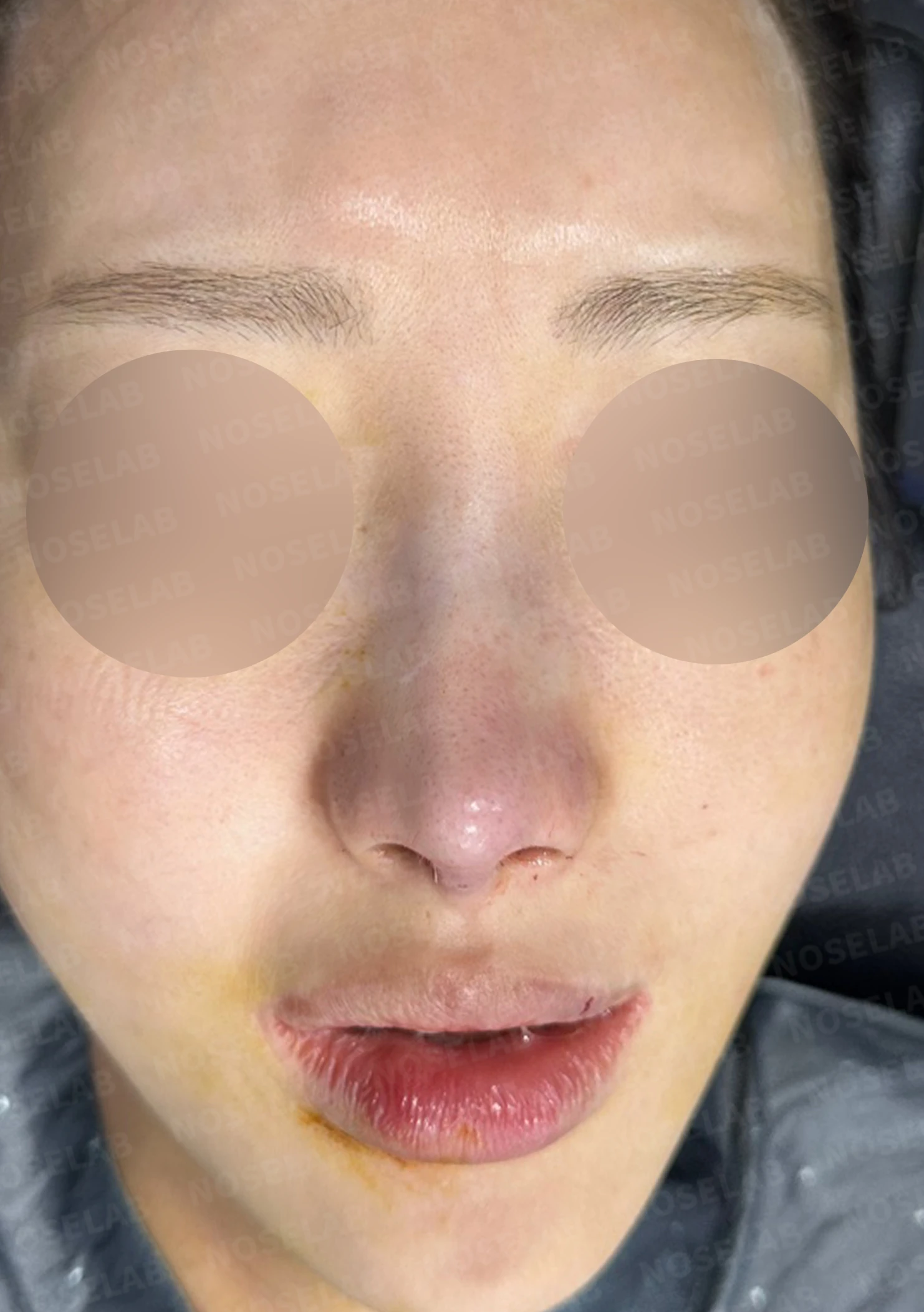
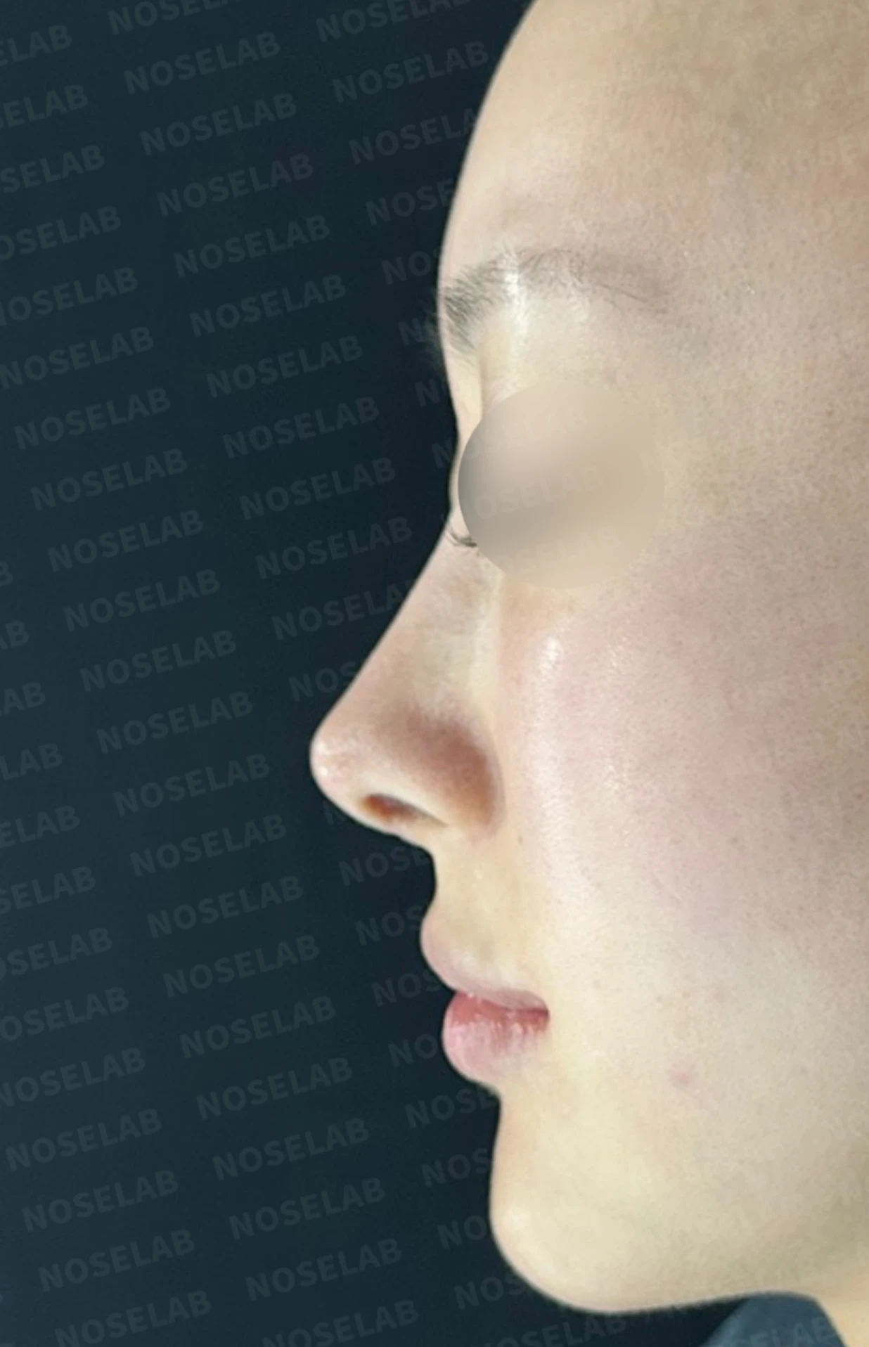
Before surgery / Day 0 immediately after surgery Photos taken immediately after surgery show swelling and may differ from the final result.
1. Front view
Natural nasal tip
Improved width
Balanced profile
Before surgery / Day 0 immediately after surgery Photos taken immediately after surgery show swelling and may differ from the final result.
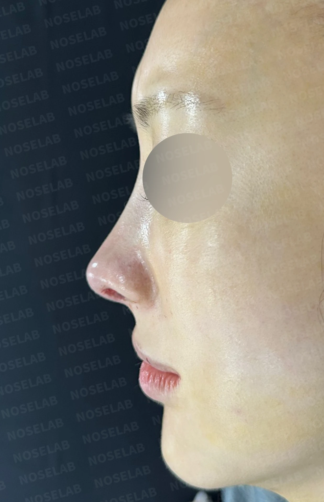
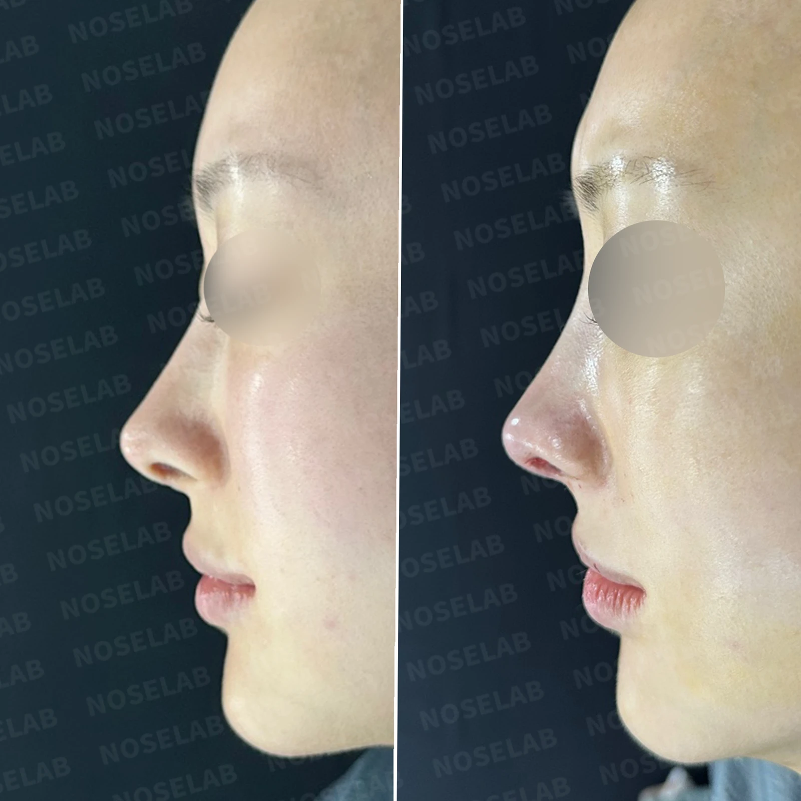
2. Profile view
Softened nasal tip
Stable columellar position
Natural nasolabial angle
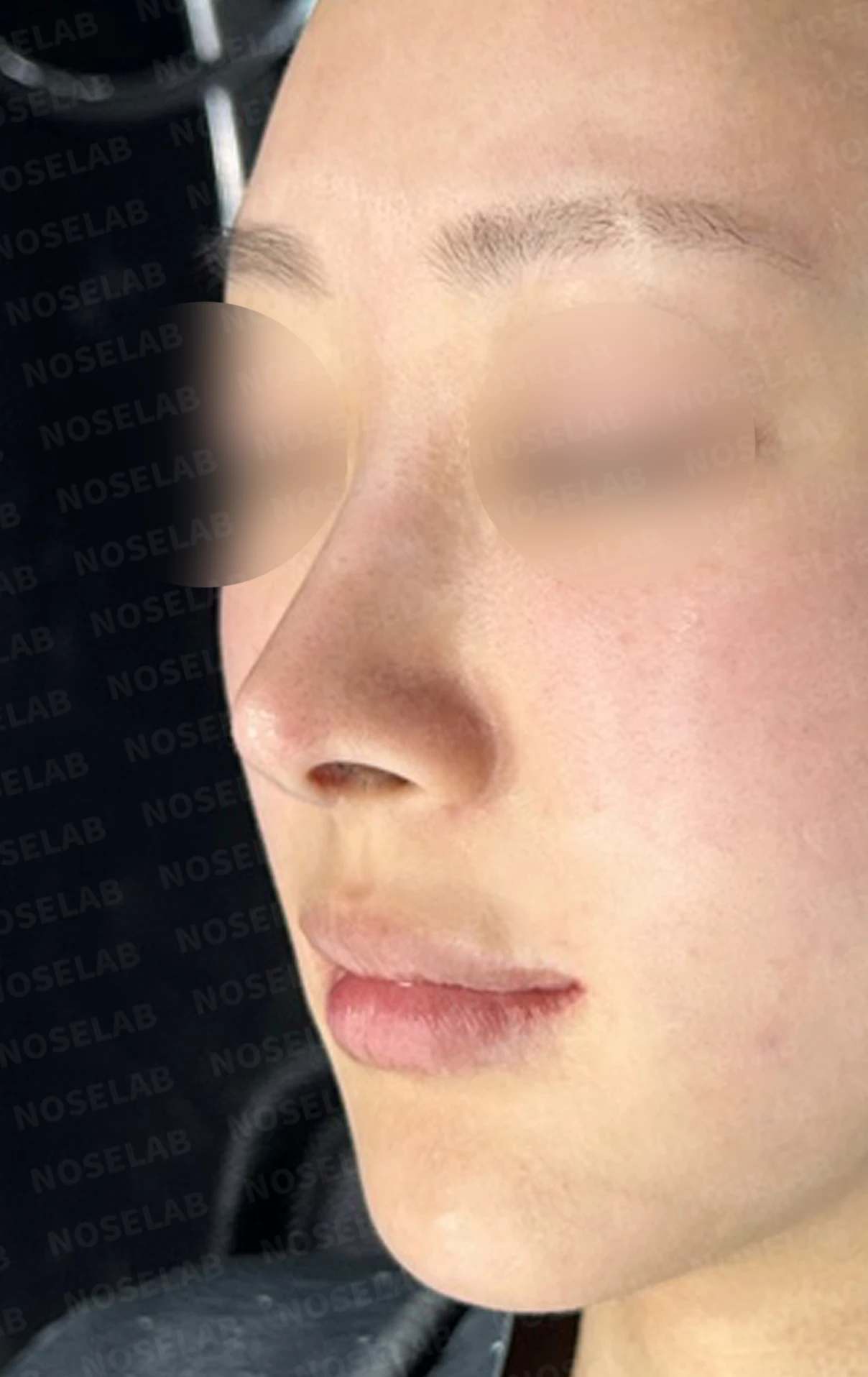
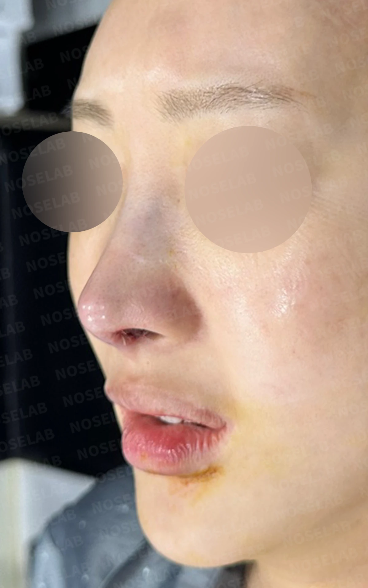
Before surgery / Day 0 immediately after surgery Photos taken immediately after surgery show swelling and may differ from the final result.
3. Function
Improved breathing
Reduction of rhinitis symptoms
Structural stability
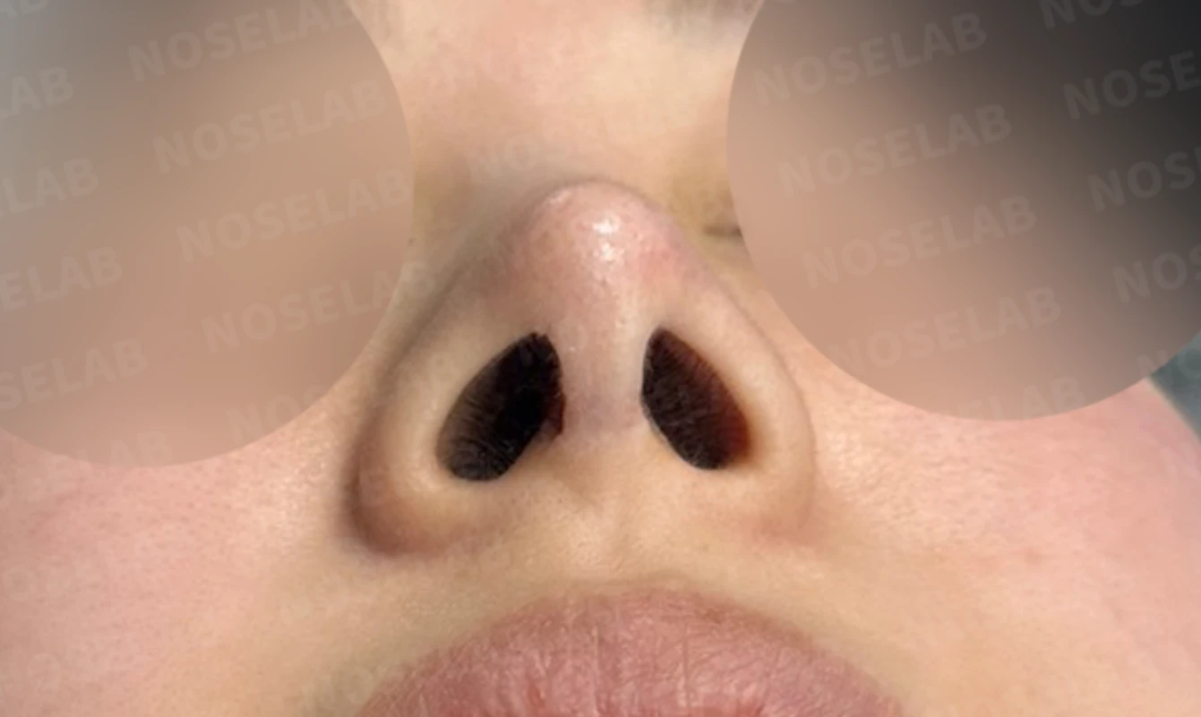
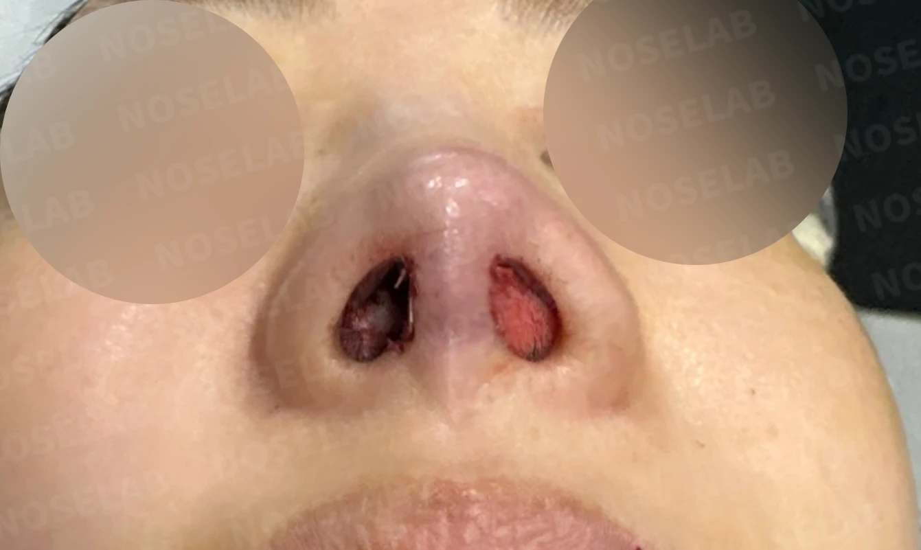
Before surgery / Day 0 immediately after surgery Photos taken immediately after surgery show swelling and may differ from the final result.
👨⚕️ Specialist's assessment
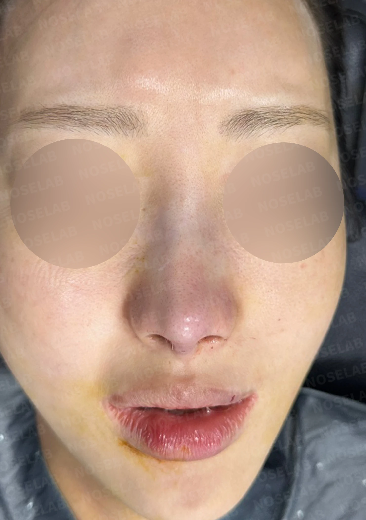
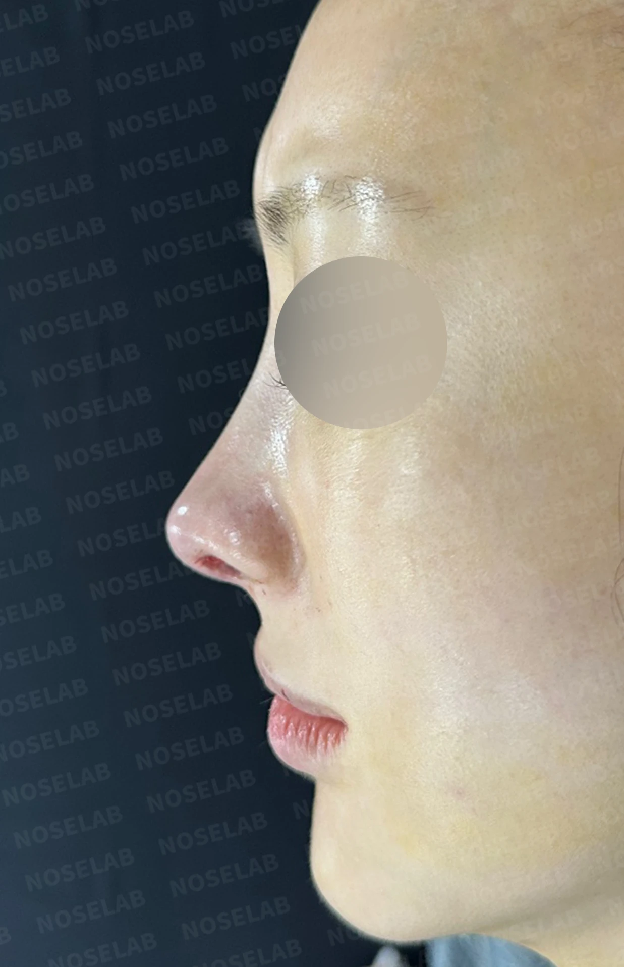
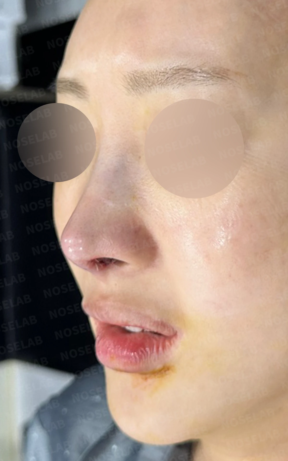
Front, profile, and oblique views taken immediately after surgery (day 0).Immediate postoperative photos show swelling and may differ from the final result.
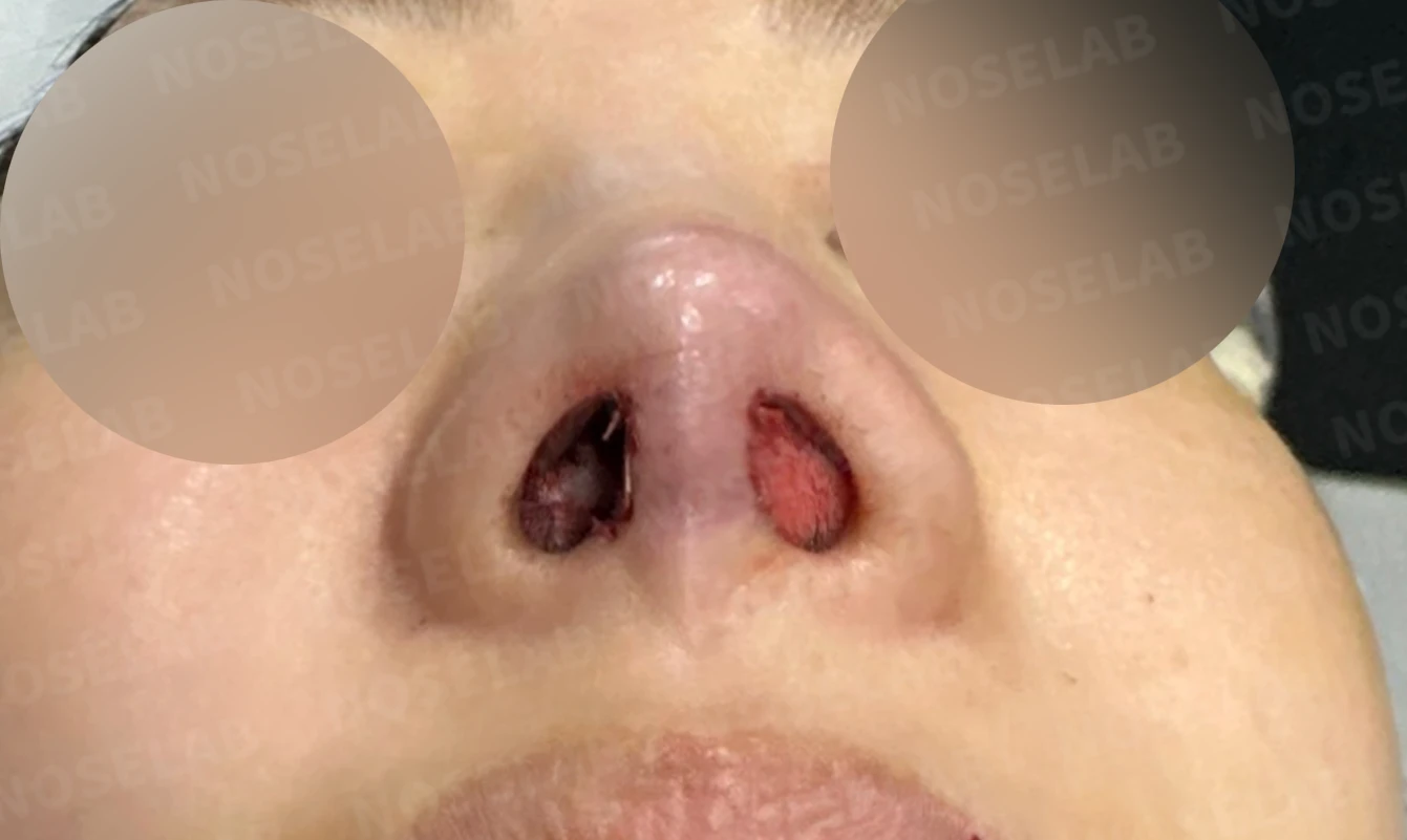
Nostril view on day 0 immediately after surgery. Photos taken immediately after surgery show swelling and may differ from the final result.
This was a case requiring correction of thinned nasal tip skin and cartilage show-through resulting from multiple surgeries. By reinforcing the skin using autologous dermis harvested from behind the ear, a natural nasal tip was restored, and overall balance was improved through lateral osteotomy and columellar correction.
This was Dr. Peter C. Kang. Thank you.
Thank you.
#closedrhinoplasty #revisionrhinoplasty #autologousdermisgraft #tiprhinoplasty #customrhinoplasty

Limitations and risks
When autologous rib cartilage is used, scarring and pain may remain at the donor site on the chest, and pneumothorax (pleural injury) can occur rarely. Over time, the grafted cartilage may warp or undergo partial resorption.
When the septum is corrected at the same time, nasal obstruction may persist or recur, and in rare cases, septal perforation or changes in smell may occur.
Common potential risks include infection, bleeding, asymmetry, scarring, skin necrosis, and insufficient improvement; results vary depending on individual skin condition and previous surgical history.