Hello, this is Dr. Peter C. Kang, medical director.
Today, I would like to introduce a nasal reconstruction case of a patient with nasal bridge skin necrosis and complex issues resulting from previous surgeries.
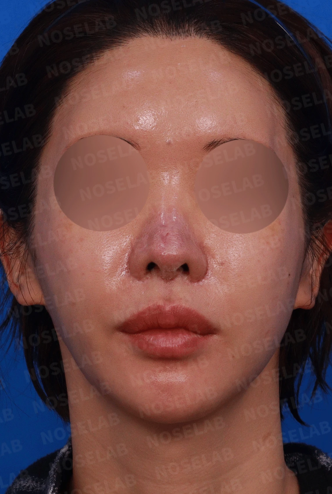
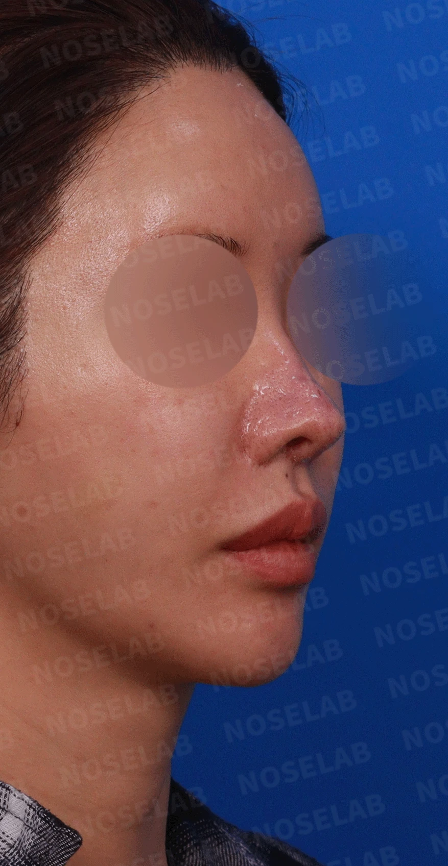
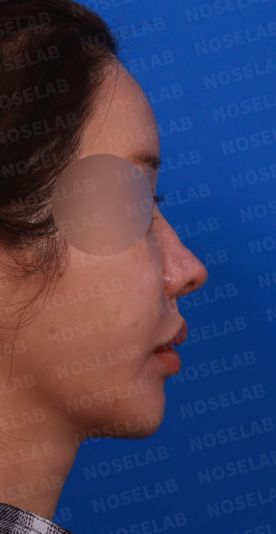
Design consultation: front view, oblique view, right profile view
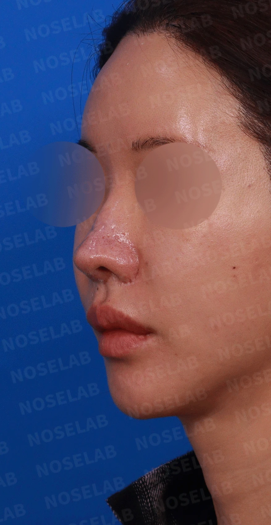
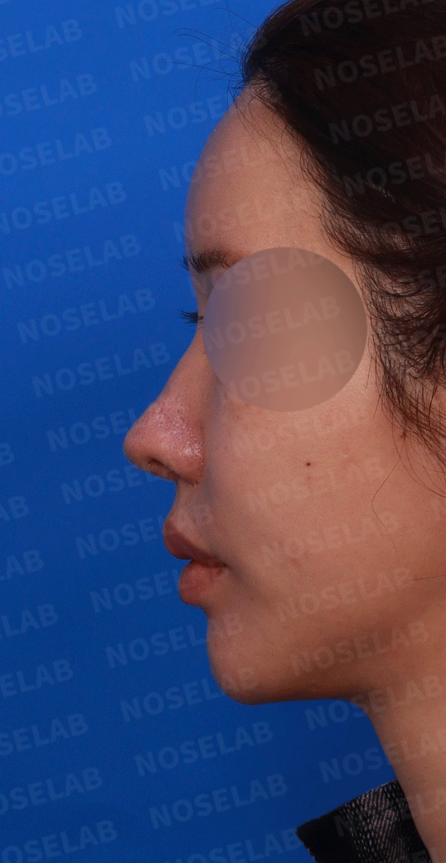
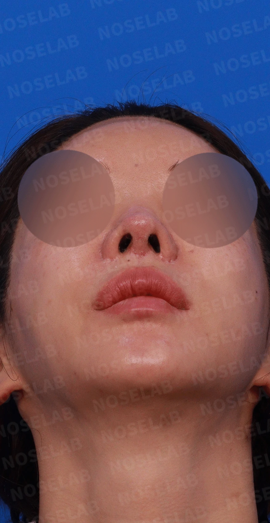
Design consultation: oblique view, left profile view, basal nostril view
📍 Surgical history
15 years ago: Silicone implant surgery
10 years ago: Ear cartilage grafting, silicone implant removal, alar reduction
9 years ago: Ear cartilage grafting → inflammation occurred
Afterward: Total removal followed by AlloDerm (nasal bridge) and autologous buttock dermis (nasal tip) procedures
Underwent various dermatological treatments (chemical peels, etc.) → skin became thin
5 years ago: Autologous rib cartilage and dermis grafting
6 to 7 years ago: Philtrum reduction, two-jaw (orthognathic) surgery (6 years ago)
🔎 Initial consultation findings
Characteristics
Traces of partial necrosis of the nasal bridge skin from previous surgeries
Thinning of the dermal layer due to skin necrosis, with re-epithelialization following shedding of the epidermis
Uneven surface on the nasal bridge due to dermis resorption
Redness of the nasal tip skin caused by vascular compromise
Asymmetry in left and right alar height
Unusual nostril shape (^ ^ shape)
Overall drooping shape of the alae
Extensive scar tissue in the nasal tip from multiple operations
History of using diverse graft materials
✨ Patient's requests
Improvement of uneven areas along the nasal bridge
Correction of left-right alar height symmetry
Improvement of nostril shape (correction of ^ ^ shape)
Alar elevation
💉 Surgical procedure details
Structural improvement
Septal extension using autologous cartilage
Autologous rib cartilage grafting in depressed areas
Tissue-preserving approach to minimize further vascular damage
Application of concentrated growth factors to facilitate recovery of damaged tissue
Alar repositioning to refine nostril shape
Correction of the nasolabial angle
Improvement in shape
Smoothing of the nasal bridge surface
Adjustment of alar height for bilateral symmetry
Correction with alar elevation
Creation of a natural nostril shape
Functional improvement
Concurrent surgery for deviated septum and rhinitis to relieve nasal obstruction
✅ Surgical results
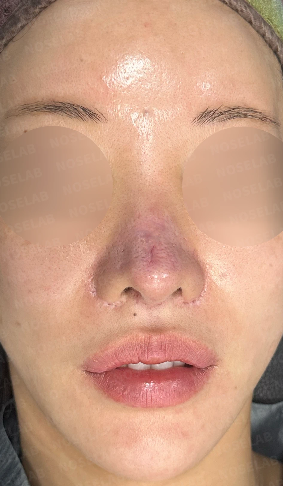
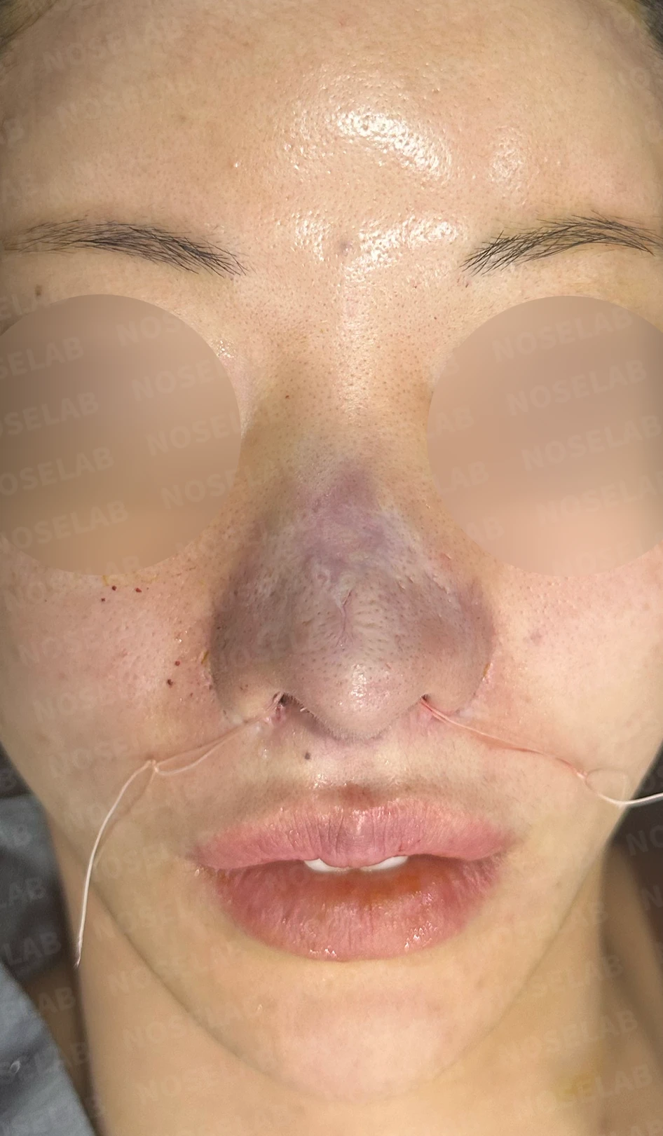
Before surgery / Day 0 immediately after surgery Photos taken immediately after surgery show swelling and may differ from the final result.
Front view
Significantly smoother nasal bridge surface
Corrected left and right alar height
Natural nostril shape
Harmonious overall proportions
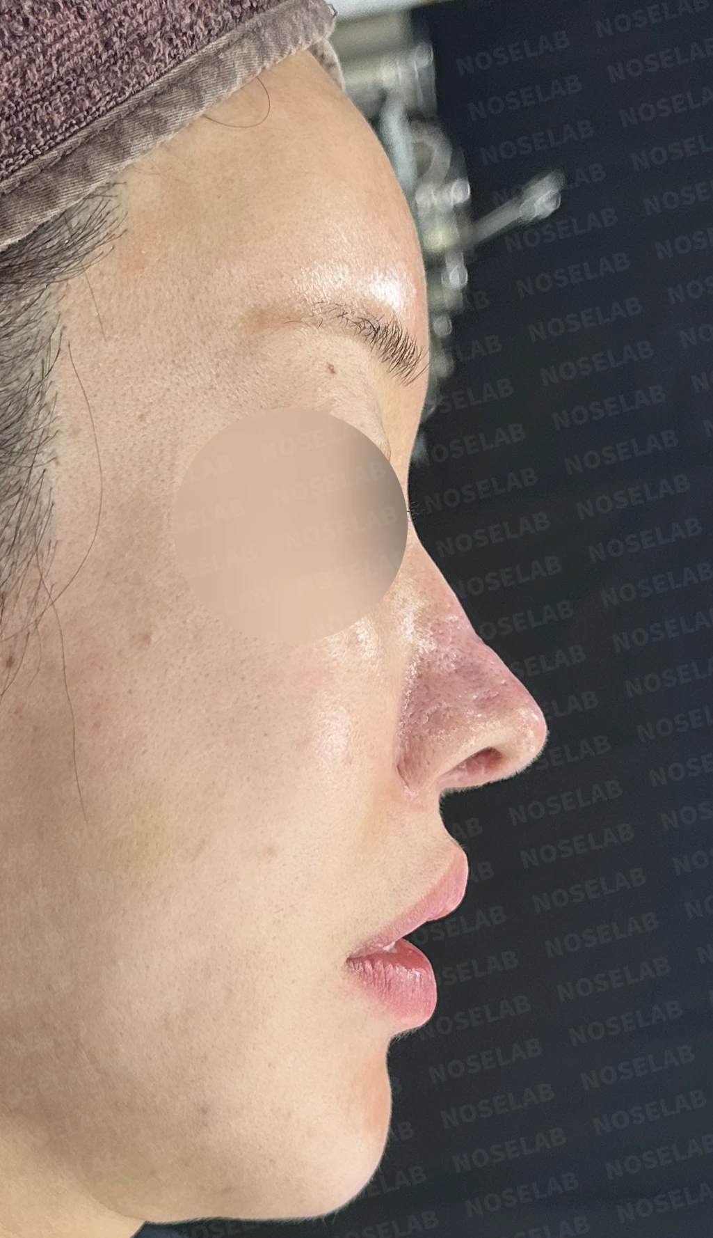
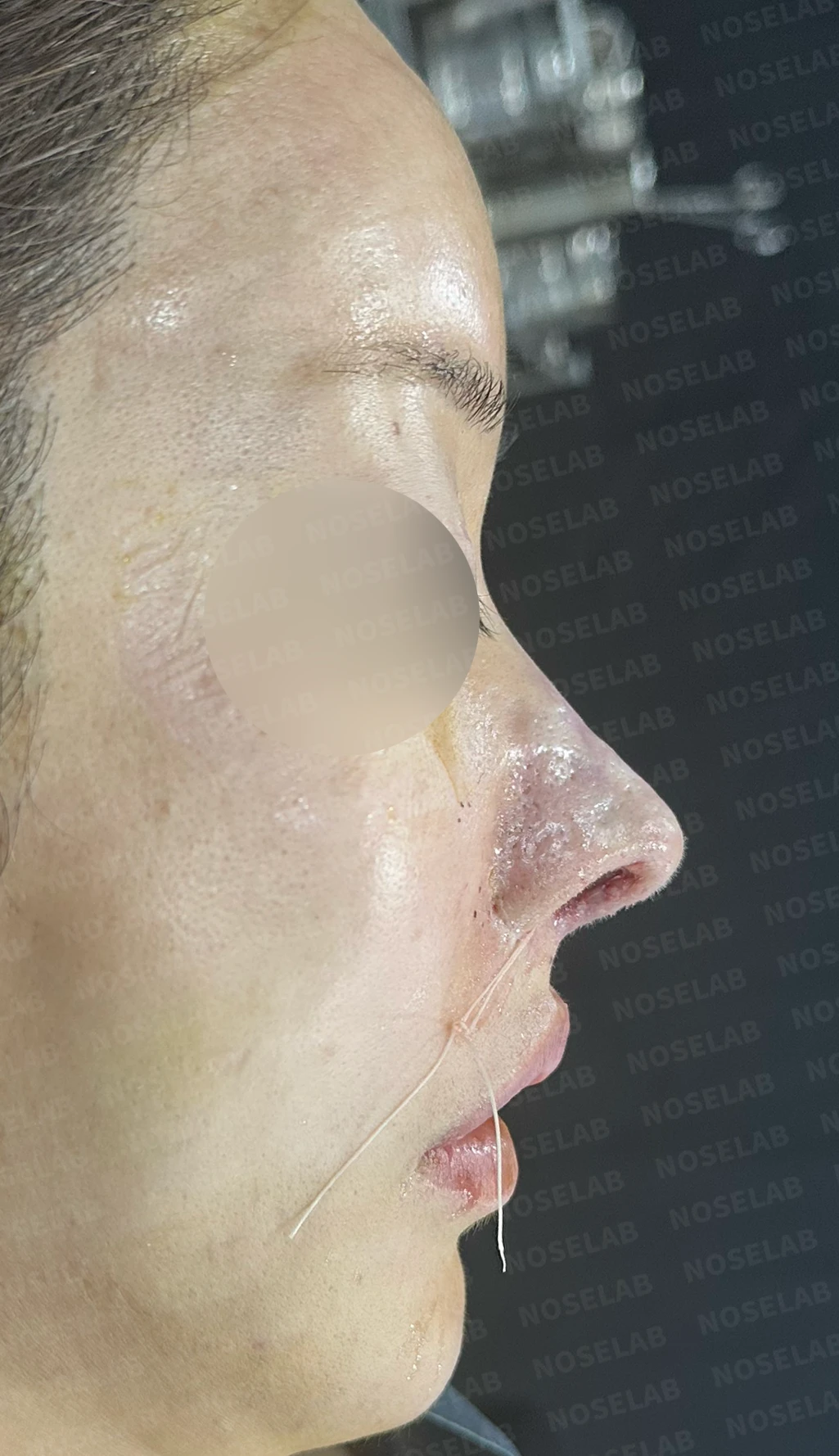
Before surgery / Day 0 immediately after surgery Photos taken immediately after surgery show swelling and may differ from the final result.
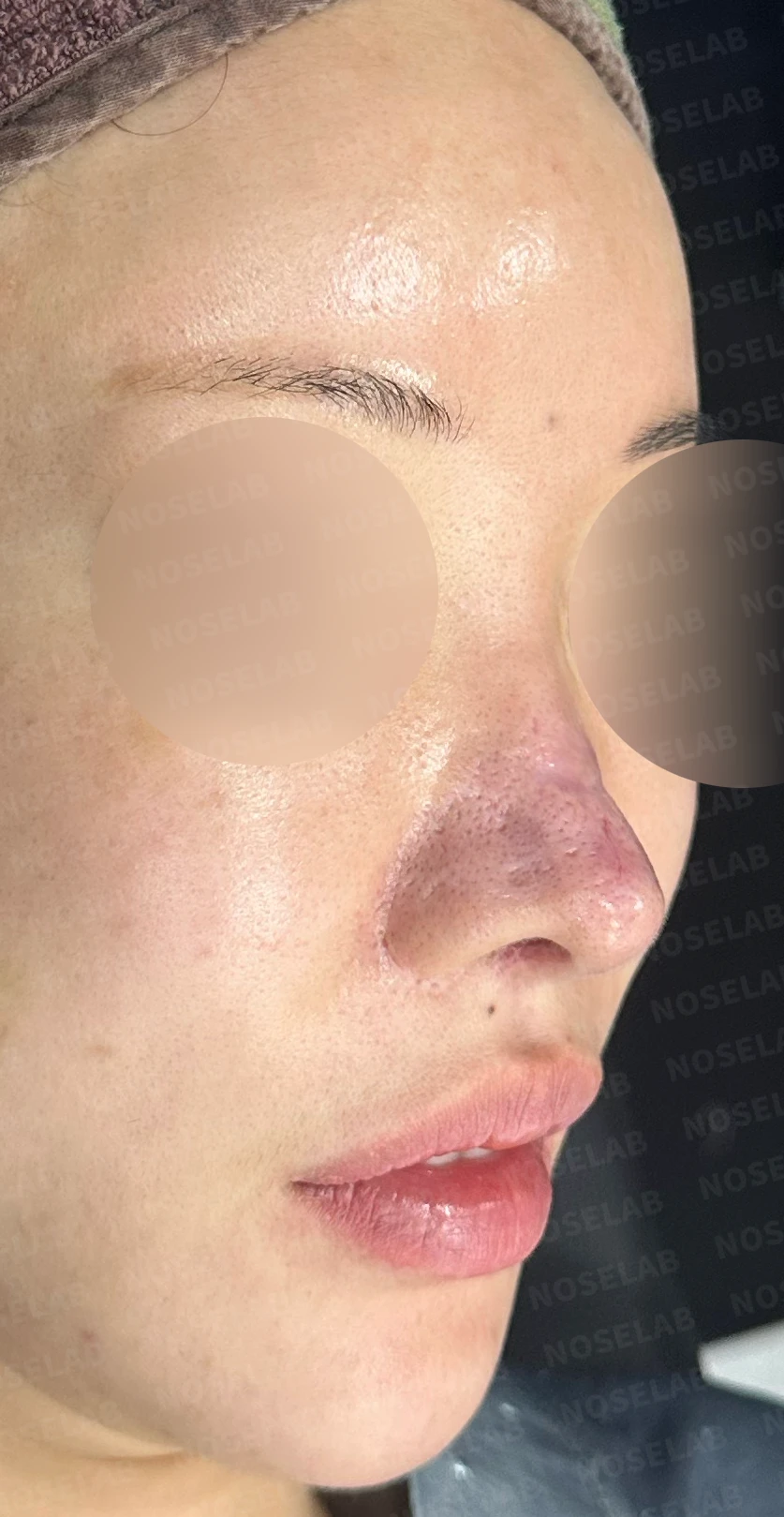
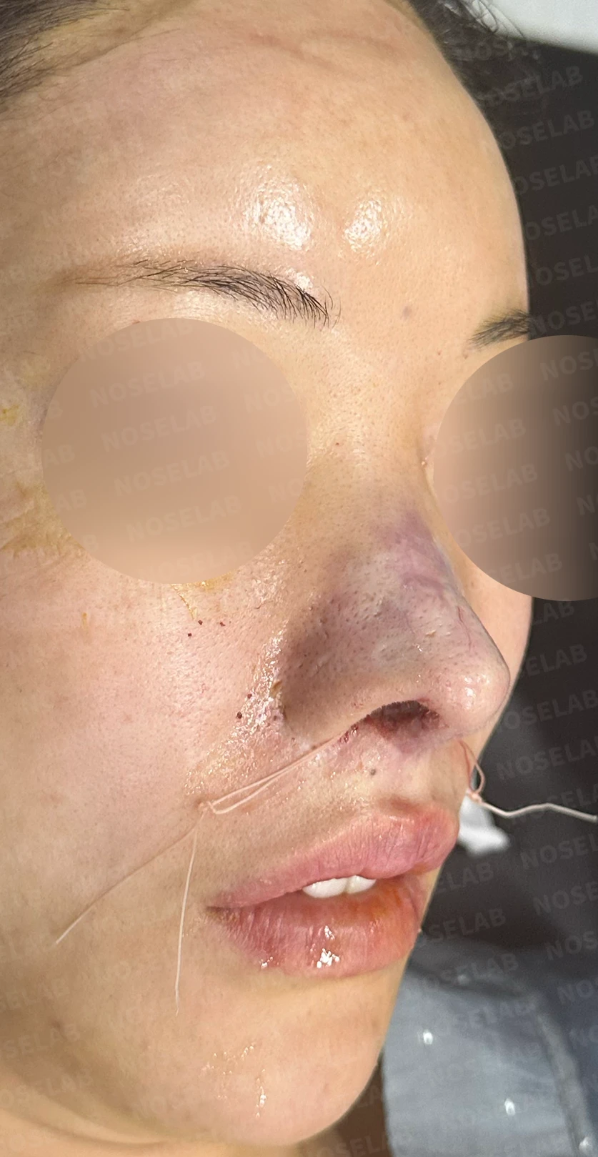
Before surgery / Day 0 immediately after surgery Photos taken immediately after surgery show swelling and may differ from the final result.
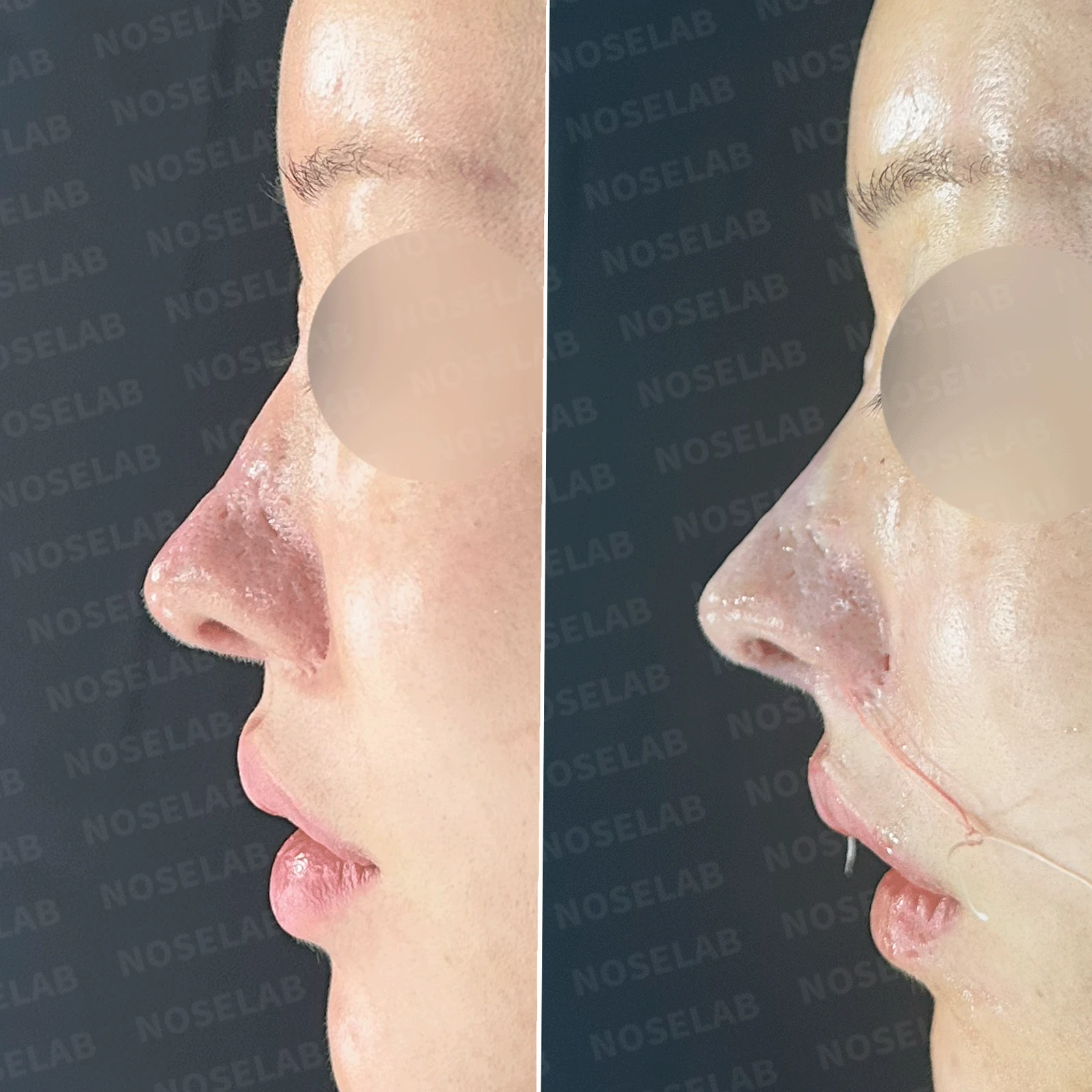
Profile view
Substantial improvement in surface irregularities
Establishment of appropriate height and length
Improved nasolabial angle
Balanced harmony with the philtrum
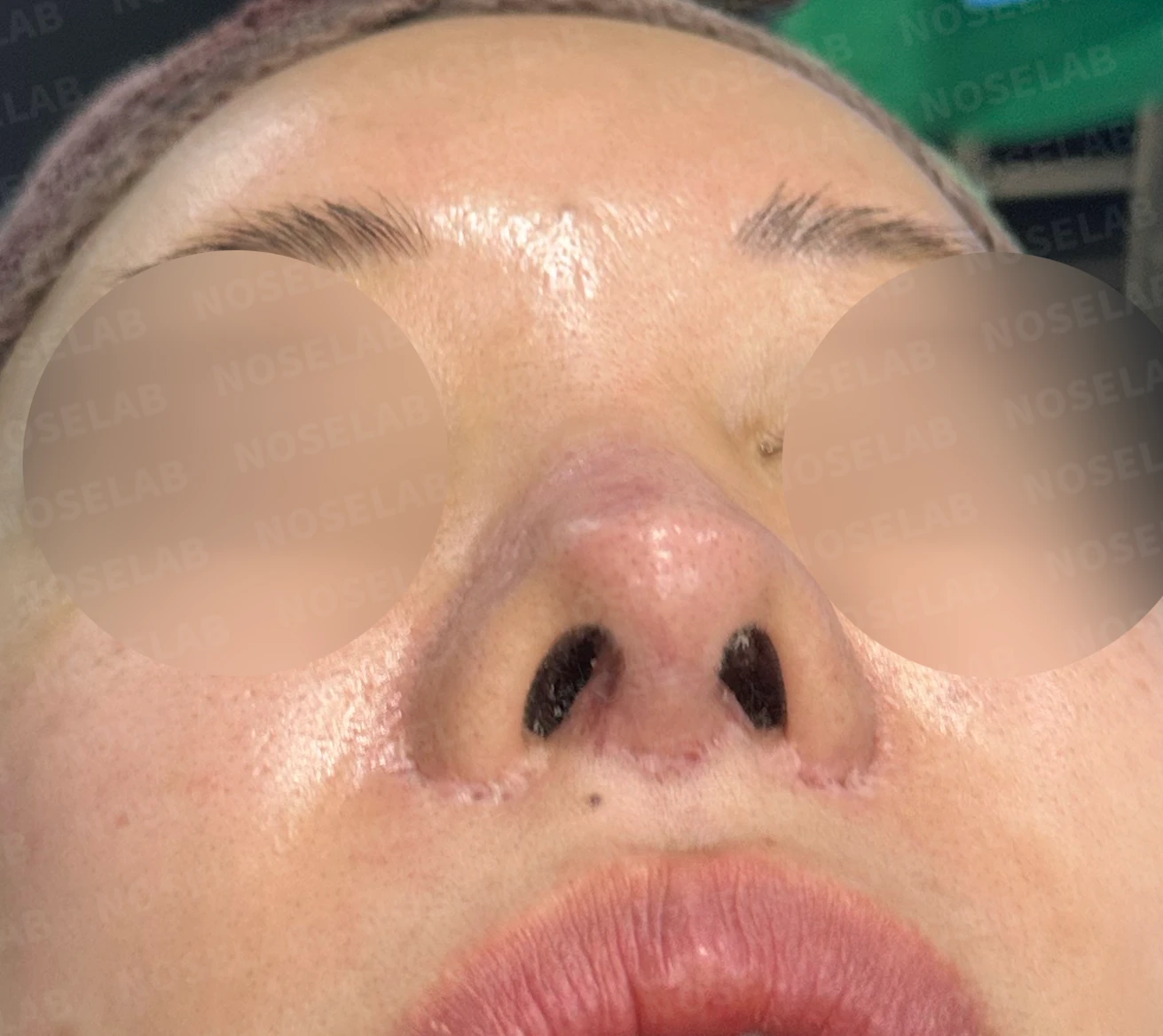
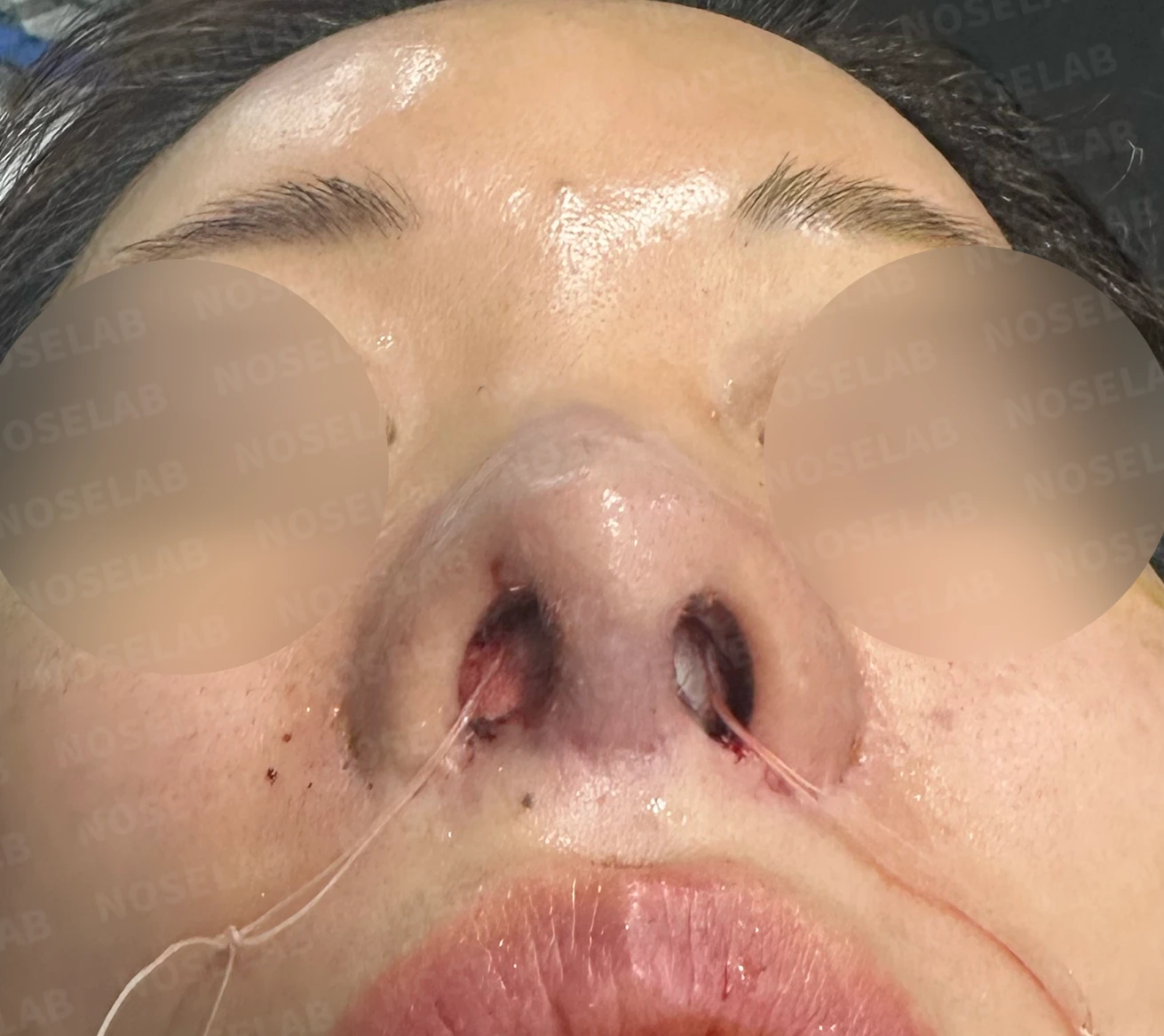
Before surgery / Day 0 immediately after surgery Photos taken immediately after surgery show swelling and may differ from the final result.
Nostrils
Refined nostril shape
Elevated alar position
Balanced nostril shape
Symmetrical nostril patency
👨⚕️ Specialist's assessment
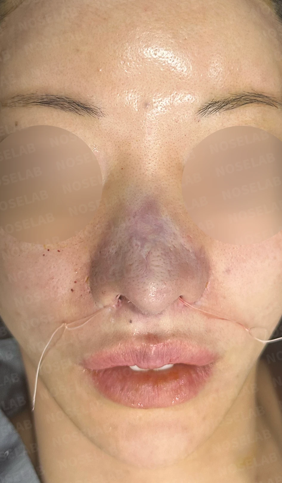
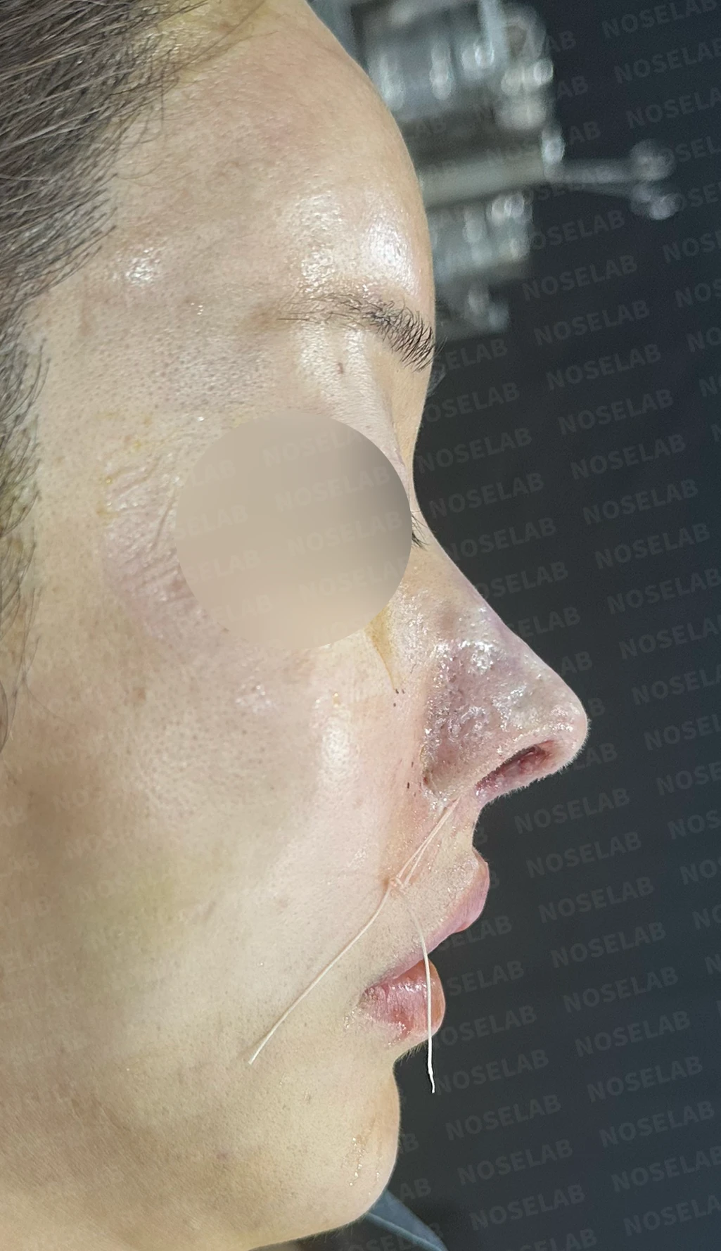
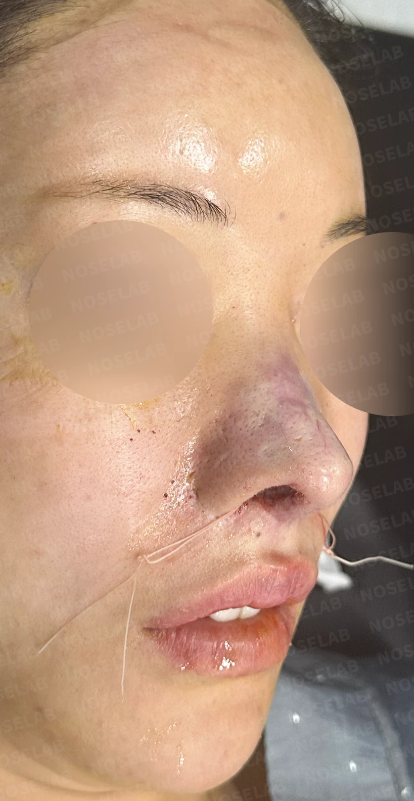
Day 0 immediately after surgery: front, profile, and oblique viewsImmediate postoperative photos show swelling and may differ from the final outcome.
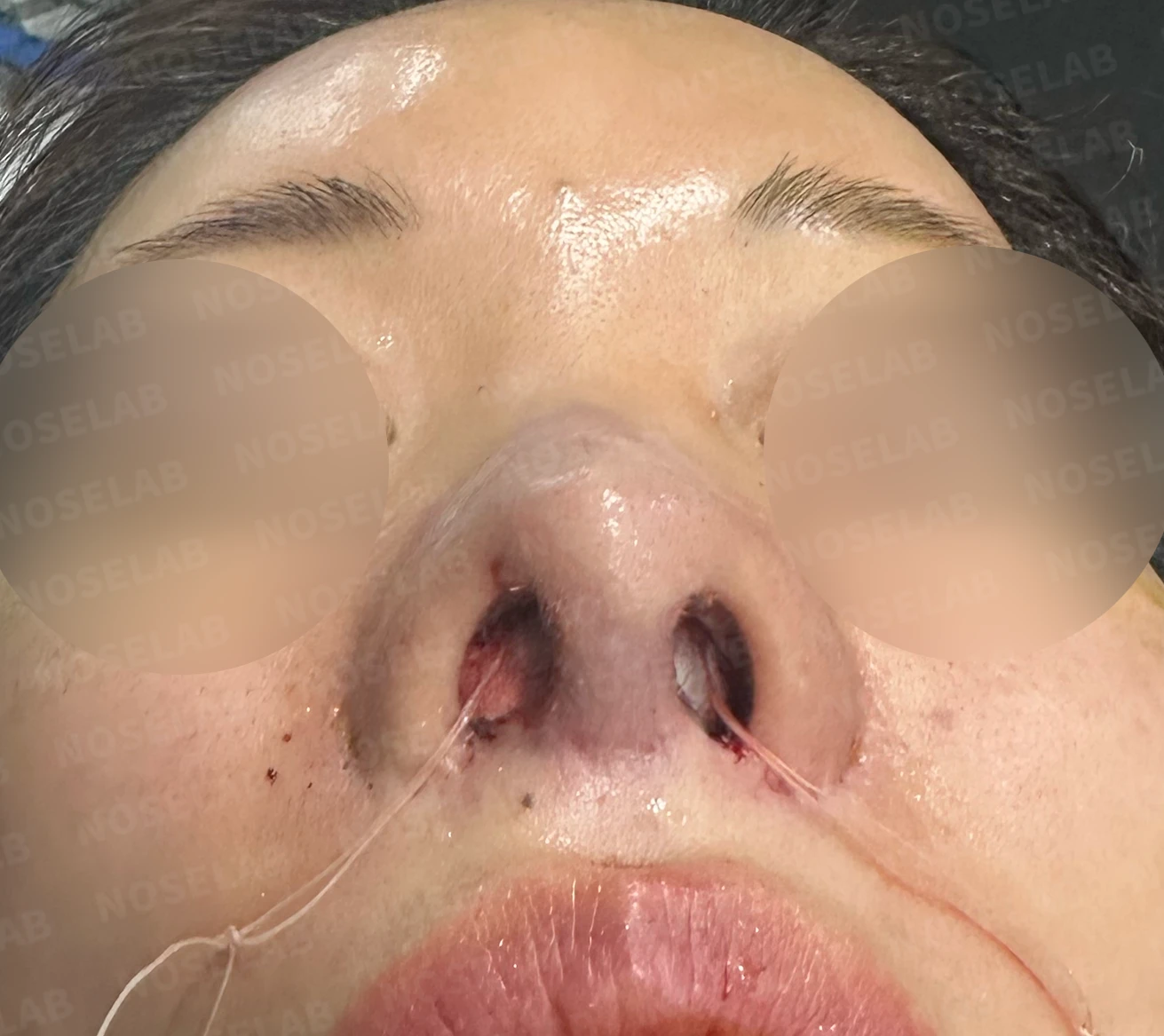
Nostril view on day 0 immediately after surgery Photos taken immediately after surgery show swelling and may differ from the final result.
This was an exceptionally complex case with a multi-layered history spanning more than 15 years, involving multiple rhinoplasties, various graft materials, and dermatological procedures. The most critical issue was partial necrosis of the nasal bridge skin from a past surgery. During the procedure, blanching of the nasal bridge skin confirmed that the microcirculation was severely compromised.
The irregularities along the nasal bridge resulted from the thinning of the dermis and loss of epidermis following necrosis; although re-epithelialization occurred, the overall skin thickness remained markedly reduced. Furthermore, the redness of the patient's nasal tip was attributed to impaired blood circulation stemming from vascular damage during earlier procedures.
To resolve these compounding issues, I took a meticulous approach while preserving as much normal tissue as possible. The depressed areas were carefully filled with autologous rib cartilage, and concentrated growth factors derived from the patient's own blood were applied to the compromised tissue to promote healing and support local circulation.
The asymmetry in alar height and the unusual nostril shape (arched like "^ ^") were improved naturally through precise adjustments to the lower lateral (alar) cartilages and alar elevation. In addition, without overlooking the functional aspects of the nose, 2 functional preservation procedures were performed concurrently to optimize breathing alongside the cosmetic improvements.
Even in such challenging cases involving skin necrosis and vascular compromise, a customized plan tailored thoughtfully to the patient's individual condition can successfully achieve both structural stability and aesthetic improvement.
This has been Dr. Peter C. Kang, medical director.
Thank you.
#closedrhinoplasty #revisionrhinoplasty #skinnecrosisrecovery #autologousribcartilage #growthfactortreatment #customrhinoplasty

Limitations and risks
When autologous rib cartilage is used, scarring and pain may remain at the donor site on the chest, and pneumothorax (pleural injury) can occur rarely. Over time, the grafted cartilage may warp or undergo partial resorption.
When revising after implant removal, contracture may progress again after removal, the nasal height and length may decrease, and staged surgery may be necessary depending on the skin condition.
When the septum is corrected at the same time, nasal obstruction may persist or recur, and in rare cases, septal perforation or changes in smell may occur.
Common potential risks include infection, bleeding, asymmetry, scarring, skin necrosis, and insufficient improvement; results vary depending on individual skin condition and previous surgical history.