Hello, I am Dr. Peter C. Kang.
Today, I would like to introduce a very special case. Let us look at the dramatic transformation process of a patient who presented with a complex 30-year nasal surgery history and a septal perforation measuring nearly 2 cm.
1. Patient history and initial consultation
This patient had a complex surgical history, beginning with an L-shaped silicone implant 30 years ago, silicone revision rhinoplasty in 2010, and silicone removal with gluteal autologous dermis grafting in 2012. The main concerns were as follows:
- Nostril asymmetry and abnormal shape
- An upturned, low, and wide nose
- A saddle nose deformity below the nasal bridge
- Symptoms caused by septal perforation (nasal obstruction, nosebleeds, crusting)
2. Preoperative analysis
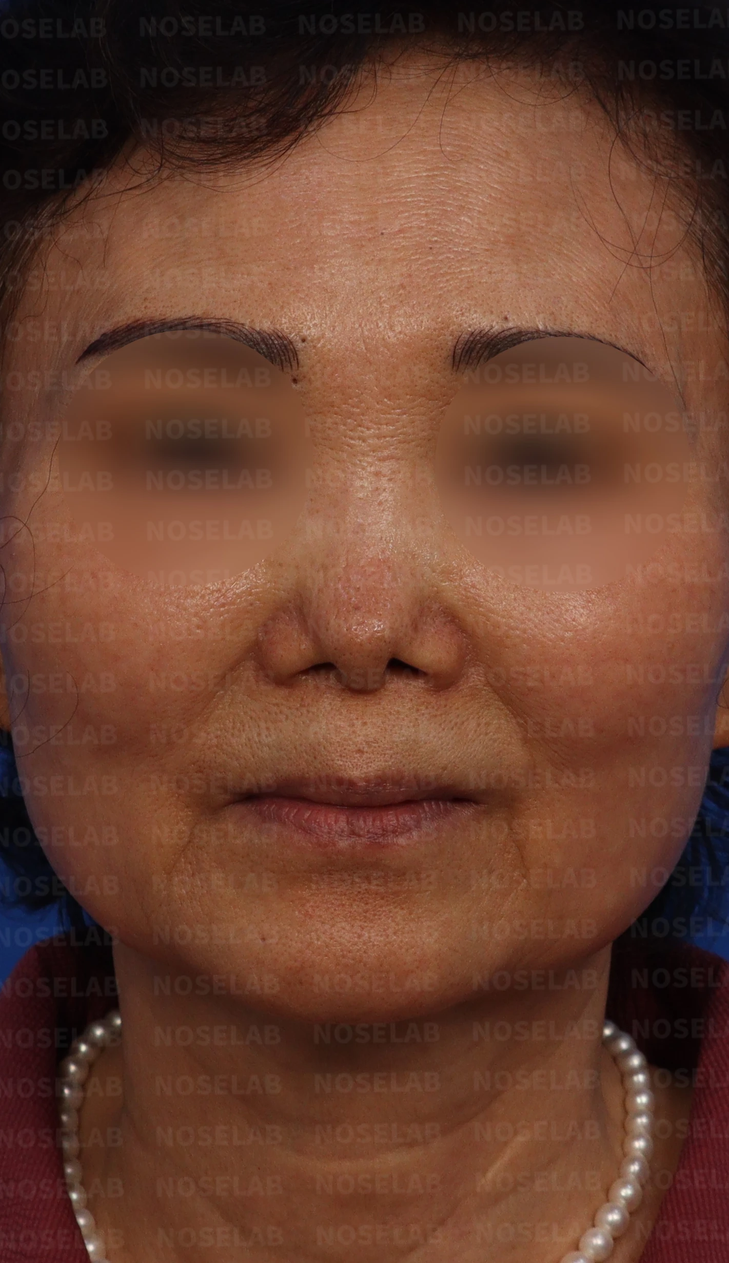
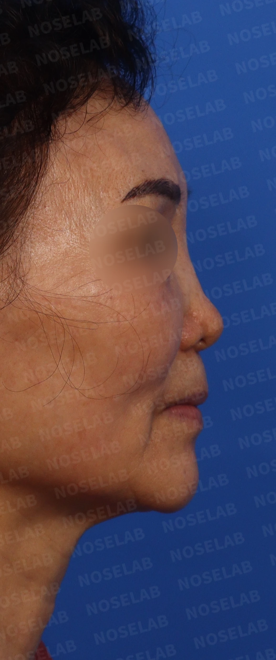
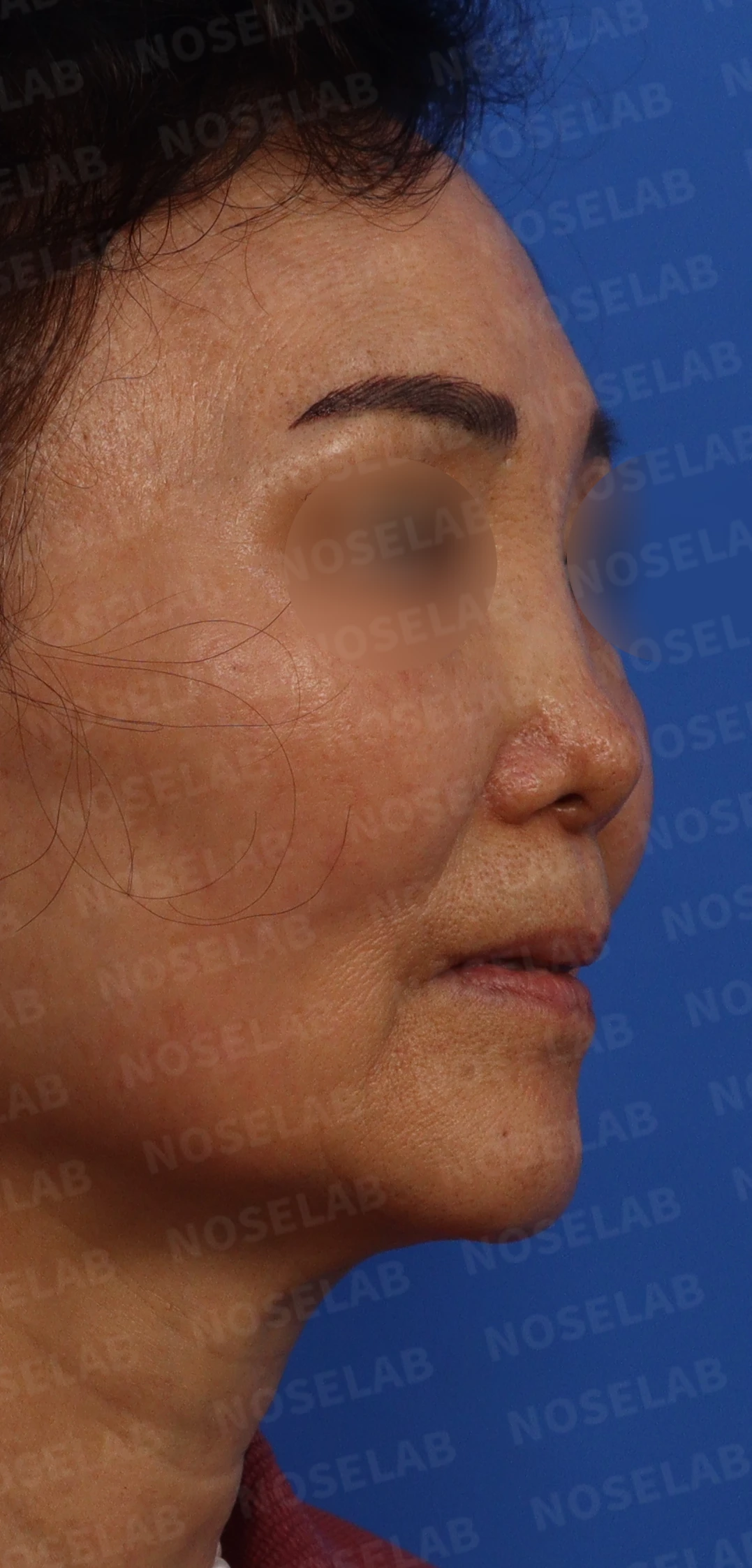
Design consultation photos: front, right profile, and right oblique views
Front view:
- Noticeable nostril asymmetry
- The nose appears broad and spread out overall
- The nasal tip is upturned, appearing unnatural
Profile view:
- The nasal bridge is low, and a saddle nose deformity is observed
- The nasal tip is upturned and looks short
- The radix is high
Oblique view:
- The overall nasal contour appears unnatural
- Skin tension due to contracture is observed
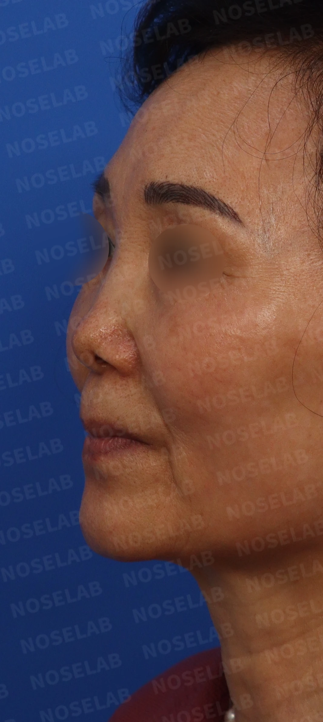
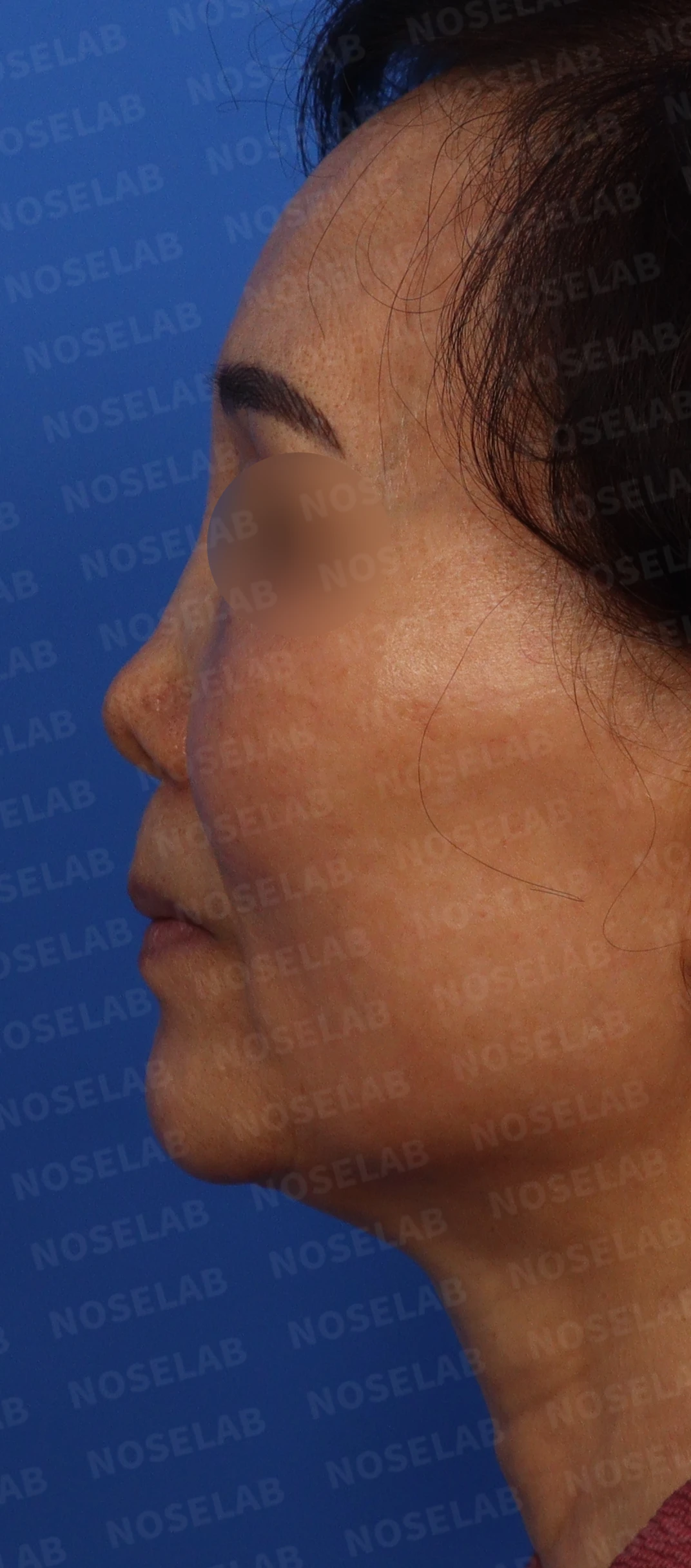
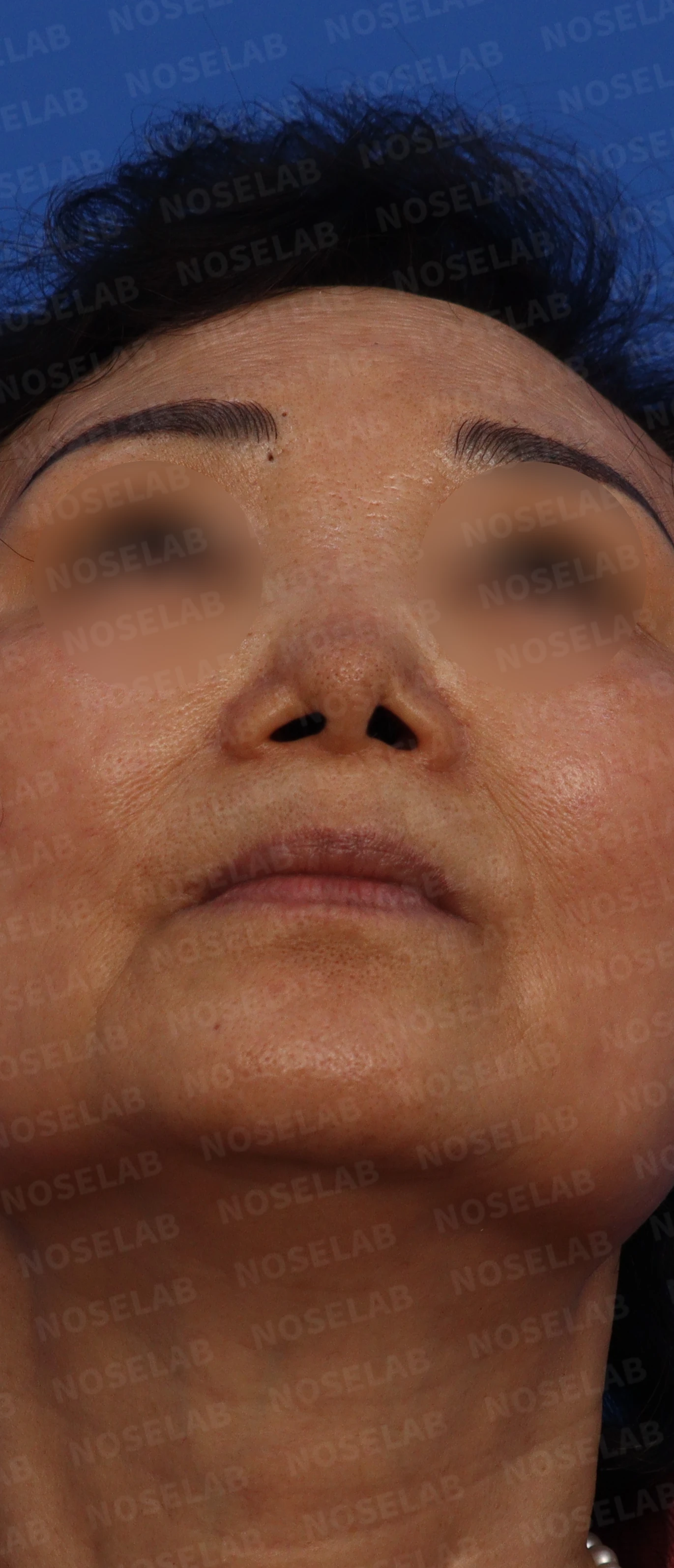
Design consultation photos: left oblique, left profile, and nostril views
Nostrils:
- Severe asymmetry
- Nostrils appear flared and widened
3. Surgical plan
Considering the complexity of this case, I developed a 2-stage surgical plan:
Stage 1 (performed 3 months prior):
- Repair of the septal perforation using a local flap of inferior turbinate mucosa
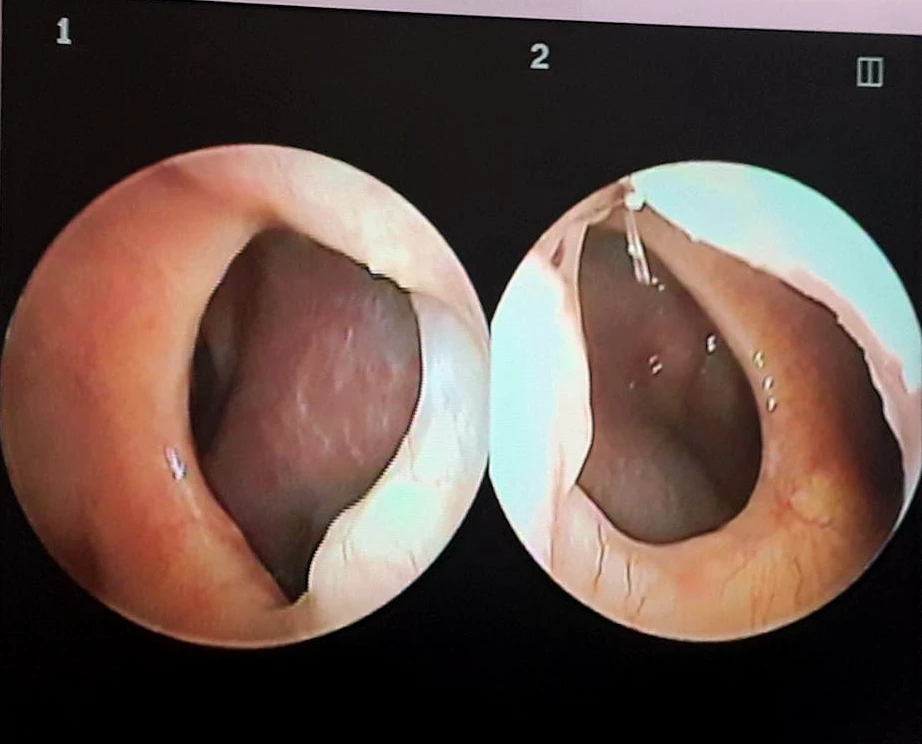
Nasal endoscopy photo showing the septal perforation.
Stage 2 (this surgery):
a) Septal reconstruction surgery to correct the saddle nose
b) Septal extension using autologous rib cartilage to correct the short and upturned nose
c) Alar lowering surgery to correct nostril asymmetry
d) Lower lateral cartilage repositioning and cartilage suturing to correct the blunt nasal tip
e) Removal of the existing implant to lower the radix
f) Overall nasal structural reconstruction using autologous rib cartilage
g) Removal of scar tissue to release and lengthen the contracted nasal tip skin
4. Procedure details
The following procedures were performed using a closed approach:
a) Septal reconstruction and extension:
- The deficient septum was completely reconstructed using autologous rib cartilage.
- Septal extension was performed to reinforce tip support and correct the upturned nose.
b) Nostril shape correction:
- Alar lowering surgery was performed.
- The lower lateral cartilages were repositioned and sutured to correct asymmetry.
- Both lower lateral cartilages were reinforced with autologous rib cartilage to elongate the nostrils.
c) Implant removal and reconstruction:
- An unexpectedly discovered thin Gore-Tex implant was removed.
- A silicone implant was used to naturally elevate the nasal bridge.
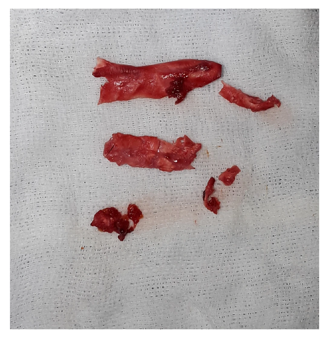
Removed Gore-Tex implant
d) Removal of scar tissue:
- Scar tissue on the nasal dorsum was removed, and the skin envelope was sufficiently released.
e) Perforation closure:
- The remaining 10% of the perforation located deep inside after stage 1 was completely closed with direct sutures.
All procedures were performed using a closed approach, completing the surgery without external scars.
5. Comparison of before-and-after photos
Comparing the photos taken immediately after surgery on day 0 shows the following noticeable improvements: Photos taken immediately after surgery include swelling and may differ from the final result.
Front view:
- Nostril asymmetry improved significantly.
- The wide and spread-out nose changed into a slender and refined shape.
- The upturned tip was lowered naturally, improving the overall balance.
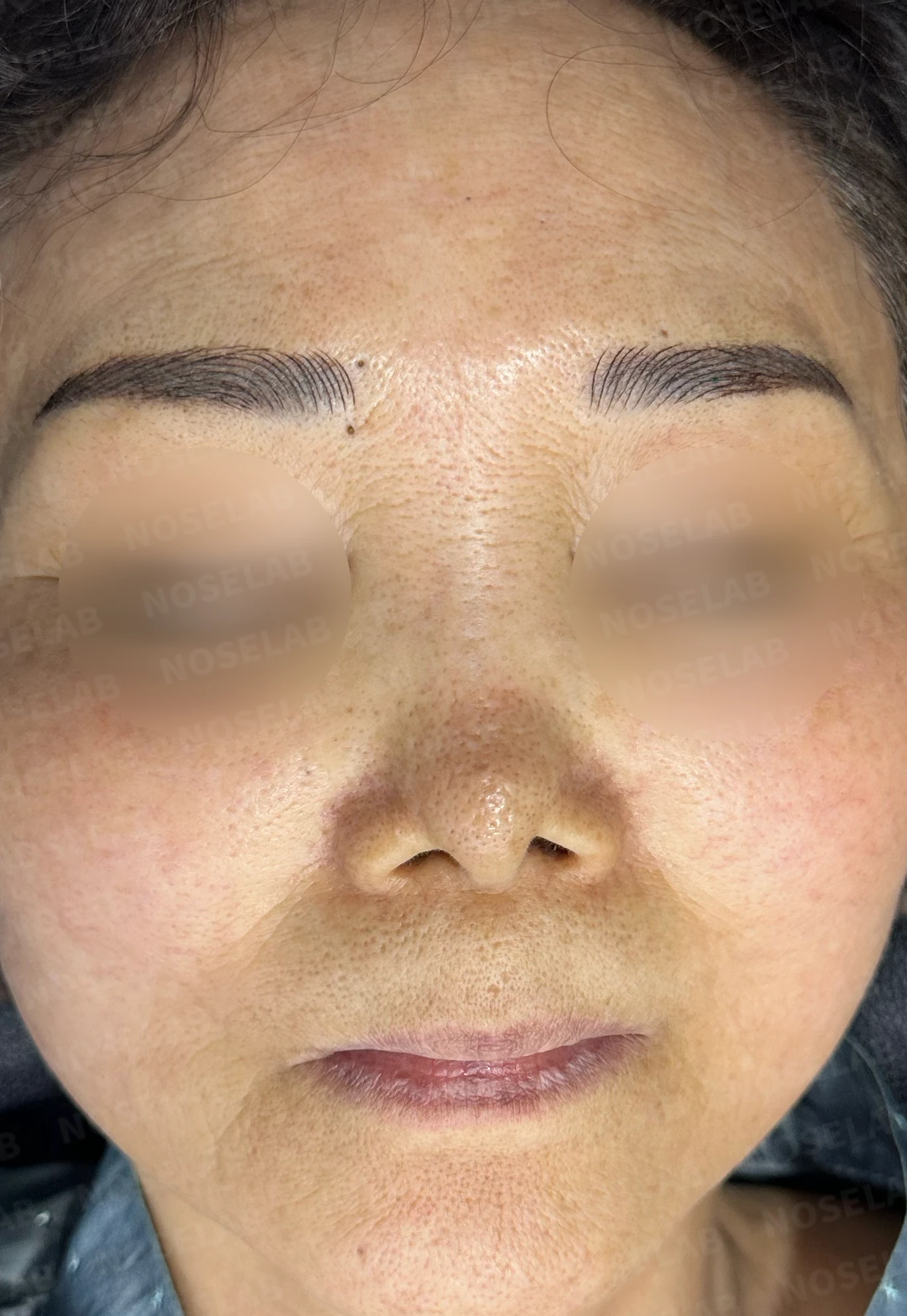
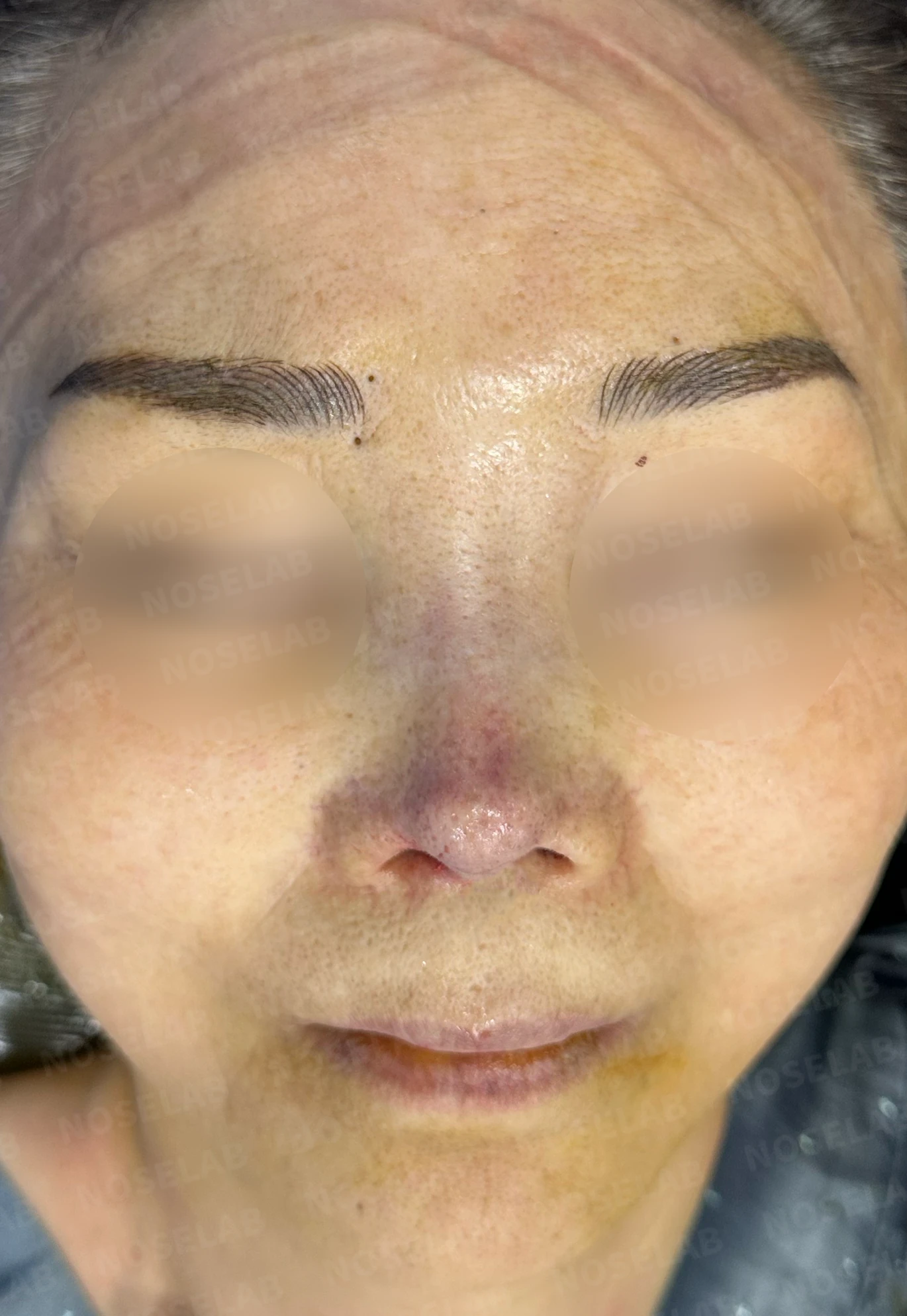
Before surgery / Day 0 immediately after surgery Photos taken immediately after surgery show swelling and may differ from the final result.
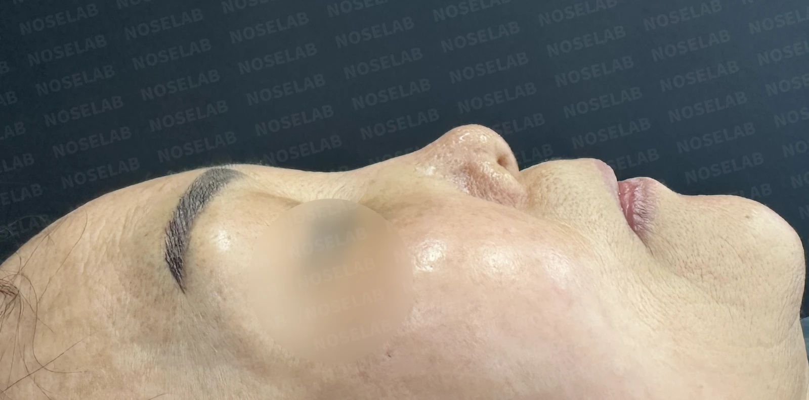
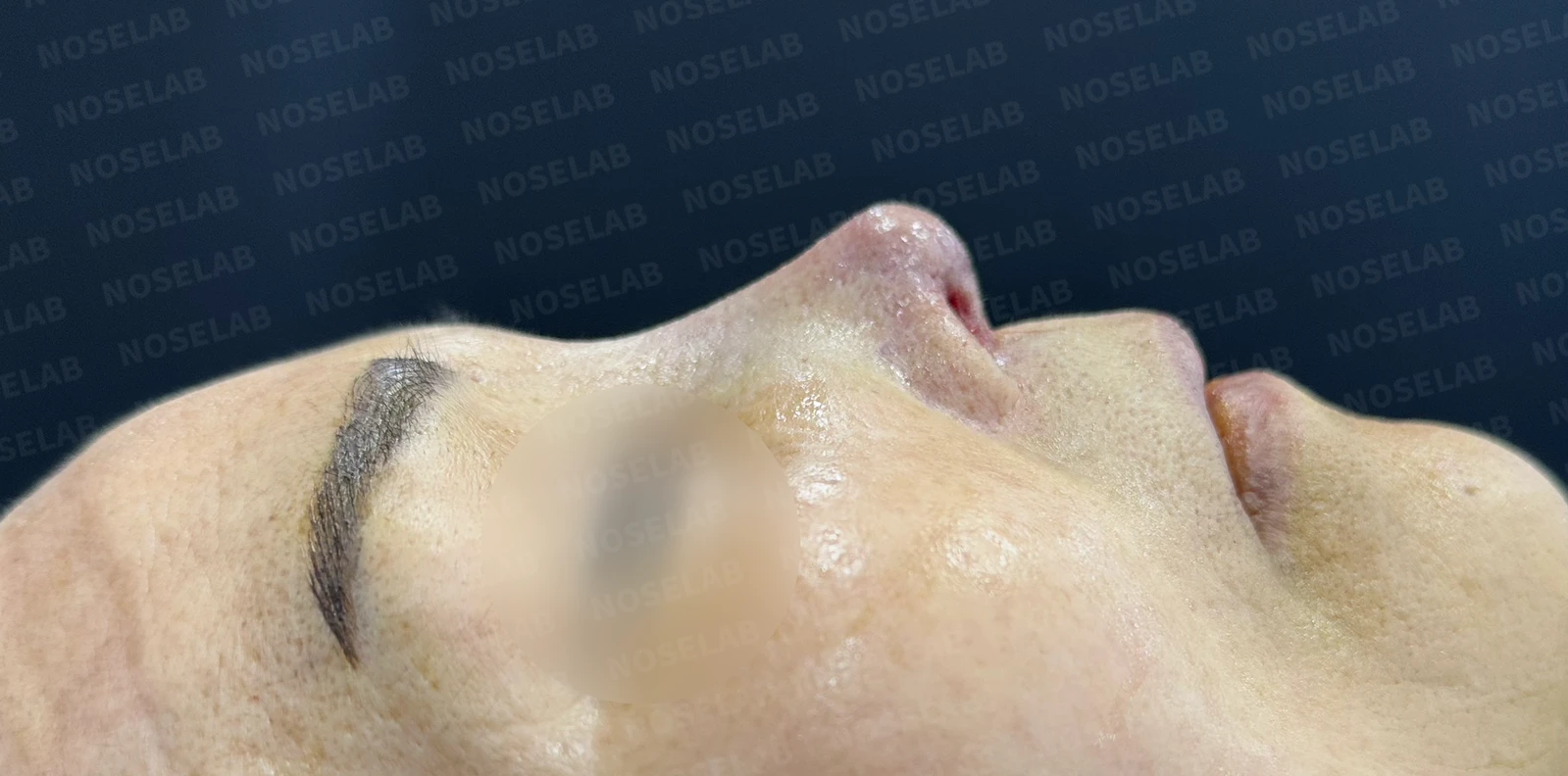
Before surgery / Day 0 immediately after surgery Photos taken immediately after surgery show swelling and may differ from the final result.
Profile view:
- The saddle nose deformity improved, forming a smooth dorsal line.
- The short-looking nose gained adequate length and projection.
- The upturned tip was lowered naturally, resulting in a harmonious nasal shape.
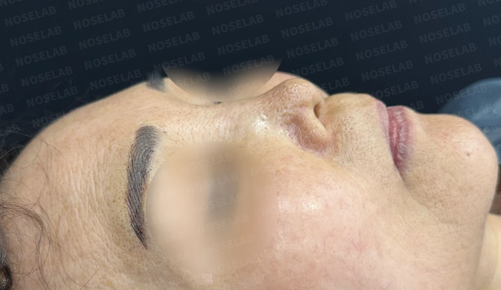
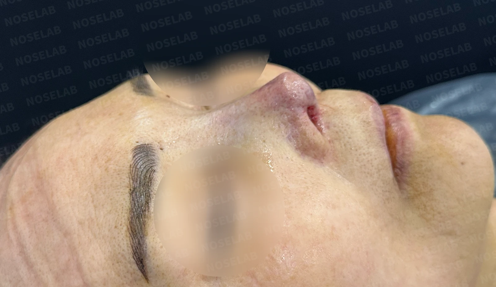
Before surgery / Day 0 immediately after surgery Photos taken immediately after surgery show swelling and may differ from the final result.
Oblique view:
- The overall nasal contour improved naturally and aesthetically.
- Skin tension from contracture was relieved, showing softer lines.
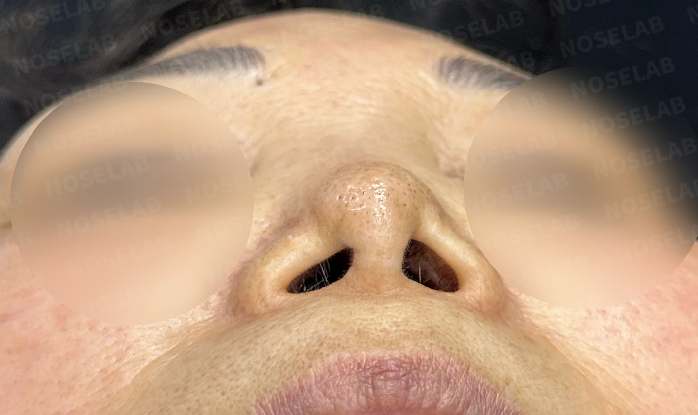
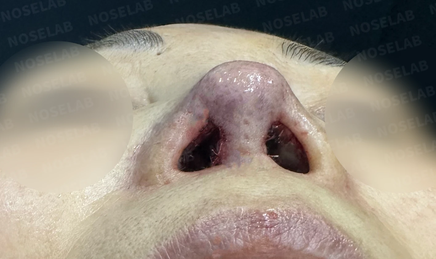
Before surgery / Day 0 immediately after surgery Photos taken immediately after surgery show swelling and may differ from the final result.
Nostrils:
- Severe asymmetry has been significantly improved.
- The flared appearance of the nostrils showed significant improvement.
6. Photos taken immediately after surgery (day 0)Immediate postoperative photos show swelling and may differ from the final result.
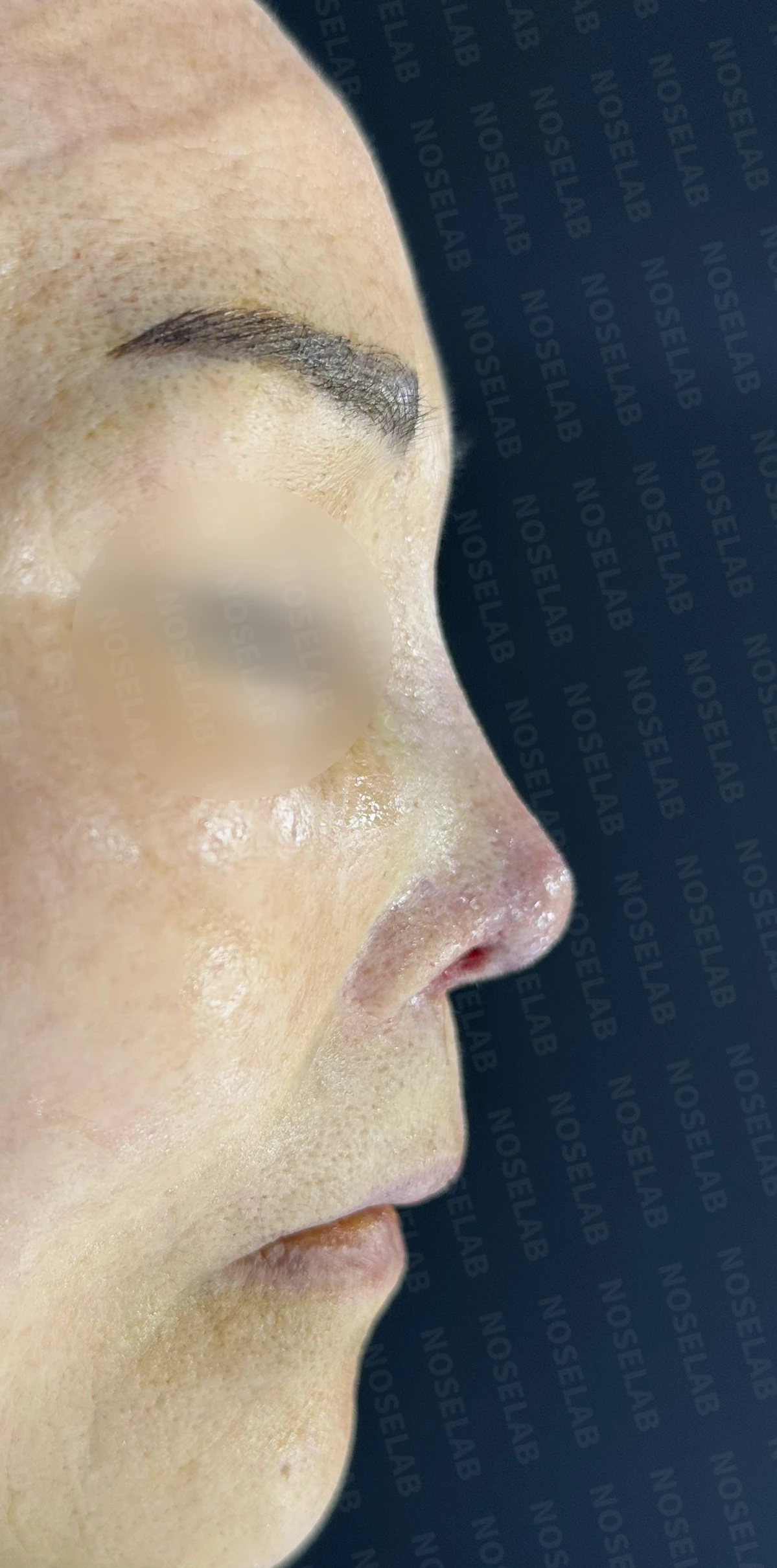
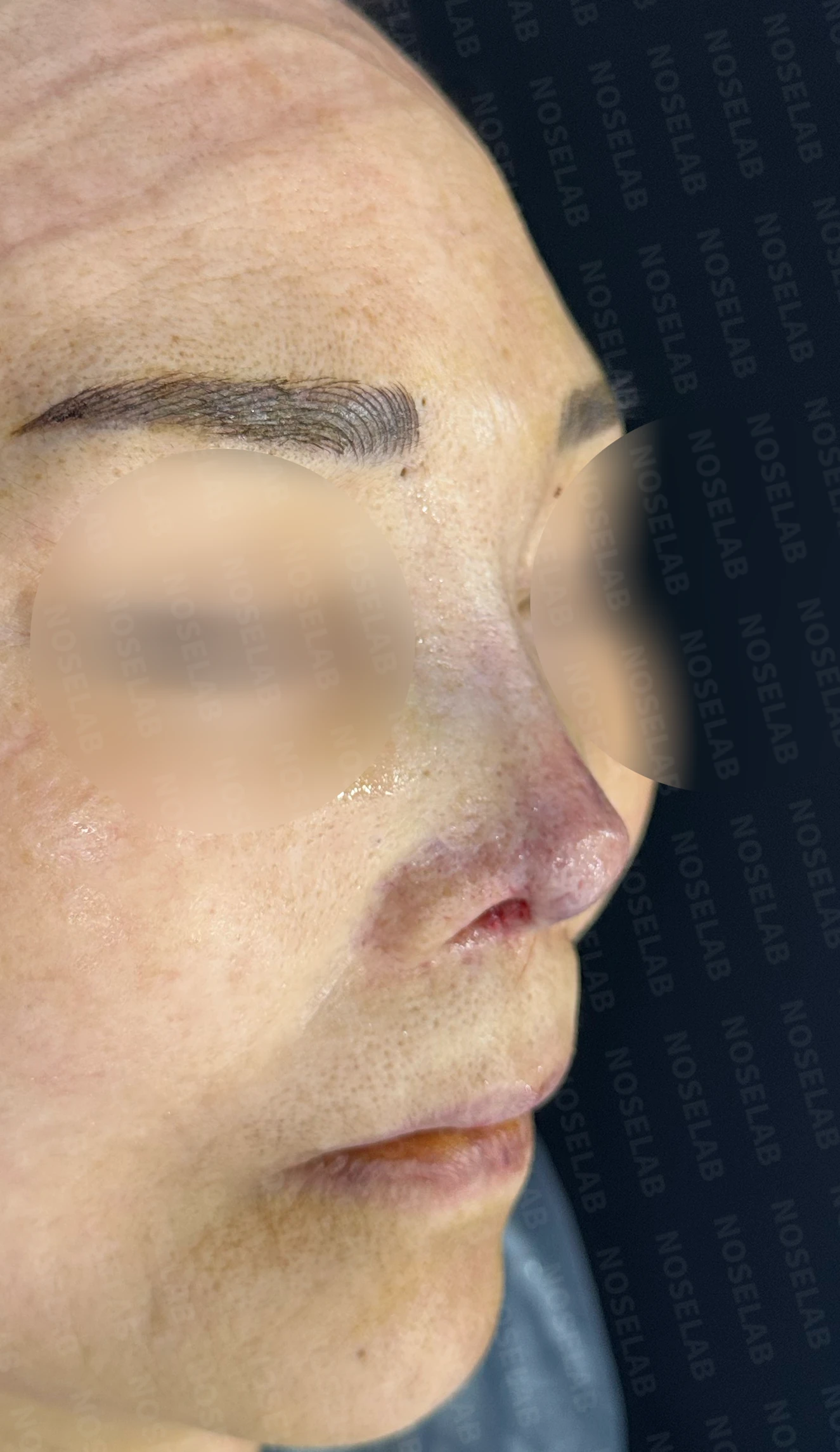
Immediately after surgery (Day 0) front, profile, and oblique viewsImmediate postoperative photos show swelling and may differ from the final result.
Basal (nostril) view immediately after surgery (Day 0)Immediate postoperative photos show swelling and may differ from the final result.
7. Overall assessment
This case was a successful correction achieved under extremely challenging conditions, including a complex surgical history and a large septal perforation. Key highlights include:
1) Treatment of the 2 cm septal perforation:
After checking the size of the septal perforation and the remaining tissue, I proceeded with reconstruction using a 2-stage approach.
2) Comprehensive resolution of combined problems:
Multiple issues—including an upturned tip, short nose, widened dorsum, and contracture—were resolved simultaneously. This was possible through an integrated approach considering both the structure and function of the entire nose, rather than addressing each issue in isolation.
3) Reconstruction using autologous tissue:
Reconstructing the septum and lower lateral cartilages with autologous rib cartilage supported stable, natural results over the long term.
4) Advantages of the closed approach:
Even in such a complex case, using a closed approach allowed surgery without external columellar scars and helped reduce the recovery period.
8. Concluding remarks
For patients with septal perforation undergoing rhinoplasty, treatment planning and surgical techniques differ greatly depending on the size and location of the perforation. In principle, an asymptomatic perforation does not necessarily require aggressive closure; however, treatment is needed when symptoms such as nasal obstruction, nosebleeds, whistling sounds, or frequent crusting are present, as in this case.
In particular, if the perforation is located anteriorly, the cartilage graft used during septal extension—which is essential for tip surgery—carries a risk of bacterial infection from the perforation site, so closure is essential. However, large perforations exceeding 2 cm have significantly lower surgical success rates, making treatment by an experienced board-certified otolaryngologist (ENT specialist) essential.
The reconstruction plan was designed taking into account the septal perforation and the condition of the surrounding tissues.
I hope this case offers hope to those struggling with septal perforation or complex nasal problems. Even in challenging cases, solutions can be found through thorough diagnosis, a structured treatment plan, and specialized surgical care. NOSELAB is here to help you achieve a healthy, well-proportioned nose.
This was Dr. Peter C. Kang. Thank you.
Thank you.
#SeptalPerforationRepair #ContractedNoseCorrection #UpturnedNoseCorrection #ShortNoseCorrection #WideNoseCorrection #AutologousRibCartilageGraft #ClosedRhinoplasty #RevisionRhinoplasty #ENTSpecialistRhinoplasty #SafeRhinoplasty #ComplexRhinoplasty

Limitations and risks
When autologous rib cartilage is used, scarring and pain may remain at the donor site on the chest, and pneumothorax (pleural injury) can occur rarely. Over time, the grafted cartilage may warp or undergo partial resorption.
When revising after implant removal, contracture may progress again after removal, the nasal height and length may decrease, and staged surgery may be necessary depending on the skin condition.
When the septum is corrected at the same time, nasal obstruction may persist or recur, and in rare cases, septal perforation or changes in smell may occur.
Common potential risks include infection, bleeding, asymmetry, scarring, skin necrosis, and insufficient improvement; results vary depending on individual skin condition and previous surgical history.