Hello, I am Dr. Peter C. Kang.
Today, I would like to share a correction case of a patient who suffered from contracture deformity resulting from multiple rhinoplasties and inflammation.
This case clearly illustrates the difficulties of complex revision rhinoplasty and the process of addressing them.
1. Preoperative design consultation
The patient wanted to correct an overall widened appearance and improve an awkward profile where the nostrils were visible. The surgical history was as follows:
1. 2009: Silicone implant + ear cartilage
2. 2010: Silicone implant + autologous dermis
3. 2012: Silicone implant + autologous rib cartilage
4. 2015: Silicone implant + autologous rib cartilage
5. 2022: Silicone implant removal due to inflammation
6. 8 months later: Autologous rib cartilage + ear cartilage used only on the nasal tip, along with stem cell injection therapy
Let us examine the condition of this patient, who had undergone multiple surgeries, in detail:
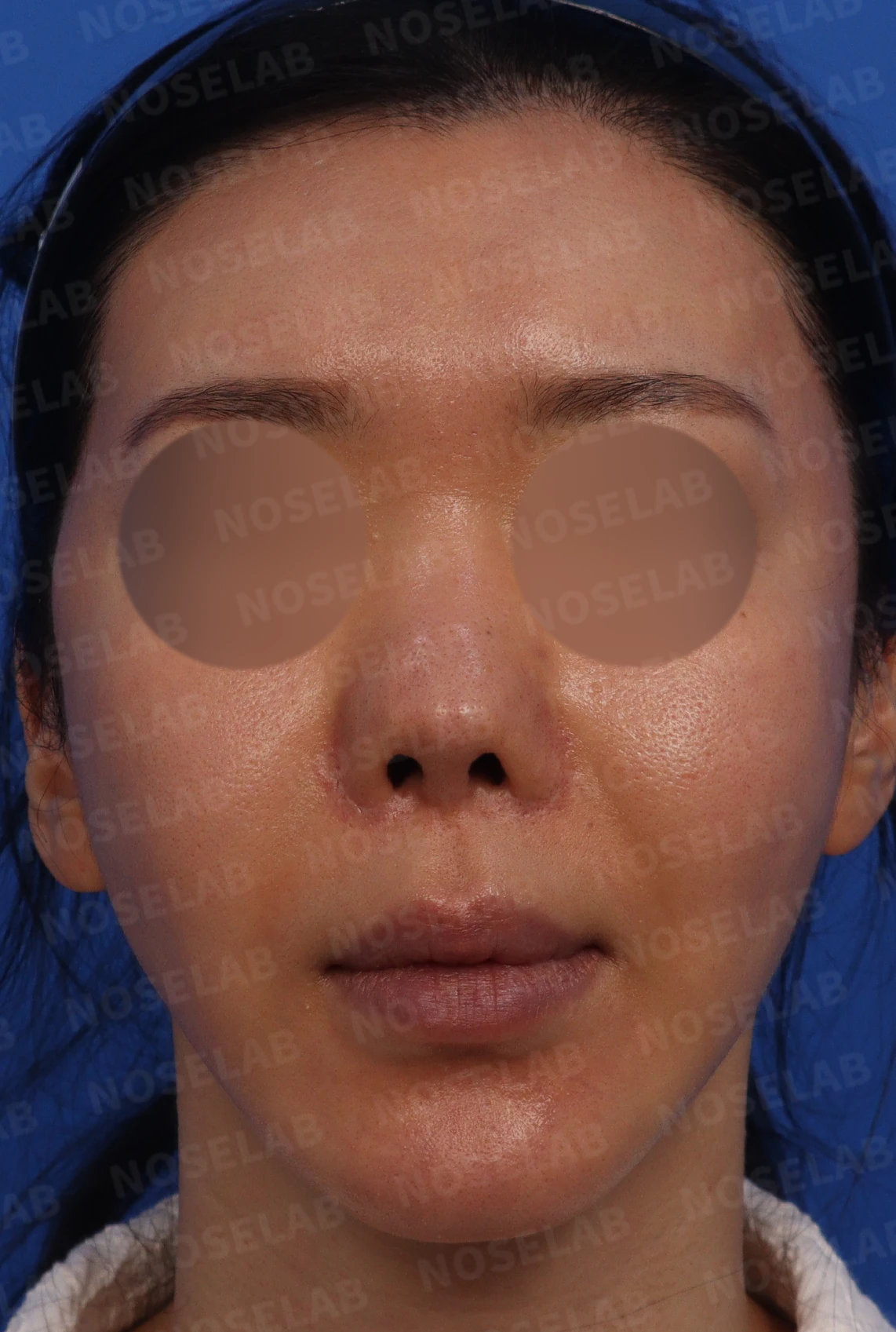
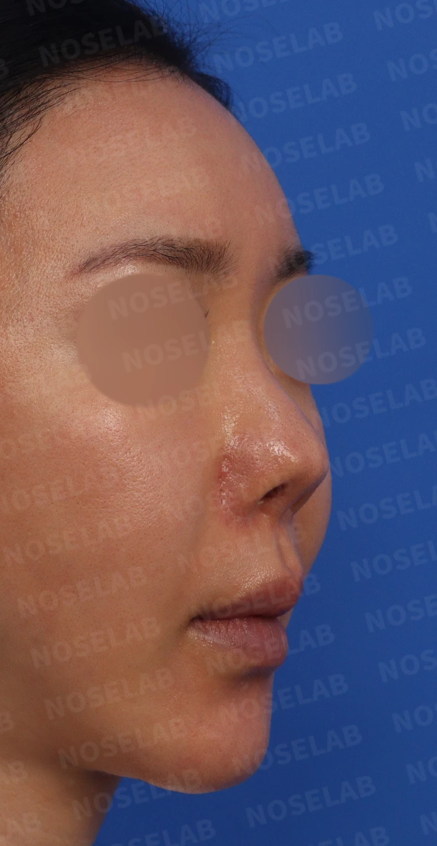
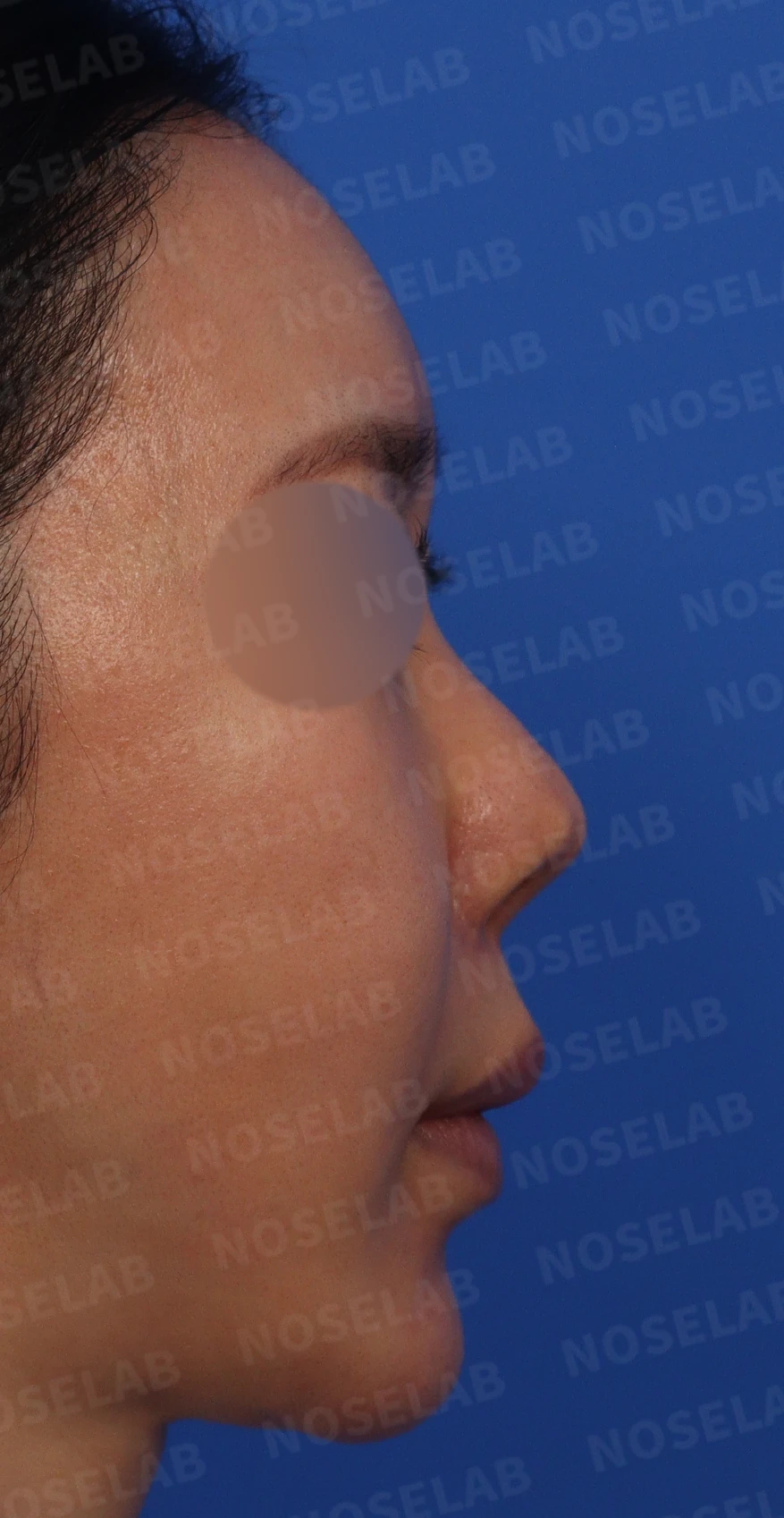
Preoperative design photos: front, oblique, and profile views
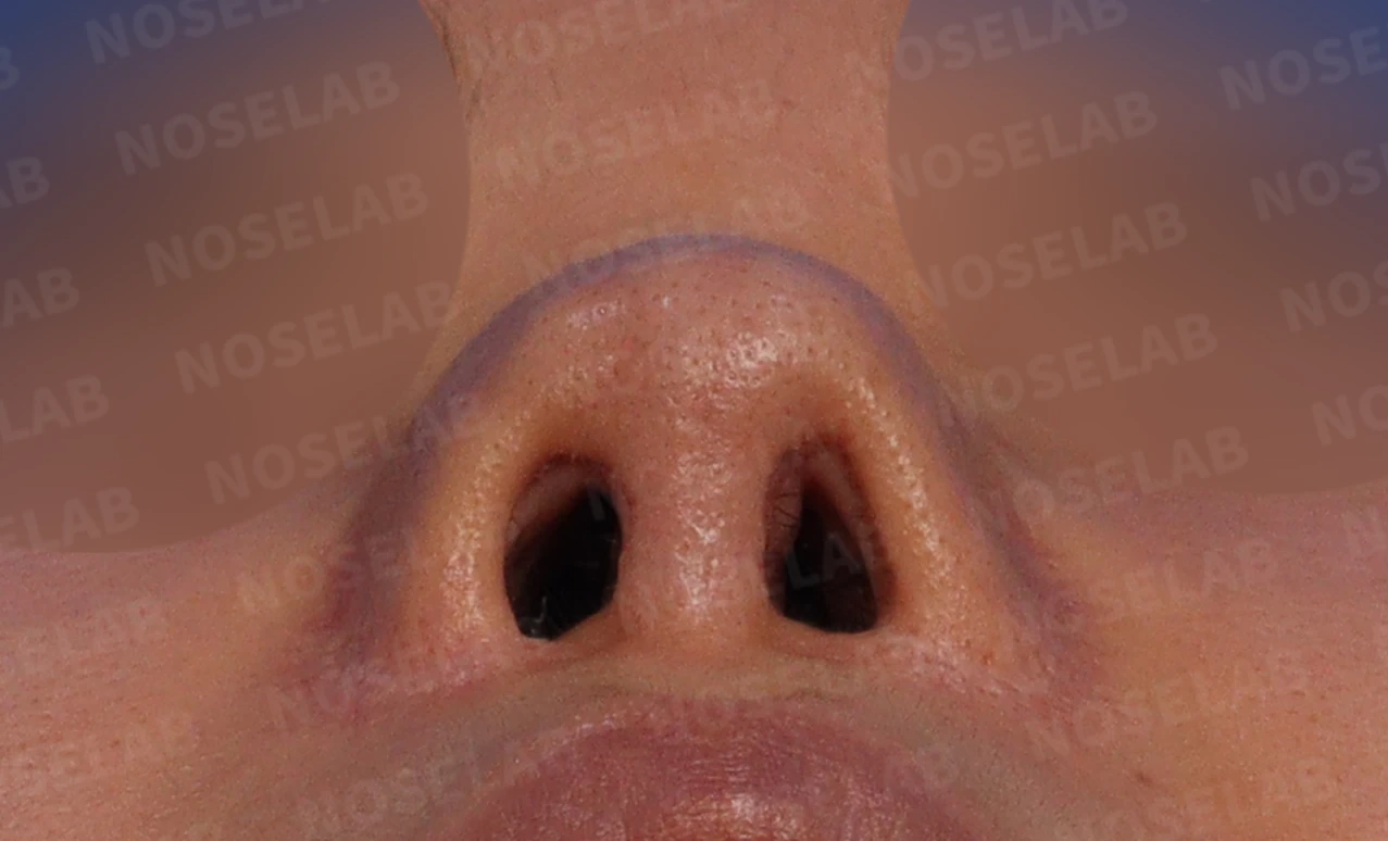
Basal (nostril) view
Front view:
- Excessive nostril show and asymmetry
- The nasal tip lacked definition, appearing wide and bulbous
- The nasal bridge was flat
Oblique view:
- The glabella was very low
- The nasal tip appears bulbous and upturned
Profile view:
- The glabella was low while the nasal dorsum was high
- The nasal tip was short and upturned
- The nasolabial angle was obtuse, with findings of a protruding mouth
Nostrils:
- Nostril asymmetry is present
- Skin grafted during a previous alar lowering surgery protruded toward the nostril
2. Surgical plan
Considering the patient's complex condition and requests, the following surgical plan was established:
a) Stem cell injection therapy: Performed 3 times at 2-week intervals to soften the nasal skin
b) Septal reconstruction and extension using autologous rib cartilage
c) Partial subcutaneous tissue excision
d) Lateral osteotomy
e) Correction of the nasolabial angle
f) Partial use of a silicone implant on the nasal bridge
g) Creating a straight bridge line
3. Surgical procedure
The following procedures were performed using a closed approach:
a) Septal reconstruction:
- As expected, most of the septal cartilage was damaged, so it was reconstructed using autologous rib cartilage.
b) Reconstruction of lower lateral (alar) cartilage:
- Bilateral lower lateral cartilage had been mostly removed.
- The ear cartilage used in previous surgery was carefully dissected and used to reconstruct both lower lateral cartilages.
c) Removal of dorsal nasal tissue:
- Tough fibrous tissue and contracture tissue on the nasal dorsum were carefully removed.
- Through this, the skin thickness over the dorsum was reduced.
d) Improvement of nasal shape:
- The bulbous and severely upturned areas were naturally refined.
e) Lateral osteotomy:
- The widened appearance and crooked look were corrected.
f) Correction of the nasolabial angle:
- The columella was naturally lowered to improve the nasolabial angle.
g) Insertion of nasal bridge implant:
- To correct the unnatural profile, a silicone implant was partially used on the nasal bridge.
All procedures were performed using a closed approach, completing the surgery without external scars.
4. Comparison of before-and-after photos
Comparing photos taken immediately after surgery on day 0 shows the following improvements: Photos taken immediately after surgery show swelling and may differ from the final result.
Front view:
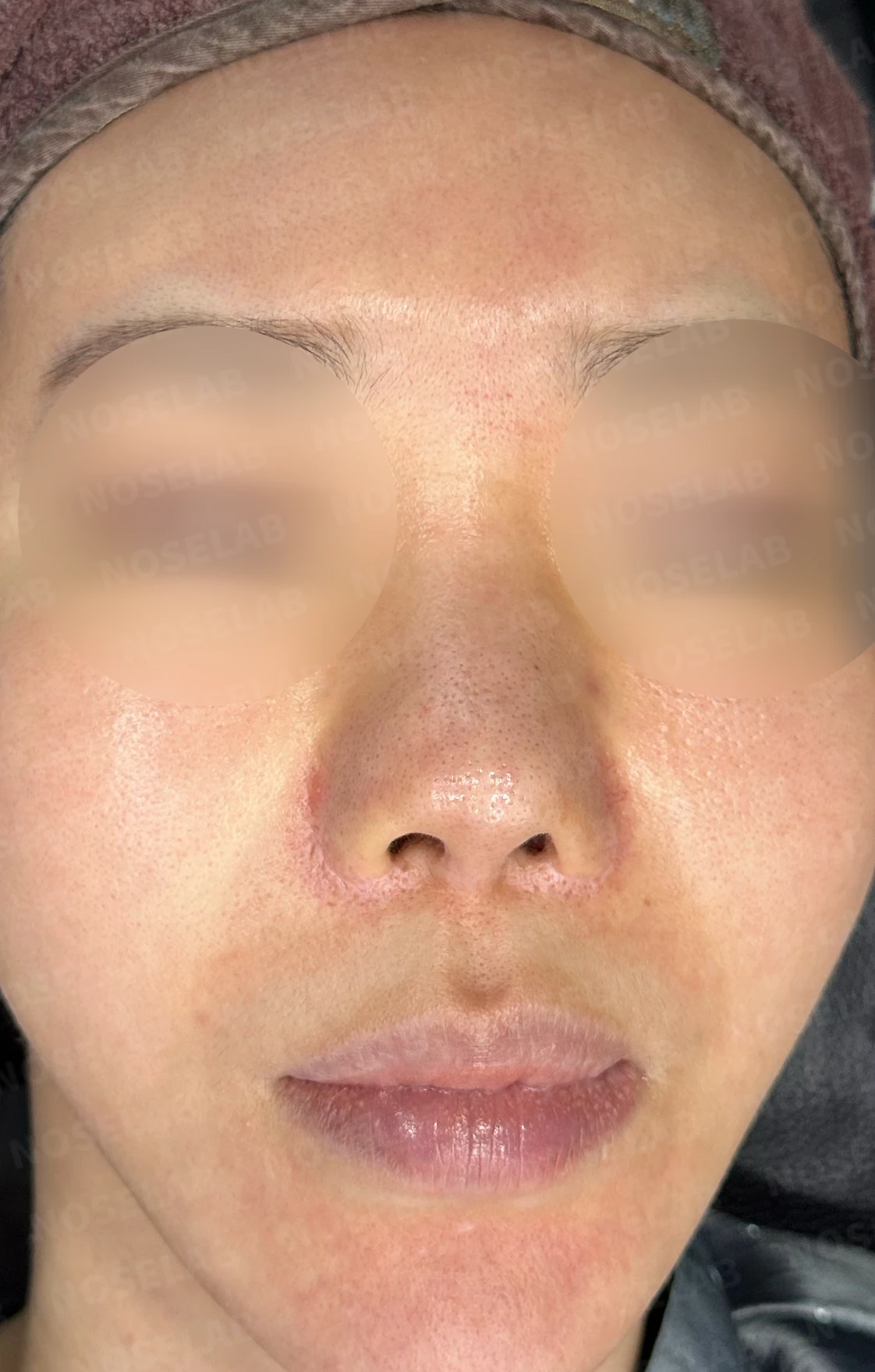
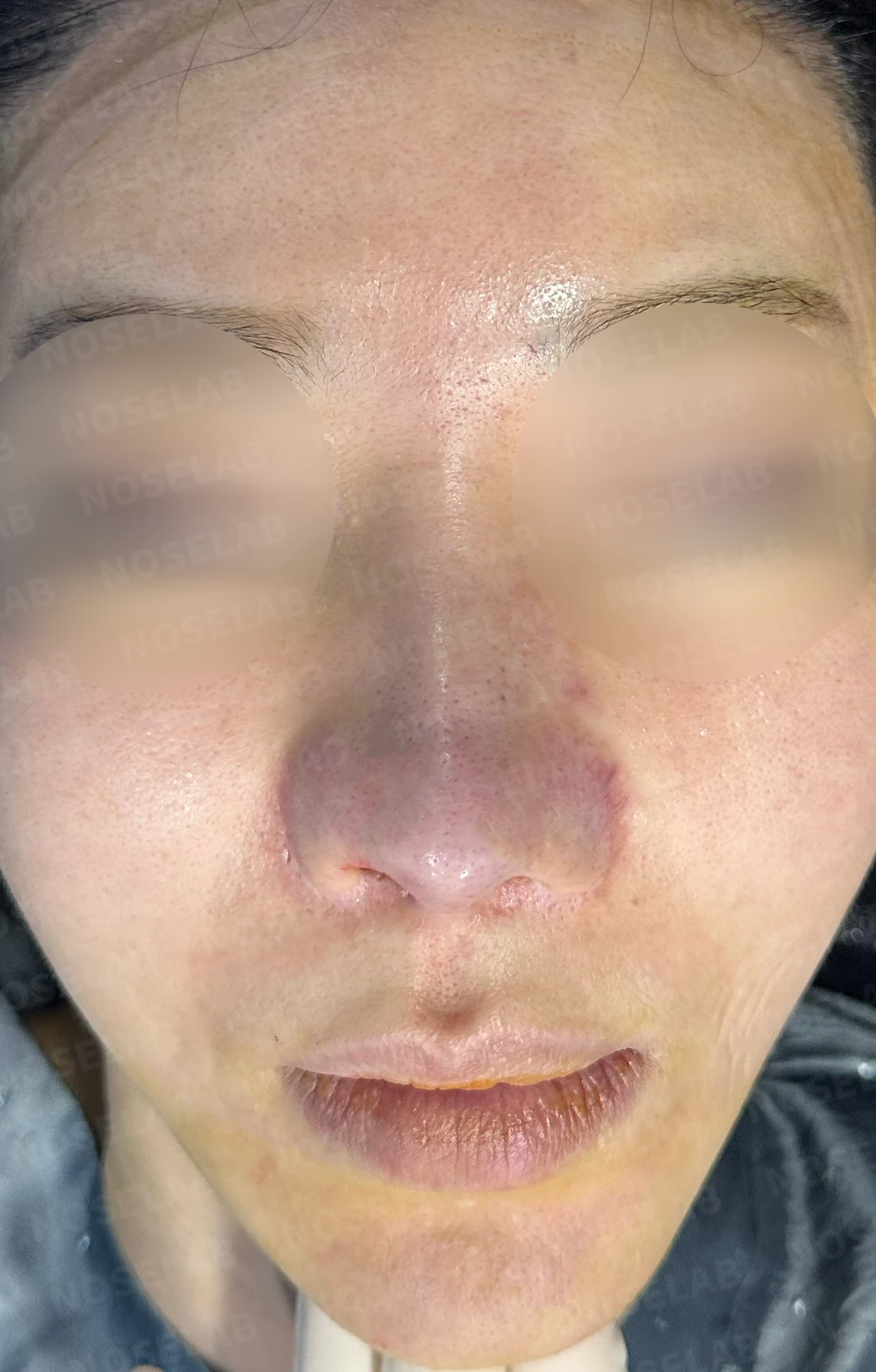
Before surgery / Day 0 immediately after surgery Photos taken immediately after surgery show swelling and may differ from the final result.
- Excessively visible nostrils are now naturally covered.
- Nostril asymmetry improved significantly.
- The widened, bulbous nasal tip was refined into a more delicate and natural shape.
- The flat nasal bridge gained appropriate height and definition.
Oblique view:
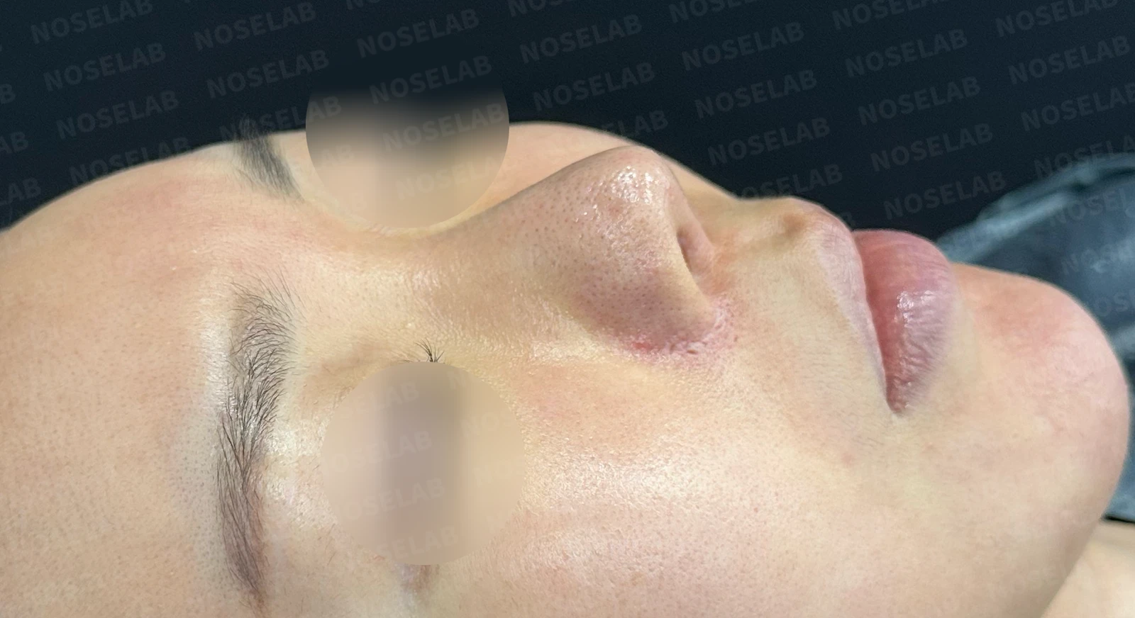
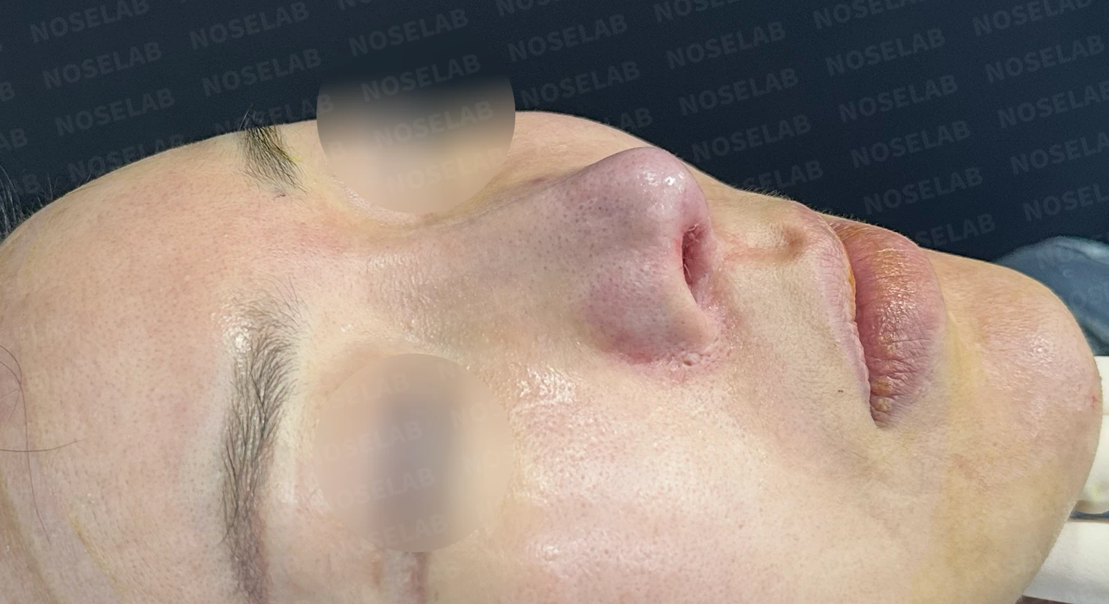
Before surgery / Day 0 immediately after surgery Photos taken immediately after surgery show swelling and may differ from the final result.
- The low glabella was naturally elevated.
- The bulbous and upturned tip was improved, resulting in a smoother overall nasal line.
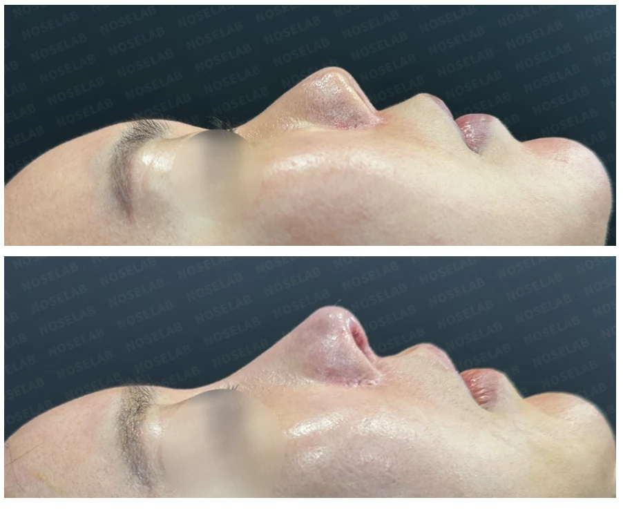
Profile view:
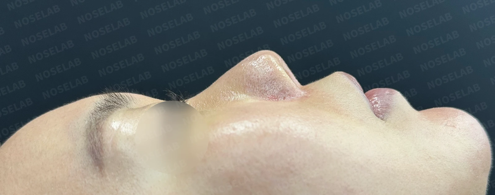
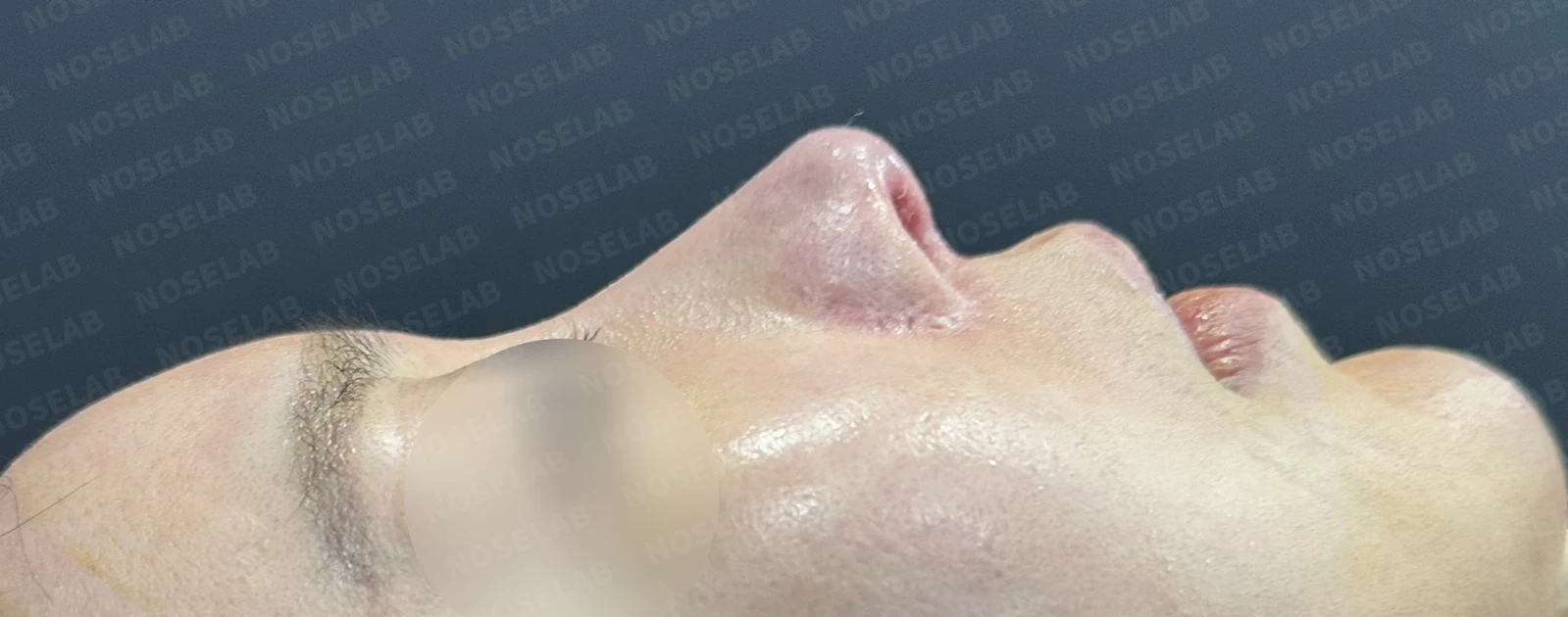
Before surgery / Day 0 immediately after surgery Photos taken immediately after surgery show swelling and may differ from the final result.
- The balance in height between the glabella and nasal dorsum was improved.
- The short, upturned tip was properly corrected, bringing the overall shape into harmony.
- The wide nasolabial angle and appearance of a protruding mouth were improved, looking more natural.
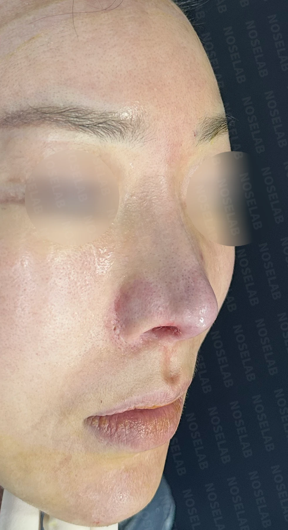
Nostrils:
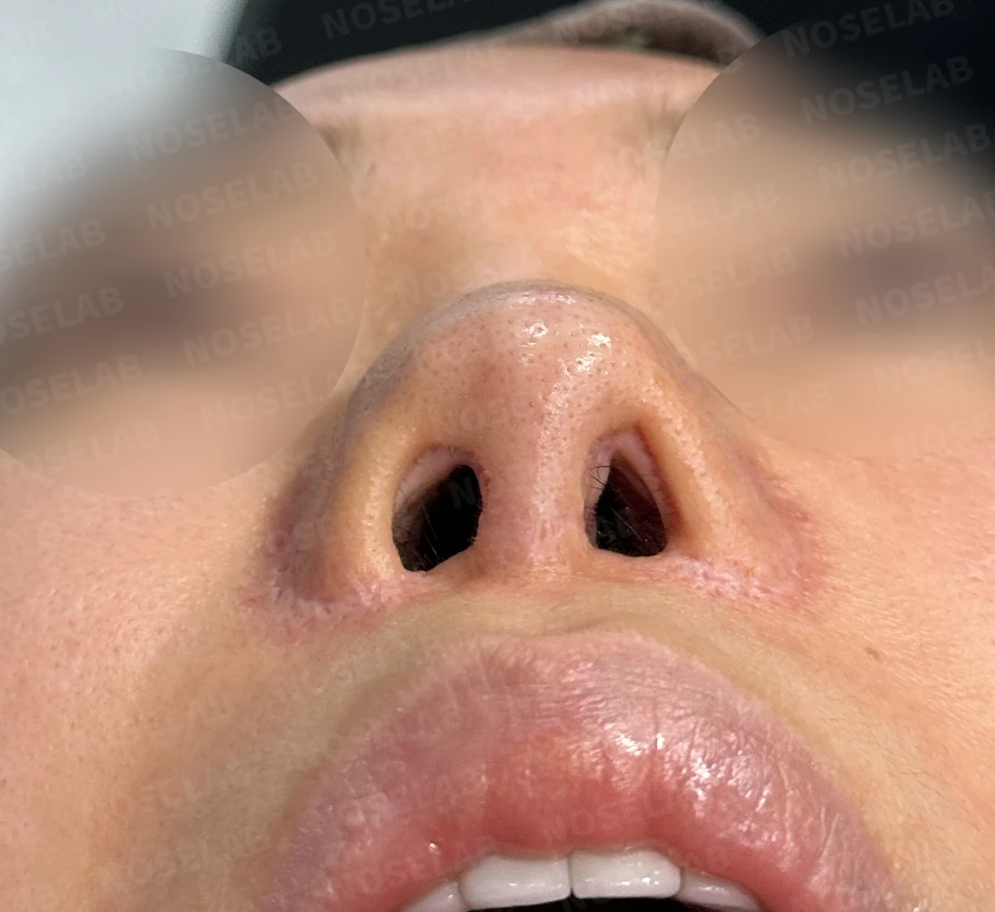
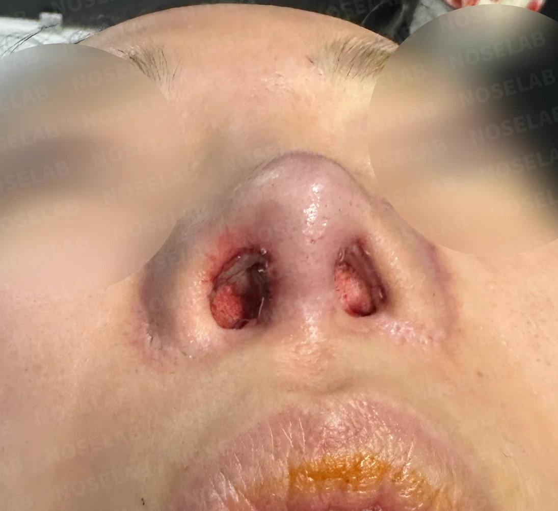
Before surgery / Day 0 immediately after surgery Photos taken immediately after surgery show swelling and may differ from the final result.
- Nostril asymmetry improved significantly.
- The skin that had protruded from the previous surgery was naturally corrected.
5. Photos immediately after surgery on day 0 Photos taken immediately after surgery show swelling and may differ from the final result.
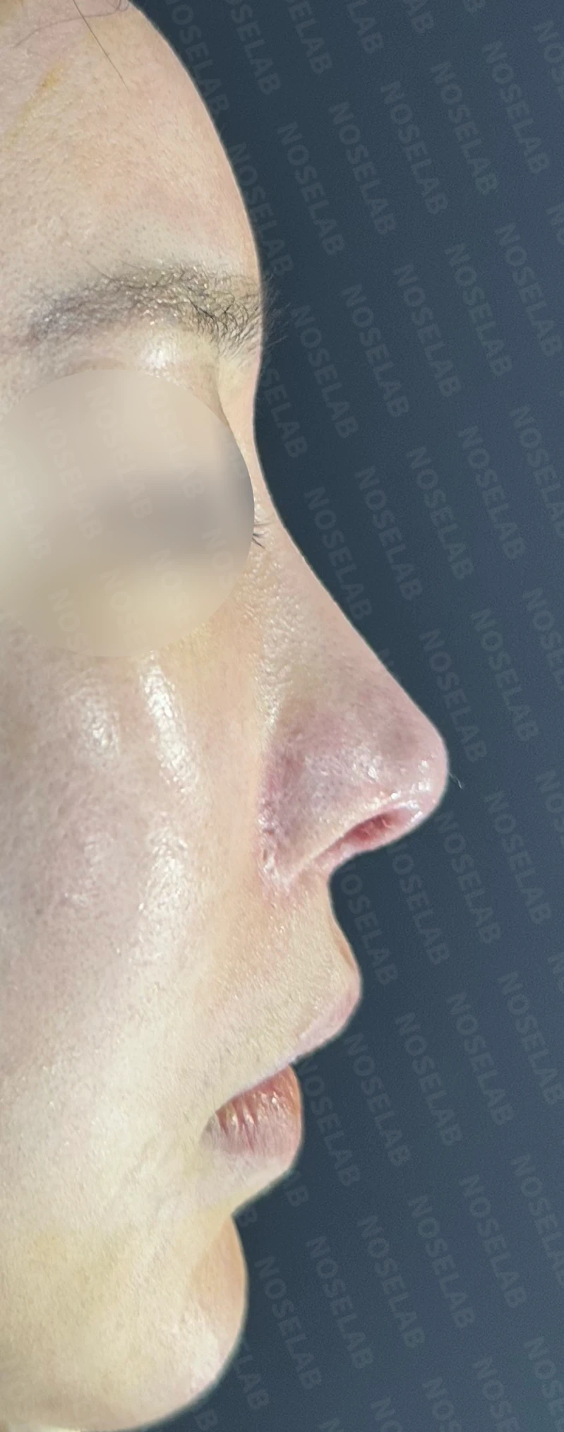
Immediately after surgery (Day 0) front, profile, and oblique viewsImmediate postoperative photos show swelling and may differ from the final result.
Basal (nostril) view immediately after surgery (Day 0)Immediate postoperative photos show swelling and may differ from the final result.
6. Overall evaluation
This was a highly complex case involving contracture deformity from multiple rhinoplasties and inflammation. In such situations, a comprehensive approach considering both cosmetic improvement and nasal function is necessary.
The greatest challenge was that almost no normal cartilage remained inside the nose. This can pose significant problems functionally as well as cosmetically. Therefore, septal reconstruction using autologous rib cartilage and reconstruction of the lower lateral cartilages were essential.
Softening the contracted nasal skin was also critical. For this purpose, stem cell injection therapy was conducted beforehand, and contracture tissue on the dorsum was carefully excised during surgery. Throughout this process, minimizing damage to normal tissue and paying particular attention to preserving blood vessels were key. This plays an important role in preventing skin necrosis and supporting postoperative recovery.
Through this surgery, the overall shape of the patient's nose was naturally improved. The widened, upturned nasal tip was corrected, the height and contour of the nasal bridge were enhanced, and overall facial balance was improved by correcting the nasolabial angle.
7. Conclusion
Revision rhinoplasty for a nose that has undergone multiple surgeries and developed contracture deformities from inflammation is an exceptionally difficult procedure. Often, the normal cartilage structures that should exist within the nose are mostly missing, requiring full reconstruction each time. This is essential not only for aesthetic reasons but also for proper function.
As a board-certified otolaryngologist (ENT specialist), I believe these complex reconstructive surgeries represent an important mission. Especially in contracted nose surgery, softening the nasal skin is the most challenging and crucial task. This demands surgical techniques that minimize damage to normal tissue and a delicate approach toward blood vessels. These steps are essential to prevent skin necrosis and achieve a favorable result.
Successfully resolving these complex revision cases requires extensive experience and profound knowledge. NOSELAB strives to achieve the intended results even in such complex cases, drawing on our expertise and long-standing clinical experience as an ENT specialist.
I hope this case offers hope to those struggling with a contracted nose or unsatisfied results after repeated rhinoplasties. I wanted to demonstrate that even highly intricate cases can be improved through a closed approach and specialized surgical techniques.
I will always be here to support you in achieving a healthy and aesthetically pleasing nose.
This was Dr. Peter C. Kang. Thank you.
Thank you.
#contractednosecorrection #revisionrhinoplasty #septalreconstruction #autologousribgraft #closedrhinoplasty #upturnednosecorrection #bulbousnosecorrection #nostrilasymmetrycorrection #ENTspecialistrhinoplasty #functionalrhinoplasty #stemcelltherapy #contractedskinsurgicalcare

Limitations and risks
When autologous rib cartilage is used, scarring and pain may remain at the donor site on the chest, and pneumothorax (pleural injury) can occur rarely. Over time, the grafted cartilage may warp or undergo partial resorption.
When revising after implant removal, contracture may progress again after removal, the nasal height and length may decrease, and staged surgery may be necessary depending on the skin condition.
When the septum is corrected at the same time, nasal obstruction may persist or recur, and in rare cases, septal perforation or changes in smell may occur.
Common potential risks include infection, bleeding, asymmetry, scarring, skin necrosis, and insufficient improvement; results vary depending on individual skin condition and previous surgical history.