Hello, I am Dr. Peter C. Kang.
Preoperative design consultation
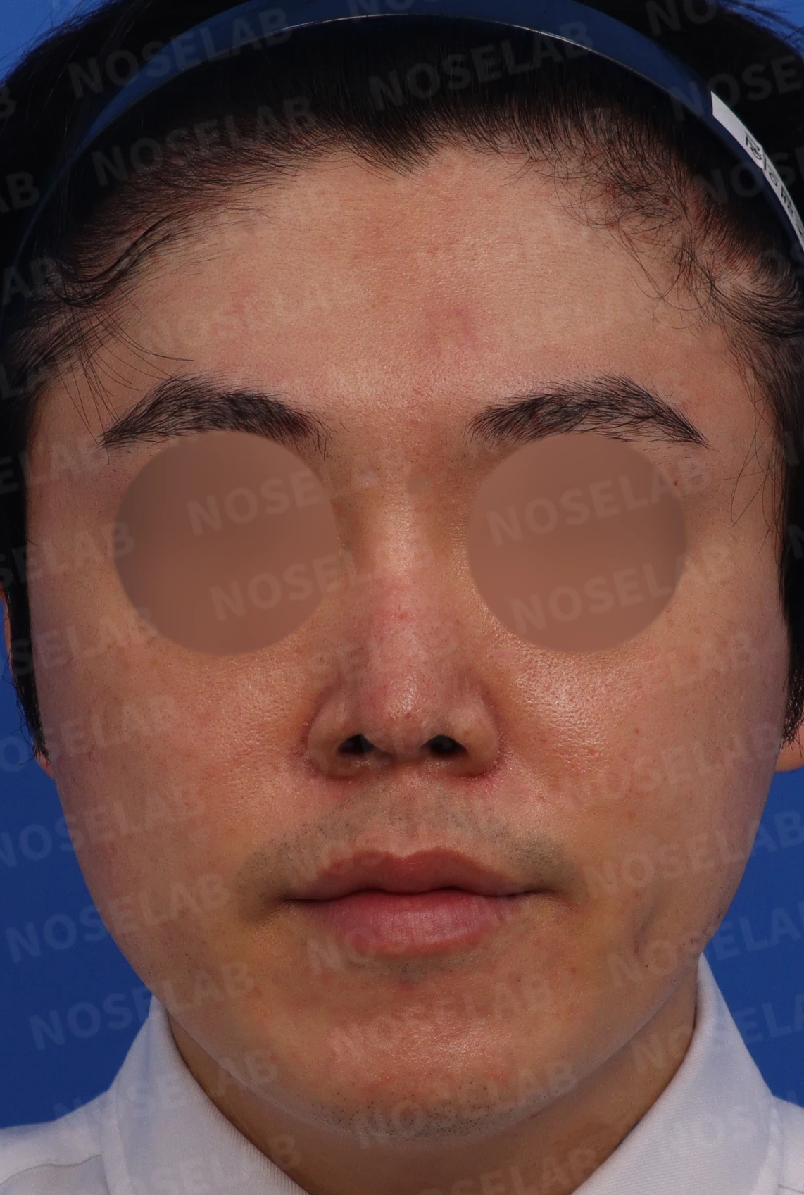
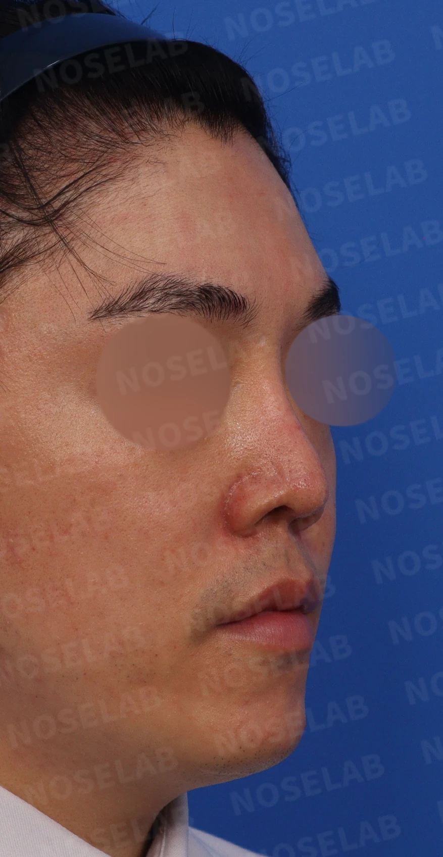
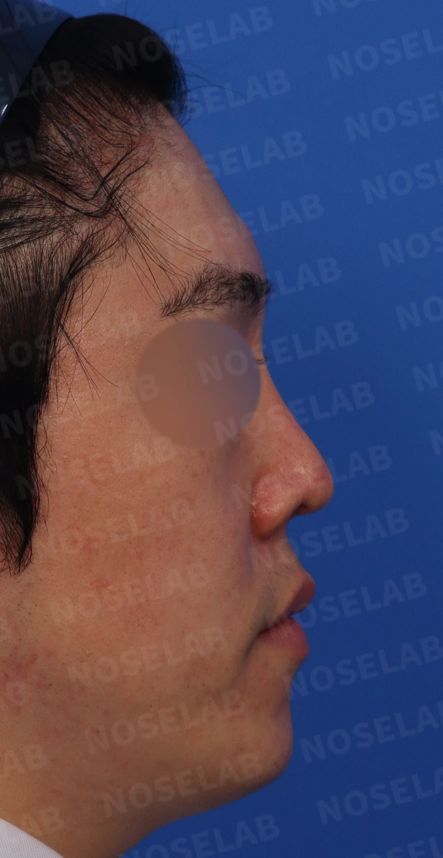
Photographs taken before surgery for the design consultation: front view, oblique view, and profile view.
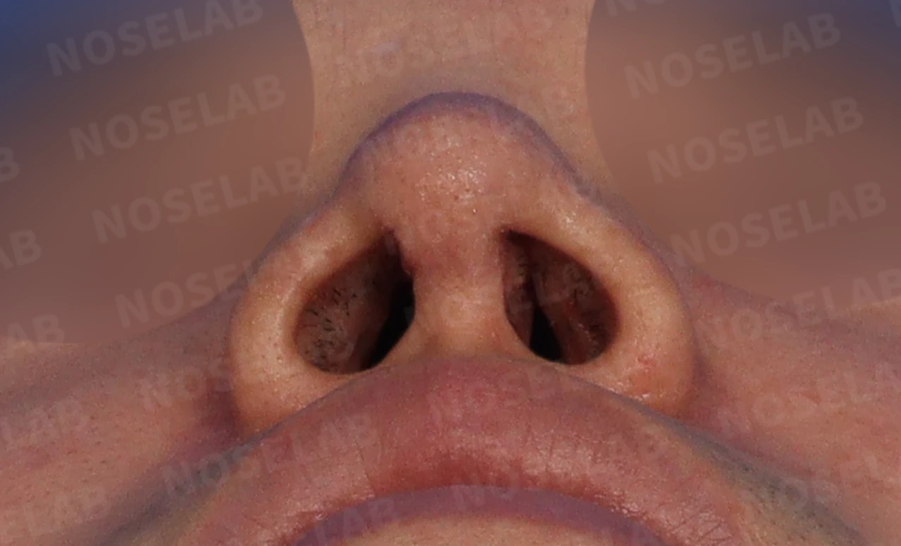
This is the basal (nostril) view.
The patient visited NOSELAB wanting to correct an upturned nose. The main concerns were as follows:
Left-sided nasal obstruction
Drooping tip
Nose deviated to the right
High starting point at the glabella and an appearance suggestive of a dorsal hump
Visible nostril show from the front and asymmetry
Retracted columella leading to an acute nasolabial angle and the appearance of a protruding mouth
The patient's requests were as follows:
Overall nasal elevation
Correction of upturned nose
Correction of wide nose
Lifting the alae if possible
An important point was that the patient had already undergone rhinoplasty twice before. This was a critical factor affecting the surgical plan.
Surgical plan
Taking the prior surgical history into account, we established the following surgical plan:
Reconstruction of the septal cartilage
Lower lateral cartilage reconstruction
Removal of contracture tissue along the nasal bridge
Maximally expanding the nasal skin envelope
Reconstruction of the lower lateral cartilages to improve nostril appearance
Nostril sill reduction and alar base elevation
Correction of the nasolabial angle
Nasal height elevation
Surgical procedure
The surgery was complex and took about 2 hours longer than anticipated. The main surgical steps were as follows:
a) Dissection of the alar cartilages
Both lower lateral cartilages had already been removed.
b) Assessment of the septal cartilage status
Most of it had been removed, leaving only a small portion along the dorsum and columella.
The tip support framework was severely weakened.
Cartilage added to the left side of the septum during a previous surgery had narrowed the left nasal cavity, causing nasal obstruction.
c) Reconstructive surgery
Using autologous rib cartilage, we reconstructed the entire septal cartilage and both lower lateral cartilages.
Contracture tissue along the nasal bridge was excised, and the skin envelope was expanded as much as safely possible.
To improve nostril shape and symmetry, the lower lateral cartilages were completely reconstructed using autologous rib cartilage.
d) Additional corrections
We performed nostril sill reduction and alar base elevation.
The nasolabial angle was corrected.
We maximized the skin envelope expansion to improve overall nasal height.
Before-and-after photo comparison
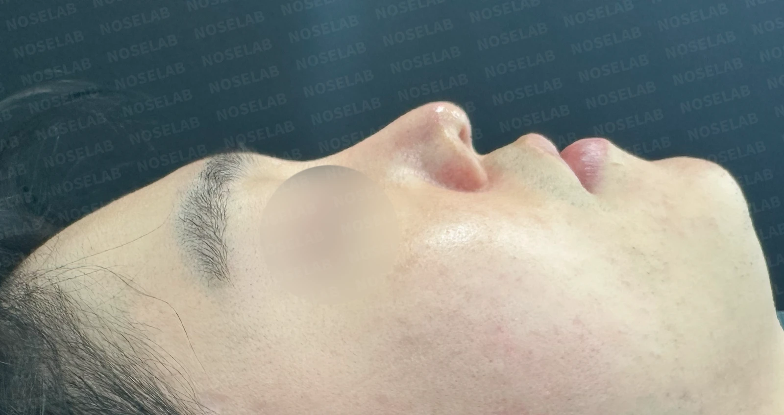
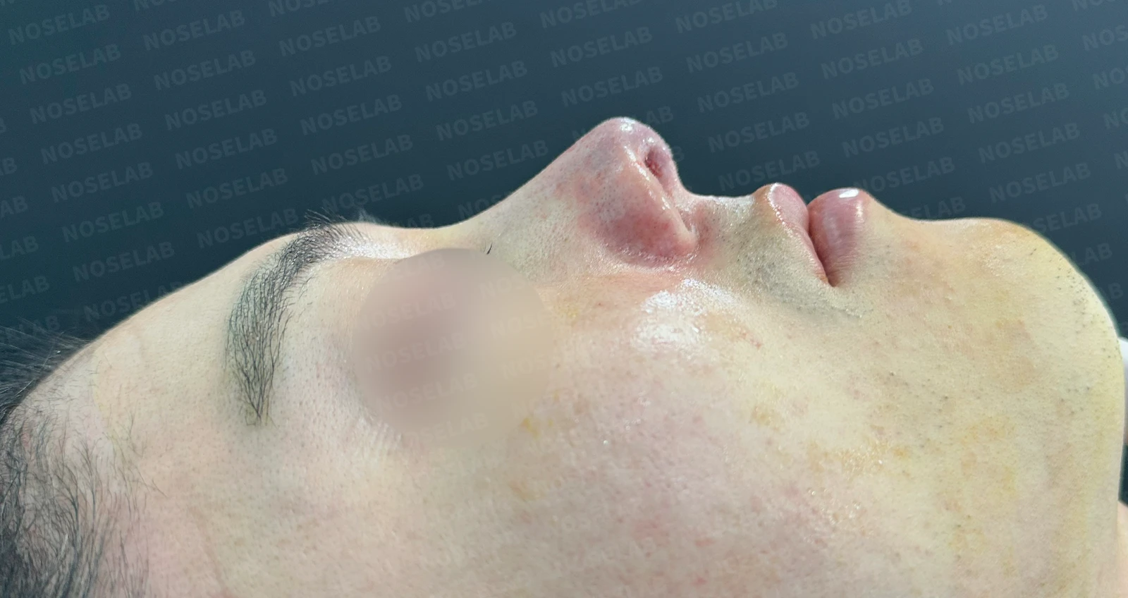
a) Profile view
The nasolabial angle has clearly been corrected.
The nasal tip projection has increased significantly relative to the forehead bone.
The drooping appearance of the tip has improved.
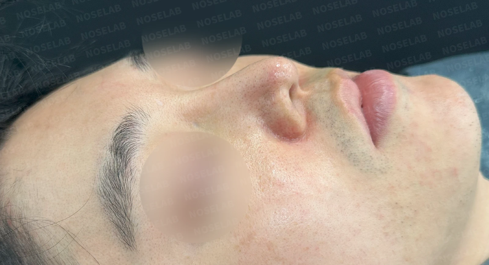
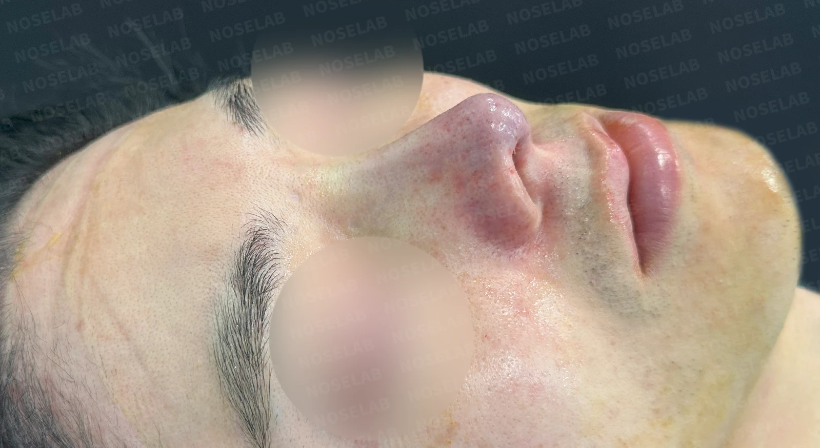
b) Oblique view
The definition and projection of the nasal tip are noticeably restored.
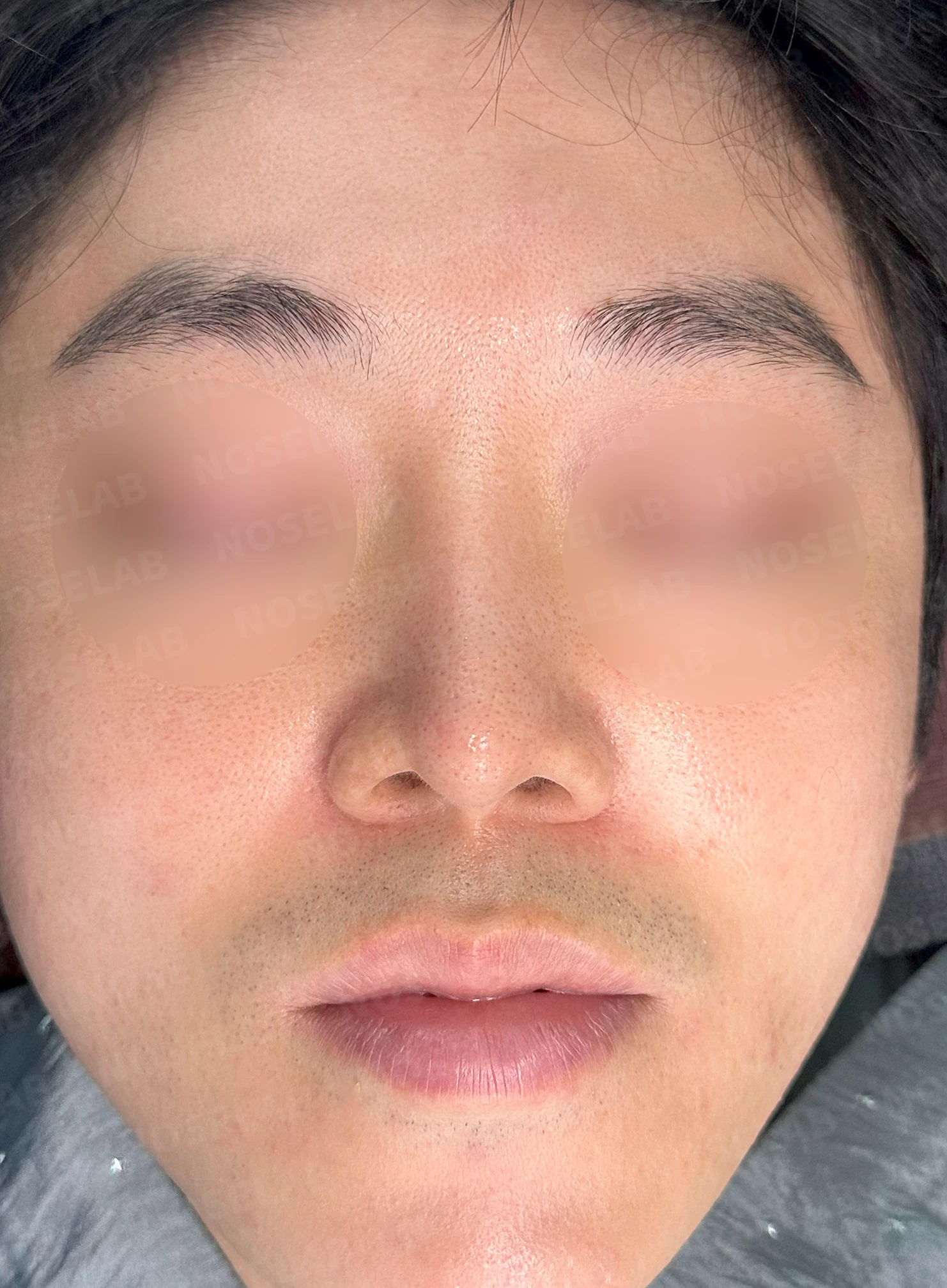
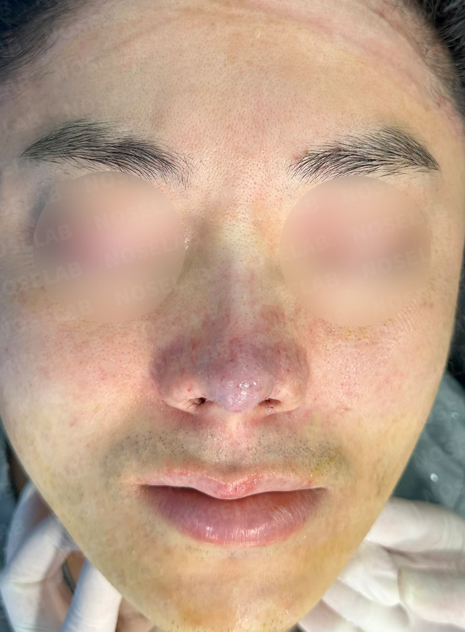
c) Front view
The previously deviated portion has significantly improved.
The thinned tip appearance and excessive nostril show have been corrected.
The elevated position of the alae can be confirmed.
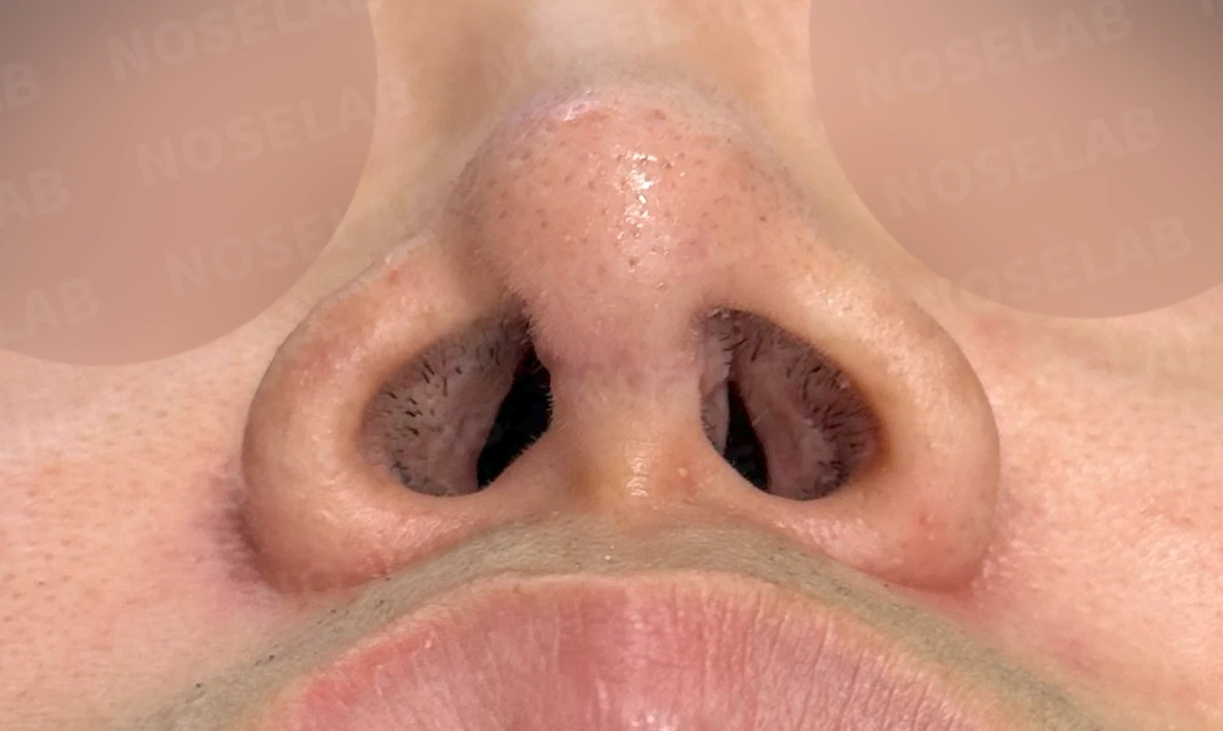
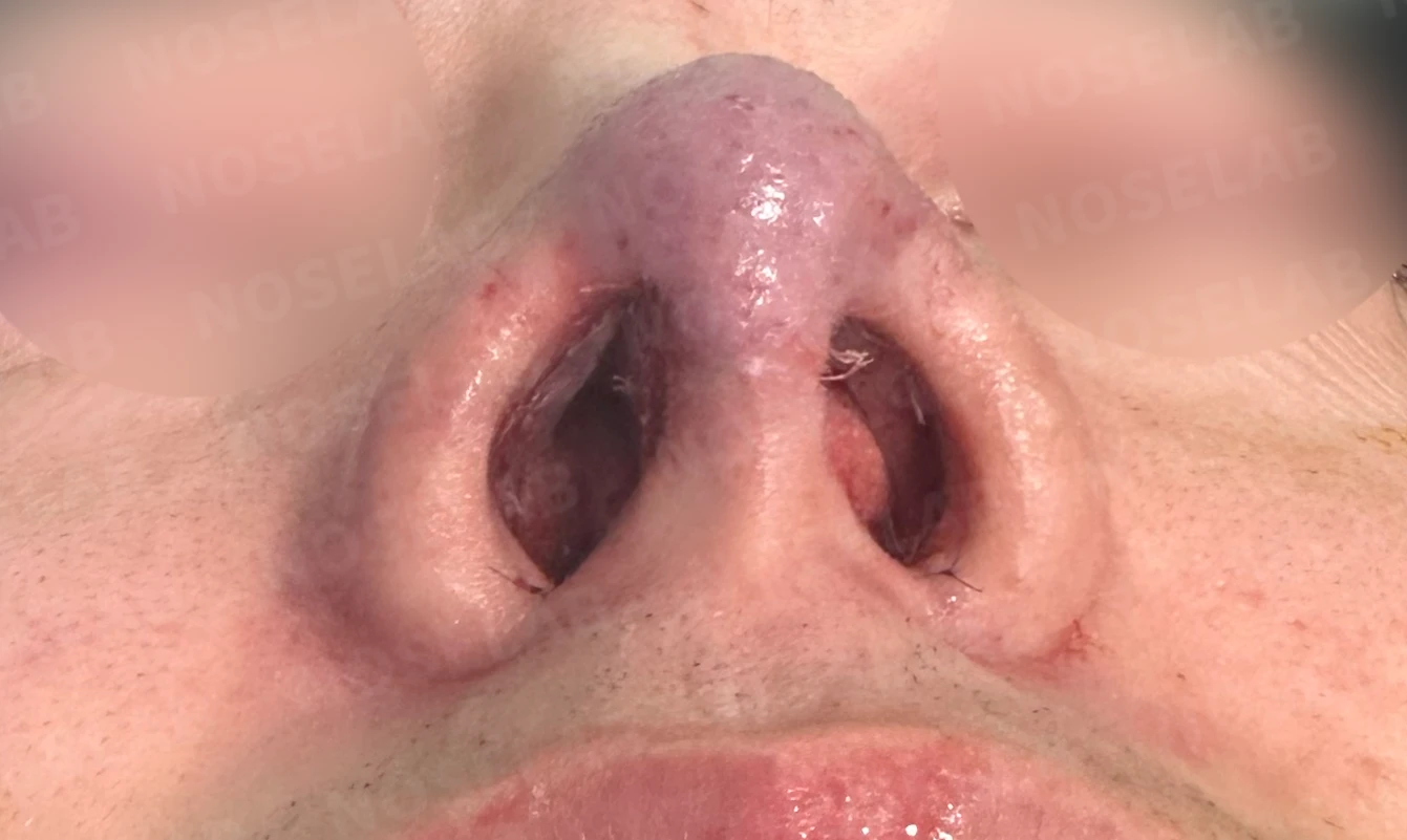
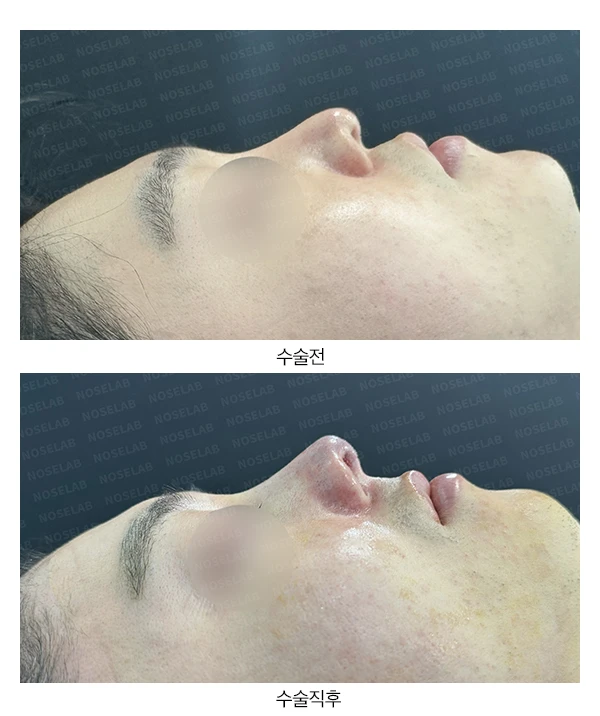
d) Nostril view
The tilted columella has been straightened.
The narrowed left nasal passage has improved substantially.
Nostril asymmetry improved significantly.
The nostril sill width has been noticeably reduced.
Photos immediately after surgery
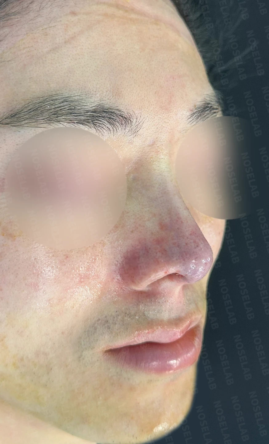
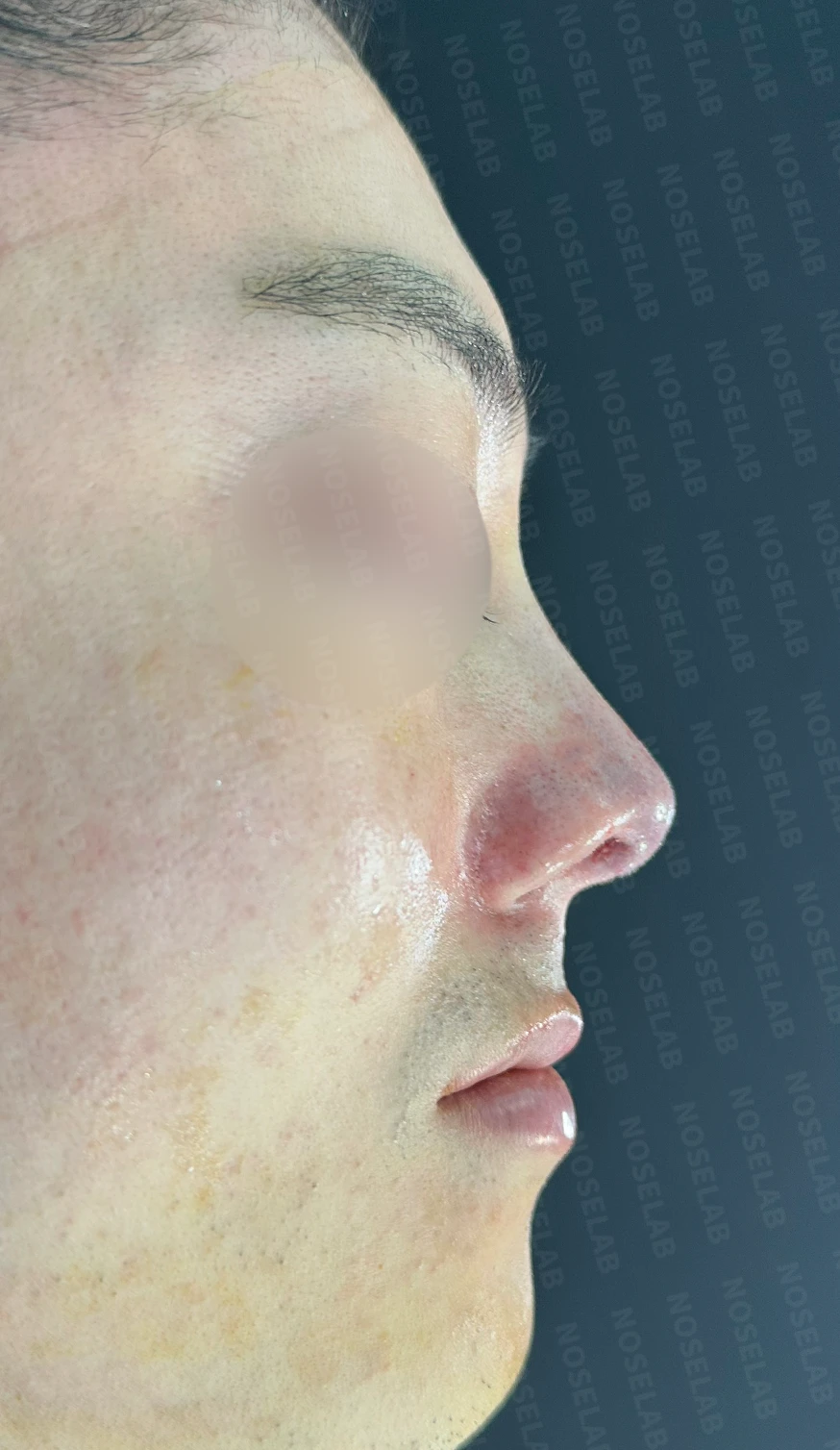
Photos taken immediately after surgery on day 0: front, oblique, profile view. Photos taken immediately after surgery show swelling and may differ from the final result.
Basal (nostril) view immediately after surgery (Day 0).Immediate postoperative photos show swelling and may differ from the final result.
Conclusion
In revision rhinoplasty patients, the internal anatomical findings frequently differ greatly from what was reported about previous procedures. At times, the actual structural damage is more severe than anticipated, and it is regrettable that this can be difficult to fully explain in advance.
In revision cases, where and how previous surgeries were performed is often more important than the simple number of operations. Even with the same number of prior surgeries, the internal anatomy and surgical outcomes can vary widely.
Whether structural reconstruction is required creates a substantial difference in operative time, outcomes, and prognosis. Because this alters the surgical plan, it is vital to share as much accurate detail as possible about your prior surgeries during the initial consultation.
Although this patient's surgery took 2 hours longer than expected, reconstructive surgery enabled us to achieve a favorable outcome.
I hope this case is informative for those troubled by septal cartilage damage or nasal deformities. Through a thorough consultation with a specialist, you can find the surgical approach best tailored to your individual needs. We will continue to work together toward healthy, well-balanced nasal results.
Frequently asked questions (FAQs)
1. How long is the recovery period after surgery?
Although recovery periods vary by individual, patients can generally return to daily activities in about 1-2 weeks.
2. Are cartilage grafts safe?
Yes, cartilage grafts are safe, and when autologous cartilage is used, the risk of rejection is minimal.
3. How is the pain after surgery?
There may be mild pain after surgery, but it can be managed with prescribed pain medication.
4. When can the surgical results be seen?
As swelling subsides, results gradually become visible after 1-2 months.
#closedrhinoplasty #closedcomplexrevisionrhinoplasty #closedautologousribcartilagerhinoplasty #closedribrhinoplasty #closednasalreconstruction #rhinoplasty #septalcartilagedamage #crookednosecorrection #nasalreconstruction #rhinoplastyreviews #tipplasty #plasticsurgery #revisionrhinoplasty #rhinoplastybeforeandafter #nasalsurgery #tipcorrection #lateralosteotomy #septalextension #nasalobstructiontreatment

Limitations and risks
When autologous rib cartilage is used, scarring and pain may remain at the donor site on the chest, and pneumothorax (pleural injury) can occur rarely. Over time, the grafted cartilage may warp or undergo partial resorption.
When the septum is corrected at the same time, nasal obstruction may persist or recur, and in rare cases, septal perforation or changes in smell may occur.
Common potential risks include infection, bleeding, asymmetry, scarring, skin necrosis, and insufficient improvement; results vary depending on individual skin condition and previous surgical history.