Hello.
I am Dr. Peter C. Kang.
Today, I would like to share the case of a patient who had undergone surgery elsewhere using autologous rib cartilage. Inflammation developed 7 days postoperatively, and despite active medical treatment, the acute inflammation could not be controlled, leading to the complete removal of materials from both the nasal bridge and tip 2 weeks after surgery. While being monitored, the patient visited NOSELAB.
The patient received 3 sessions of stem cell injection therapy using autologous fat at our clinic and underwent surgery 6 months after the total removal. In addition, because the patient had experienced significant discomfort breathing through the nose after the previous surgery at another clinic, we also decided to perform surgery to improve nasal obstruction at the same time.
Let us look at the design consultation conducted at NOSELAB before surgery.
Photos before stem cell therapy and preoperative design consultation
These are photos before the stem cell therapy. From the front view, the nostril asymmetry and a flared appearance were most noticeable, and considering that it was the 4th month after total removal, the skin of the nasal tip was very hard and uneven. In particular, the nostril asymmetry was severe, and the scar tissue formed at the suture area had contracted significantly, making the right nostril much smaller. Therefore, we performed 3 sessions of stem cell therapy by extracting stem cells from autologous fat and injecting them into the nasal tip.
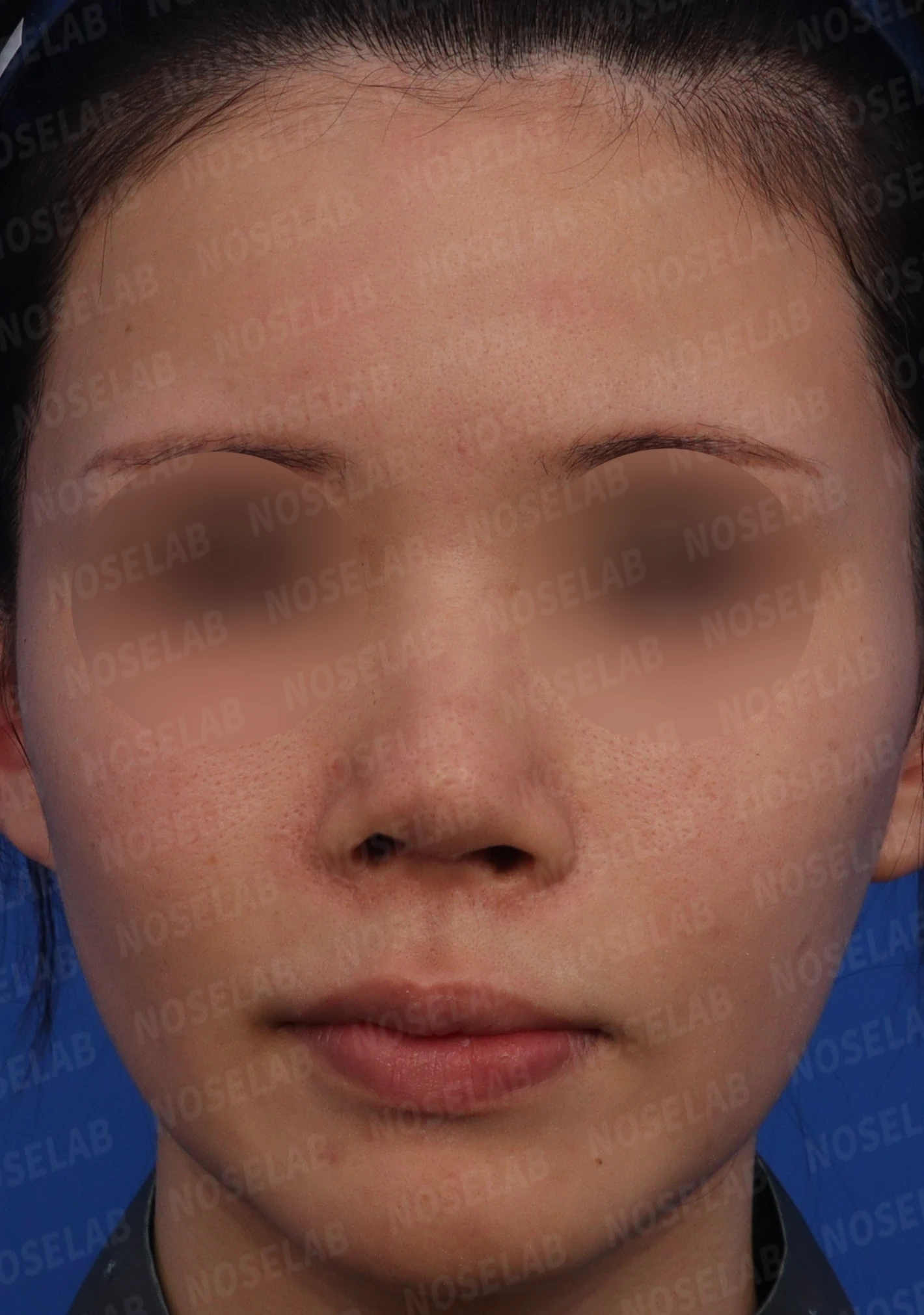
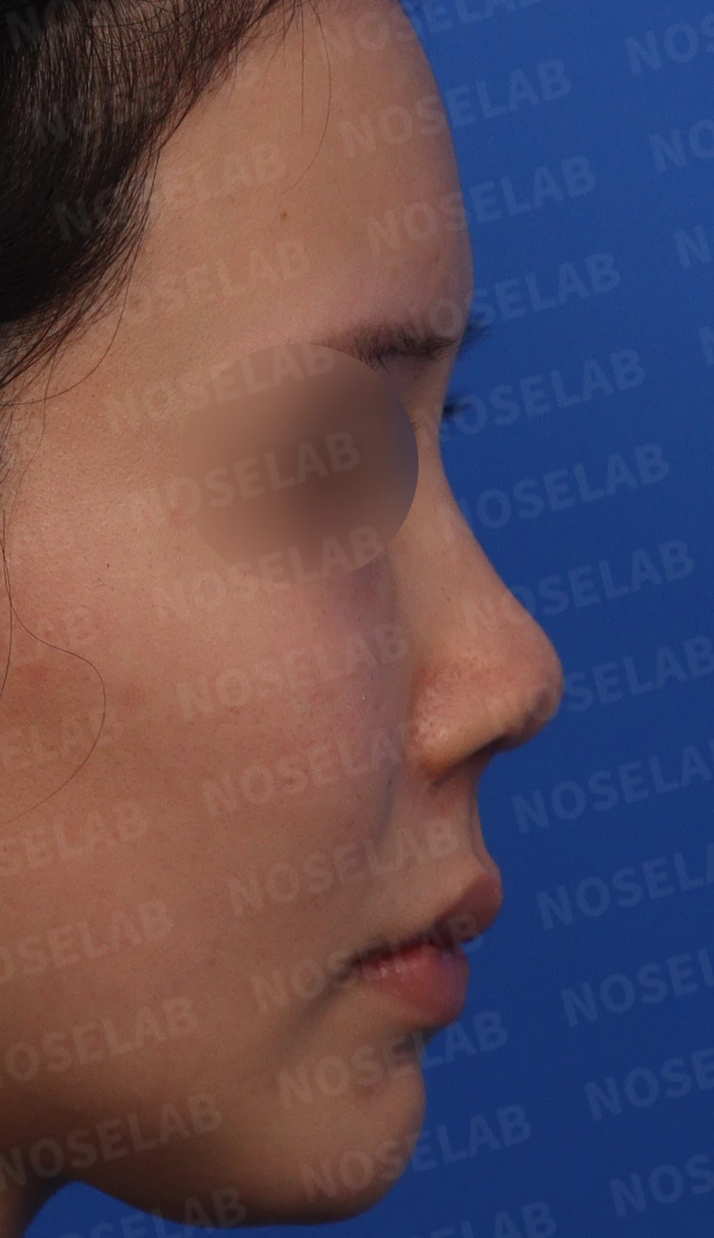
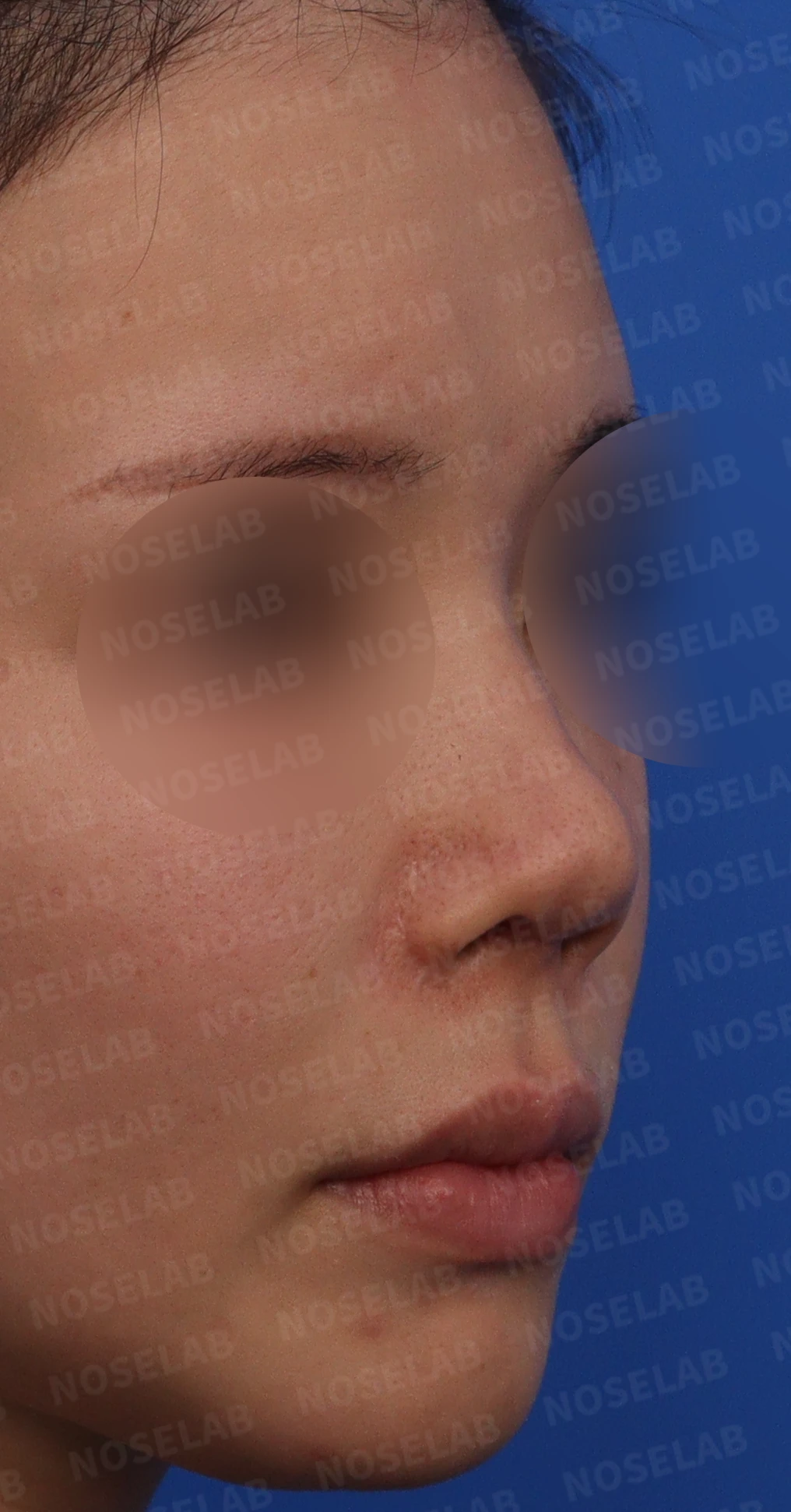
Front, 90-degree profile, and 45-degree oblique photos before stem cell therapy
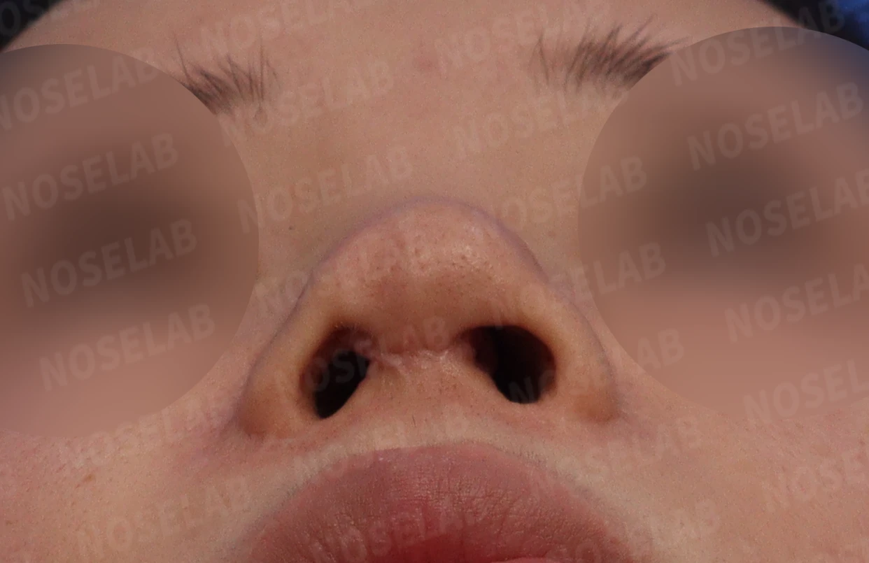
Nostril photo before stem cell therapy
These photos were taken during the preoperative design consultation after stem cell therapy. Overall, the skin of the nasal tip became much softer, and the uneven texture improved. In particular, the scar tissue at the suture site of the nostril softened substantially.
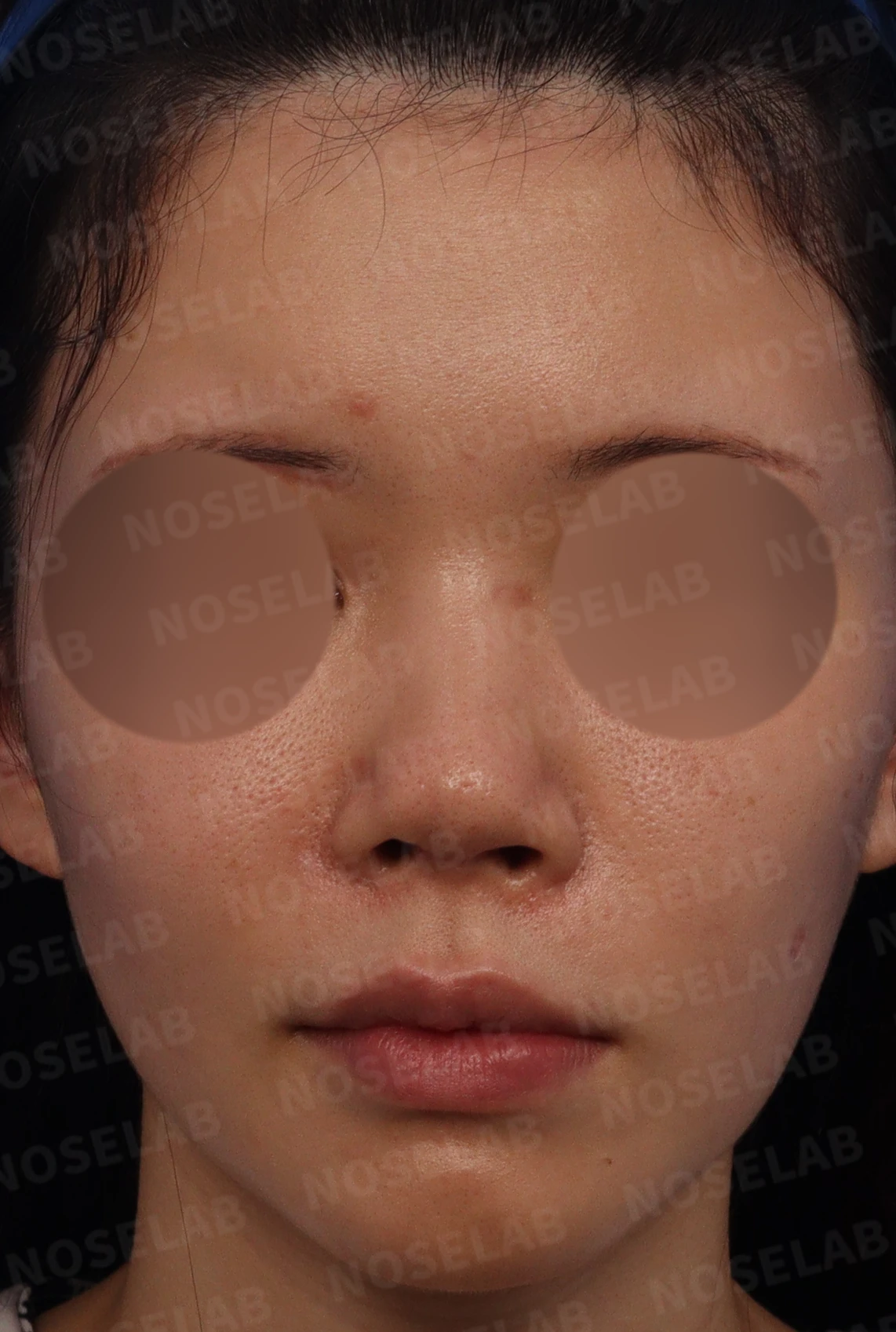
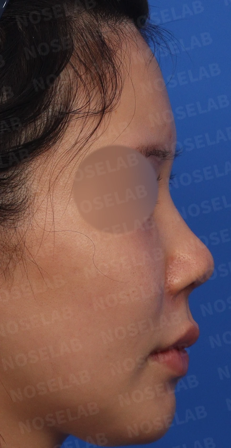
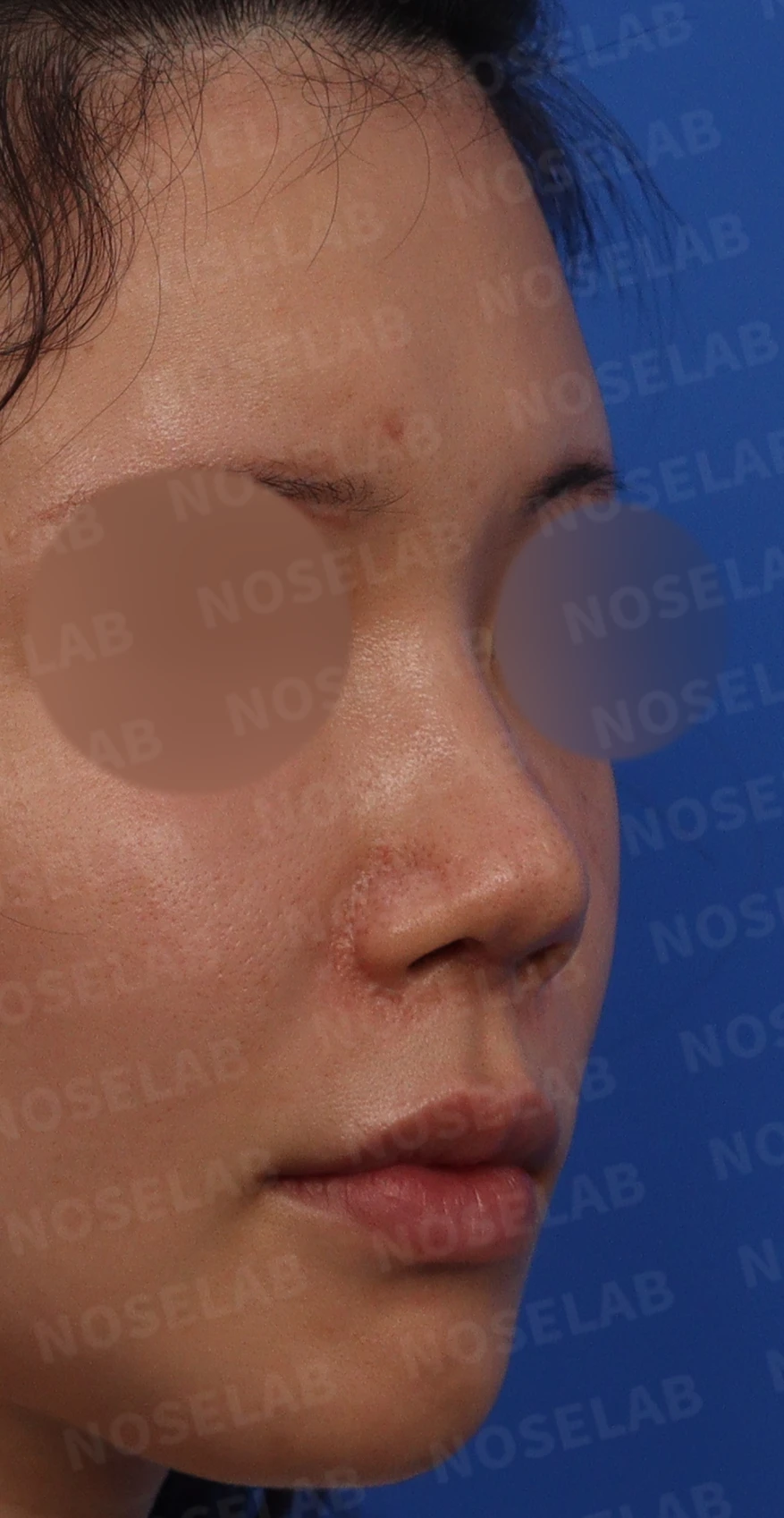
Preoperative design consultation: front view
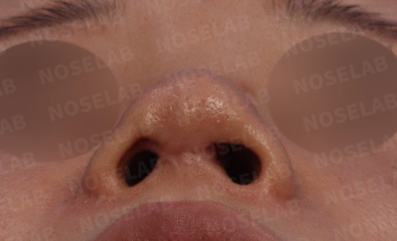
Preoperative design consultation: nostril view
Evaluations related to nasal obstruction
Because the patient reported developing nasal obstruction and experiencing significant difficulty breathing through the nose after the previous surgery, we performed basic nasal obstruction evaluations at our clinic.
On the CT scan taken at our clinic, a deviated septum and hypertrophied inferior turbinates were observed, and in particular, signs of intranasal mucosal adhesions were seen, so we examined the inside of the nose with a nasal endoscope. The nasal endoscopic findings revealed multiple adhesions between the bilateral septal mucosa and the inferior turbinate mucosa, indicating that the nasal obstruction was caused by the deviated septum and multiple intranasal mucosal adhesions.
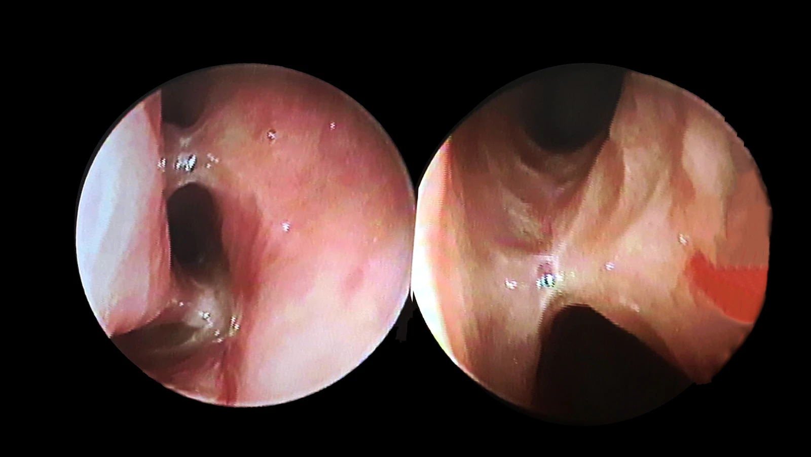
Endoscopic photo of intranasal mucosal adhesions
Surgical plan
A lateral osteotomy was planned to improve the deviated bridge and nasal bone asymmetry seen from the front.
To correct the splayed nasal tip and nostril asymmetry, lower lateral cartilage reconstruction and cartilage suturing were planned.
Lowering the columella was planned to naturally balance the alar-columellar relationship (ACR) from the front.
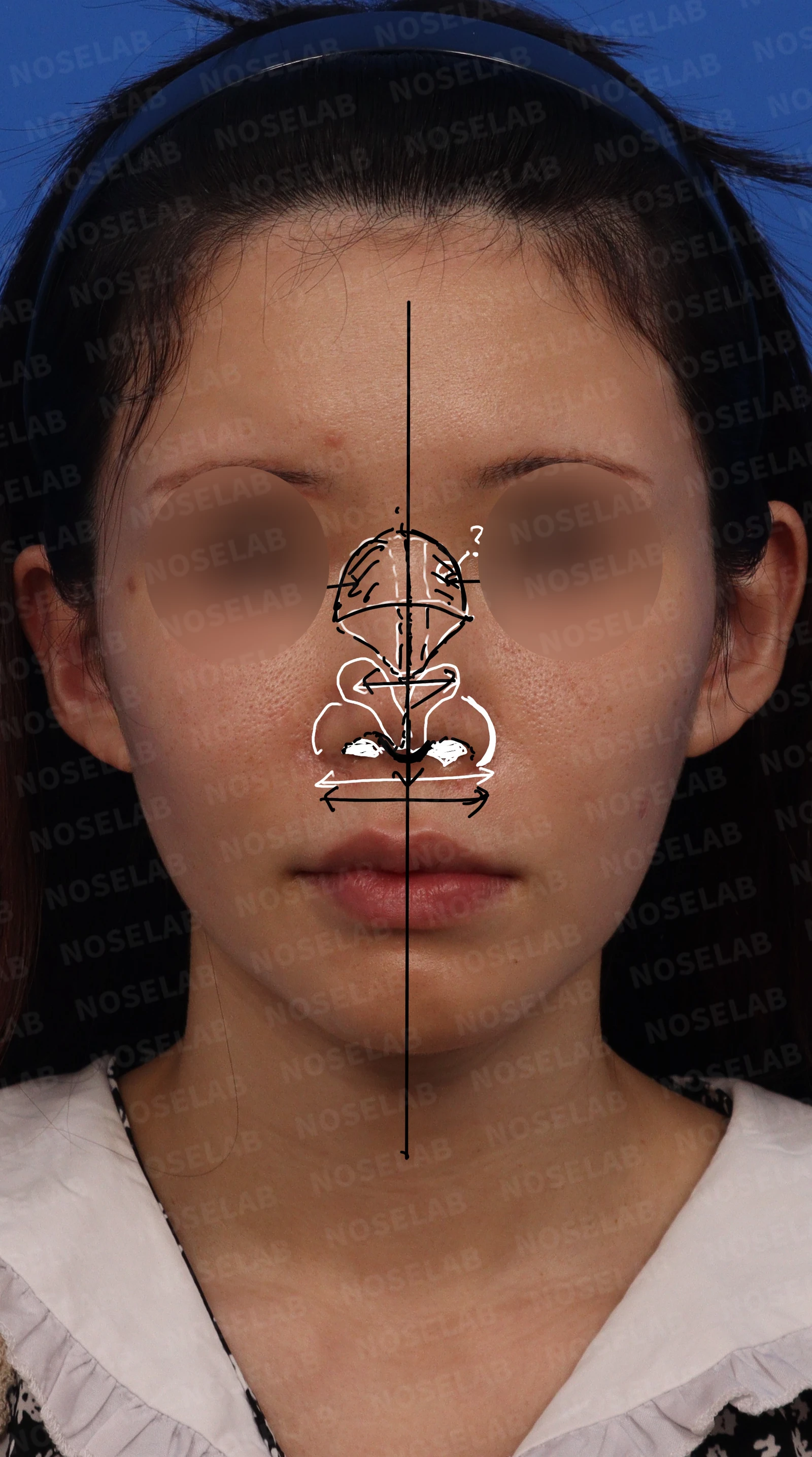
Front view
The radix was set approximately at the pupil line, and the nasal dorsum was elevated naturally using autologous rib cartilage.
The plan was to elevate the nasal tip using autologous rib cartilage without allowing it to upturn, and to adjust the nasolabial angle by gently lowering the columella.
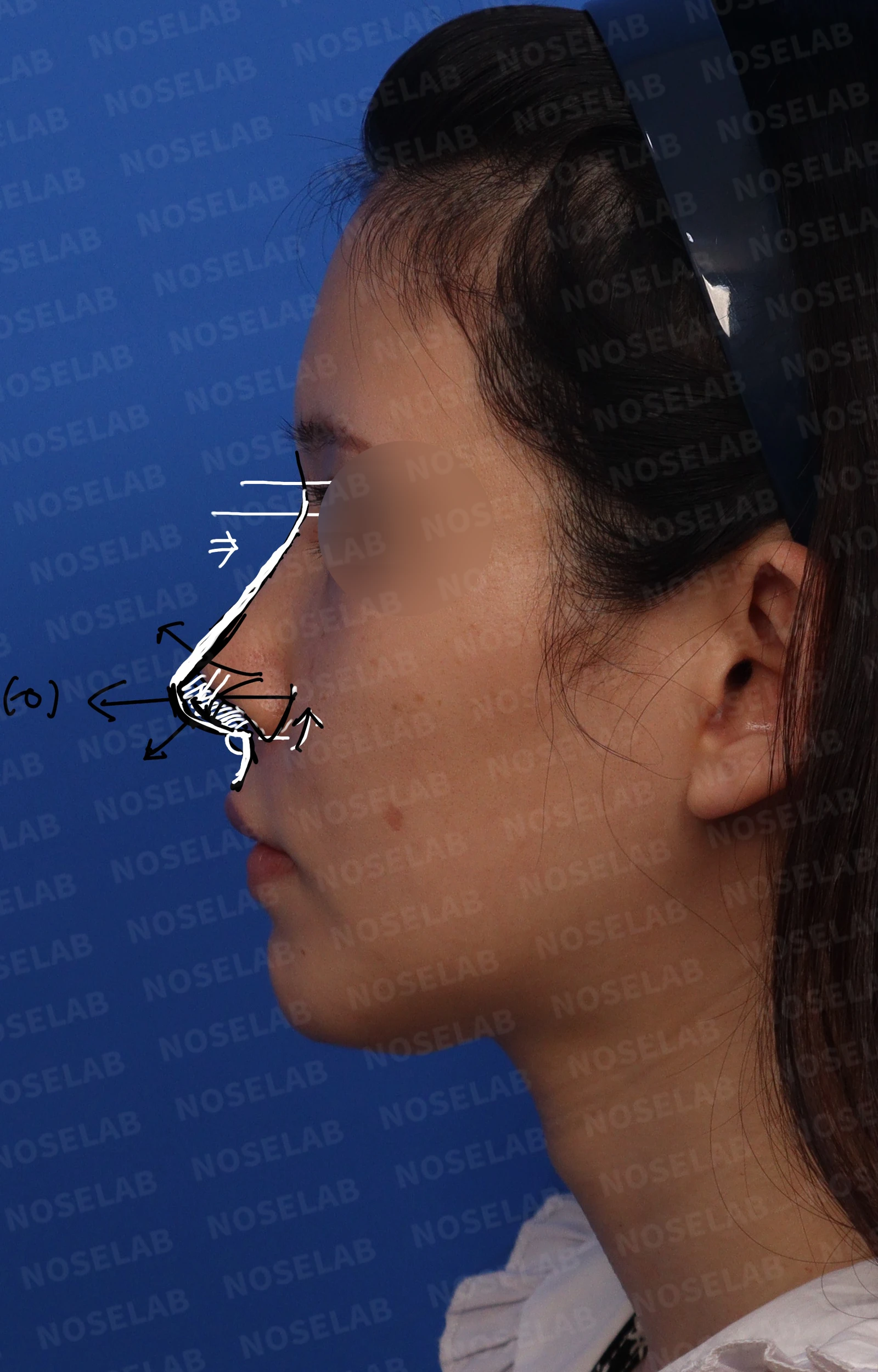
Profile view
To correct nostril asymmetry, septal reconstruction using autologous rib cartilage was planned to straighten the columella, which was tilted to the right.
We planned to excise the thickened scar tissue at the suture site that was narrowing the right nostril and perform an advancement flap advancing the internal nasal mucosa to enlarge the nostril size. In particular, shifting the septum to the left was planned to balance the nostril proportions.
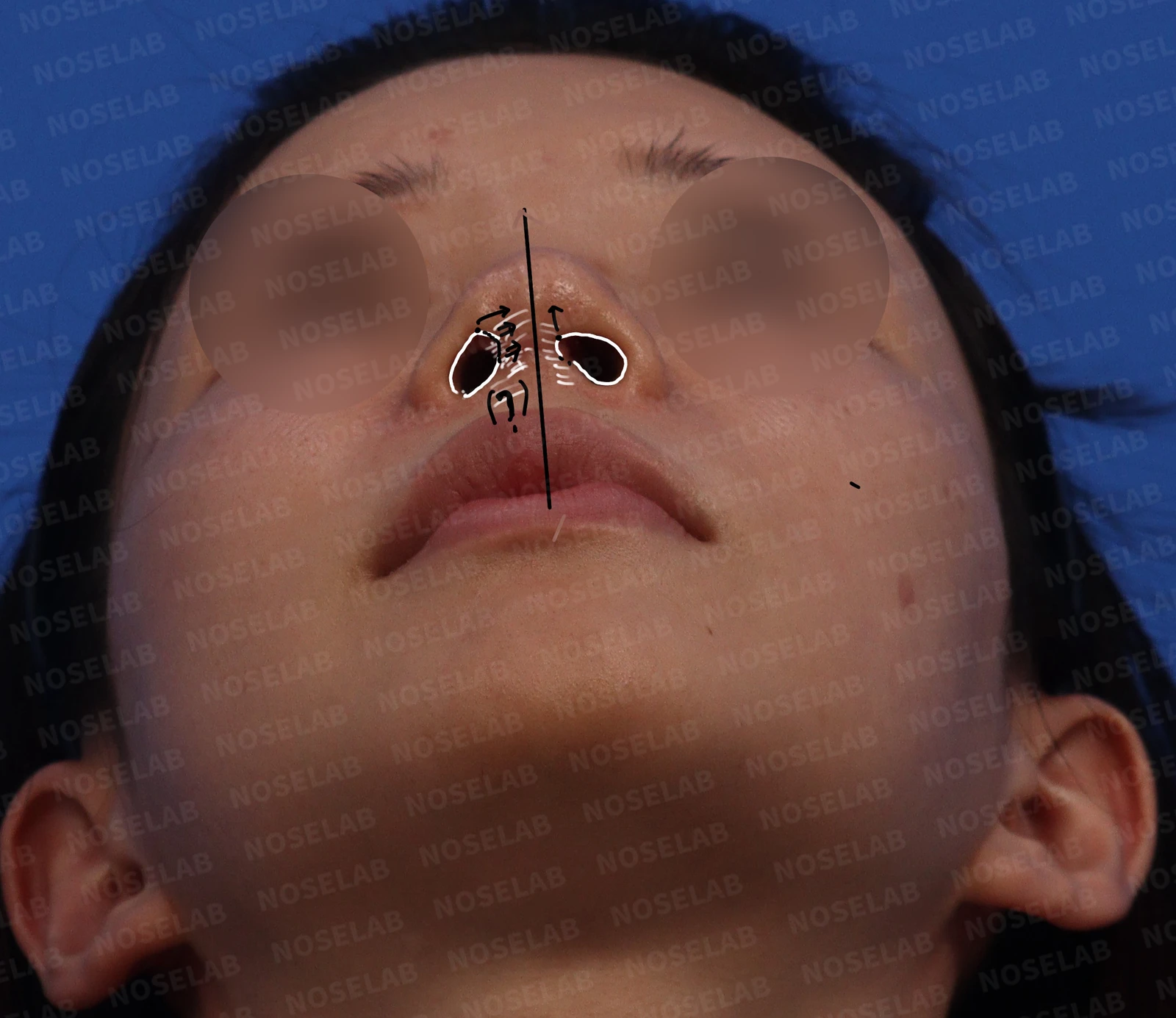
Nostrils
To relieve the patient's nasal obstruction, we planned to correct the deviated septum and release the nasal mucosal adhesions.
Before surgery vs. immediately after surgery
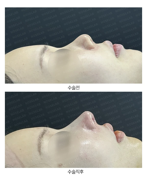
Let us compare the preoperative photos with the photos immediately after surgery (Day 0).Immediate postoperative photos show swelling and may differ from the final result.
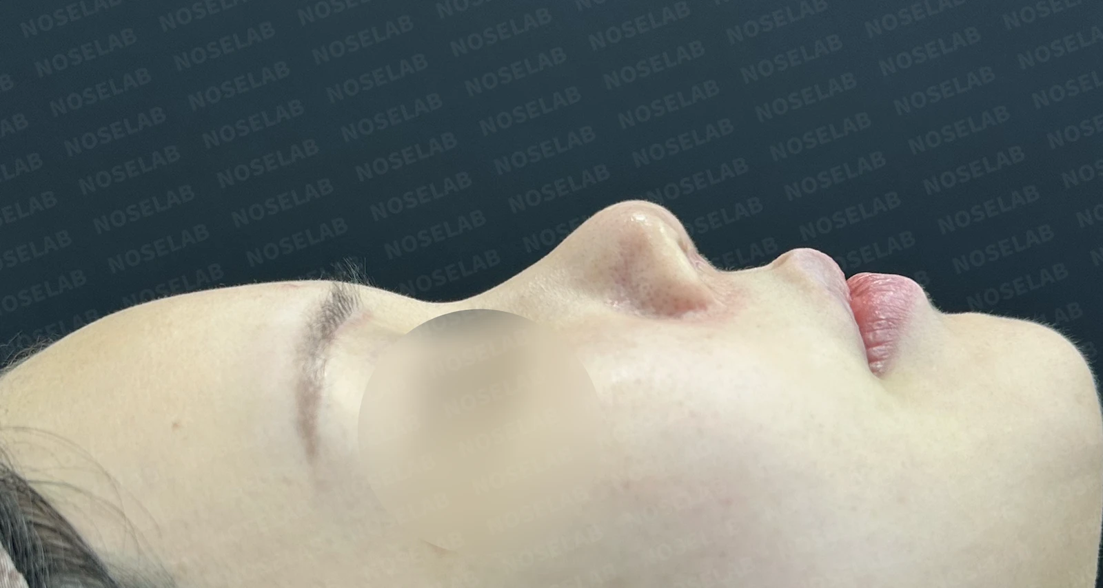
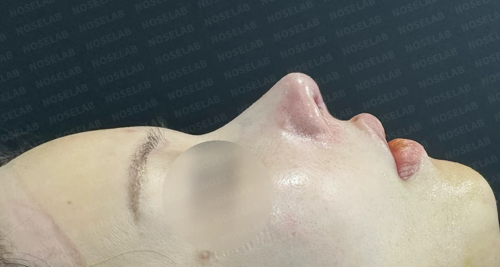
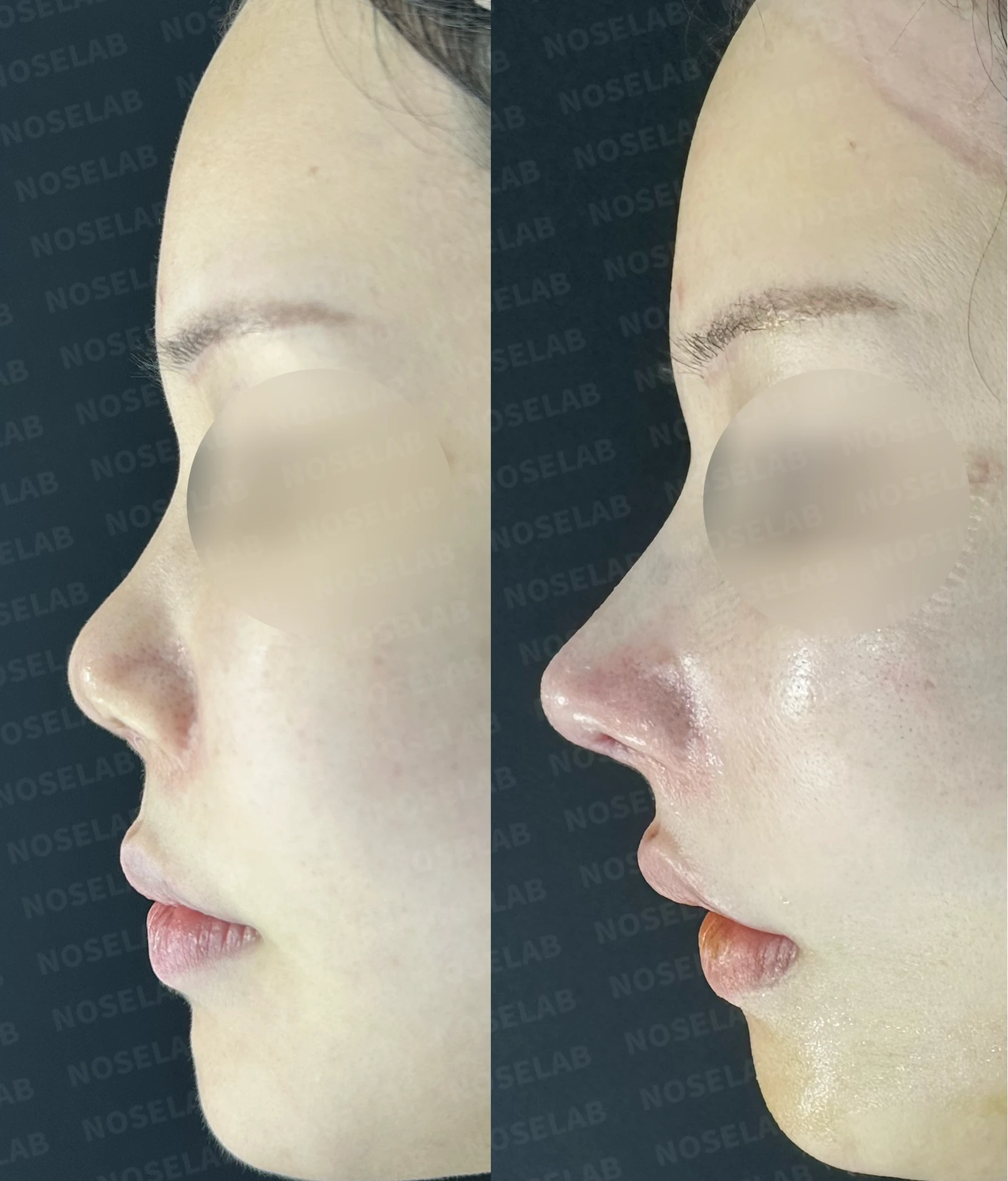
Before surgery / Day 0 immediately after surgery Photos taken immediately after surgery show swelling and may differ from the final result.
On the profile view, the blunt and upturned appearance of the nasal tip has been significantly corrected. The nasolabial angle has become natural, and the dorsum, which previously appeared uneven and low, has been smoothly elevated with the starting point set naturally at the pupil line.
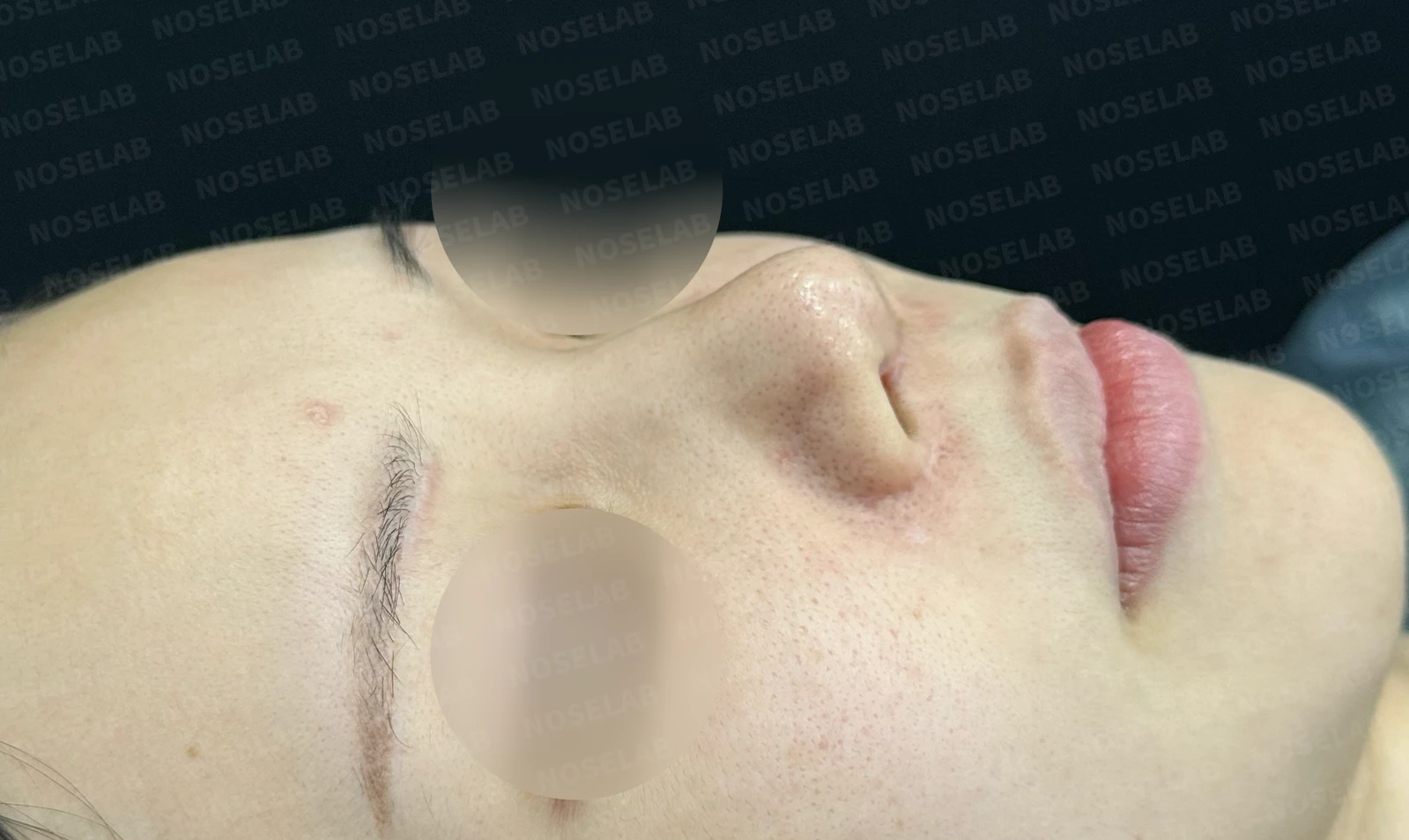
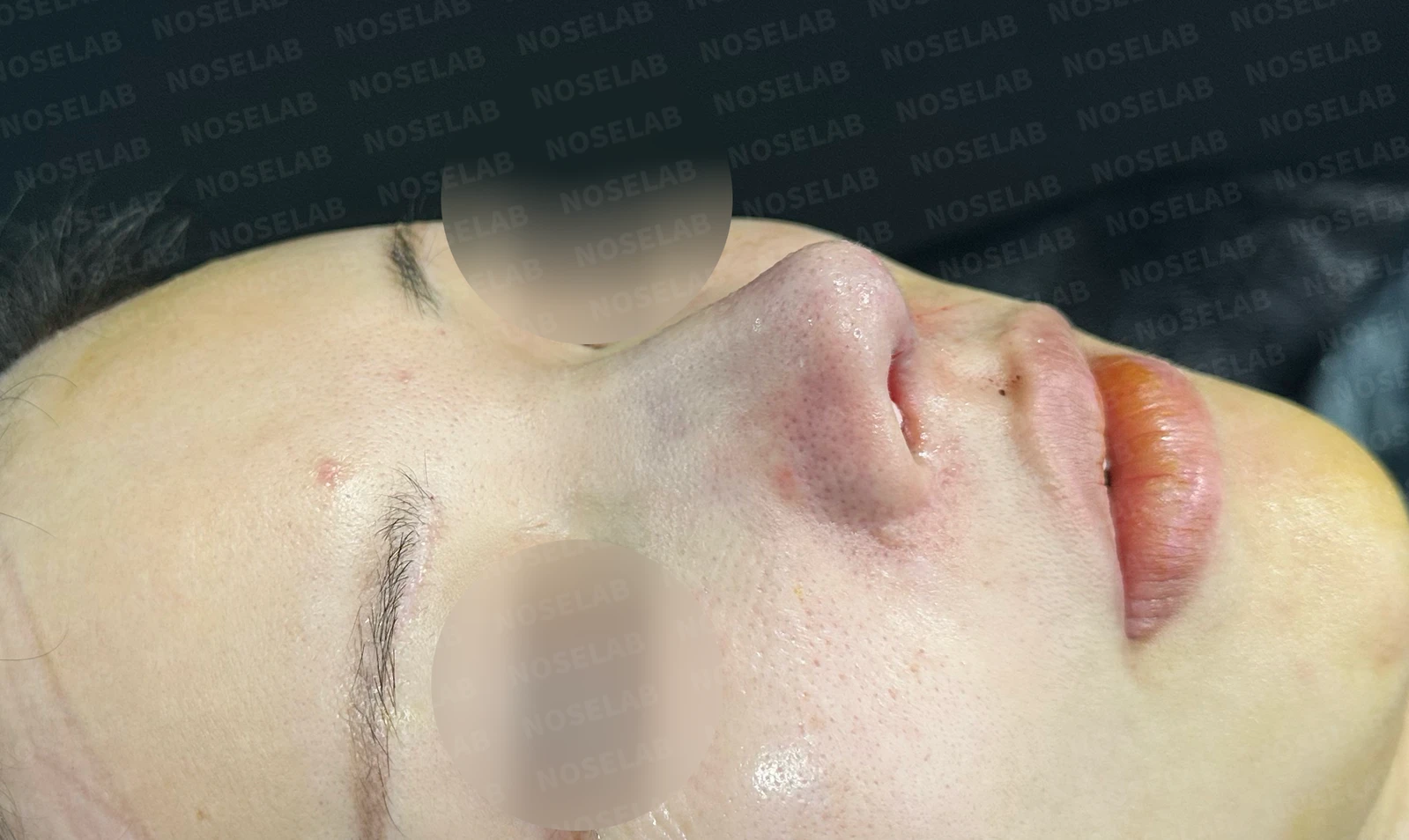
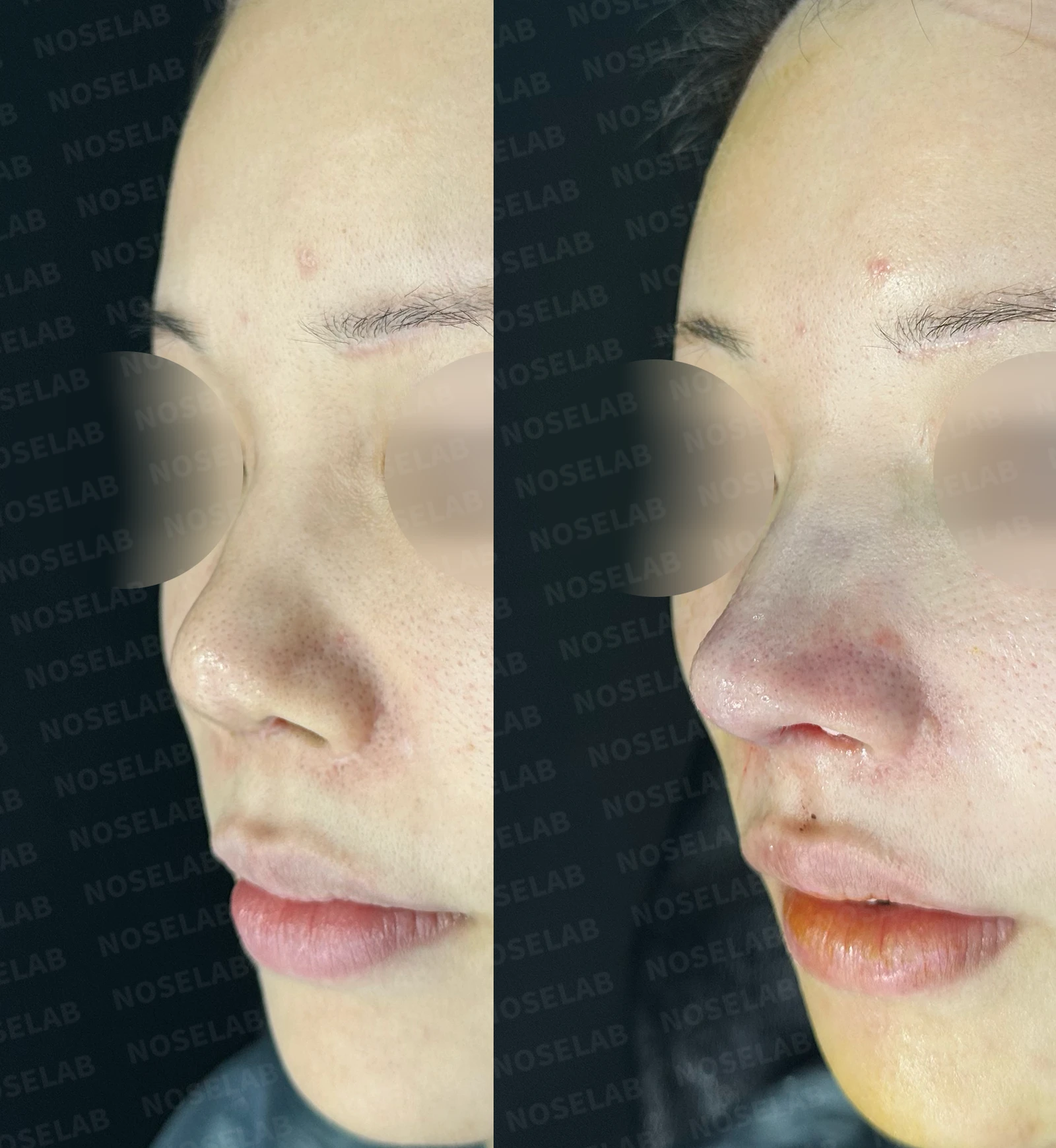
Before surgery / Day 0 immediately after surgery Photos taken immediately after surgery show swelling and may differ from the final result.
On the 45-degree oblique view, the previously short and upturned appearance shows clear improvement. In particular, it can be seen that the nostril is larger.
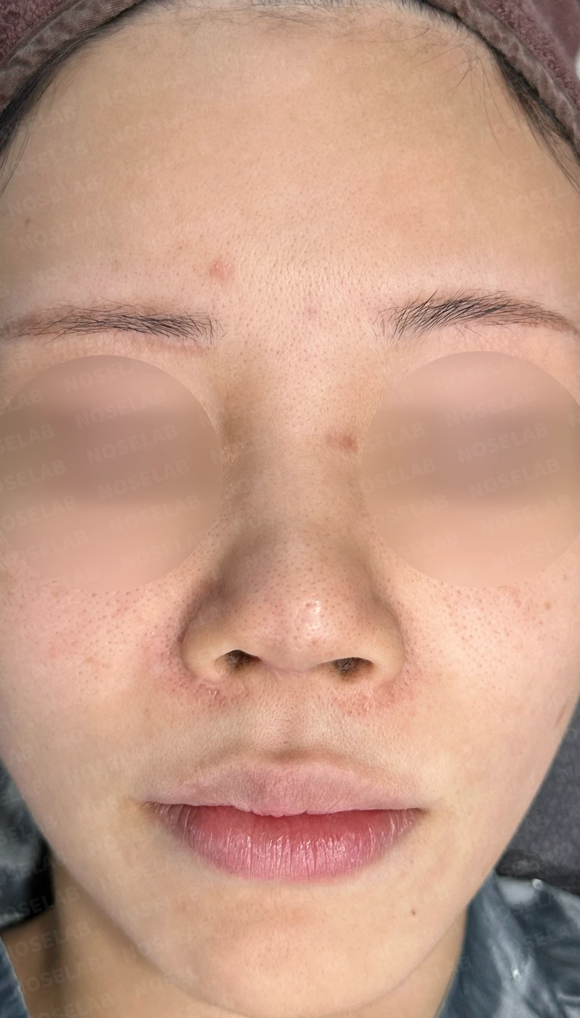
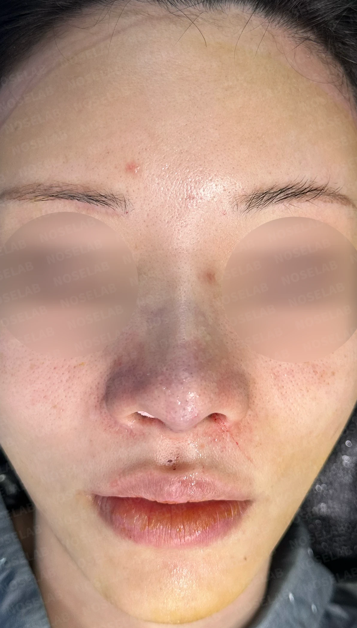
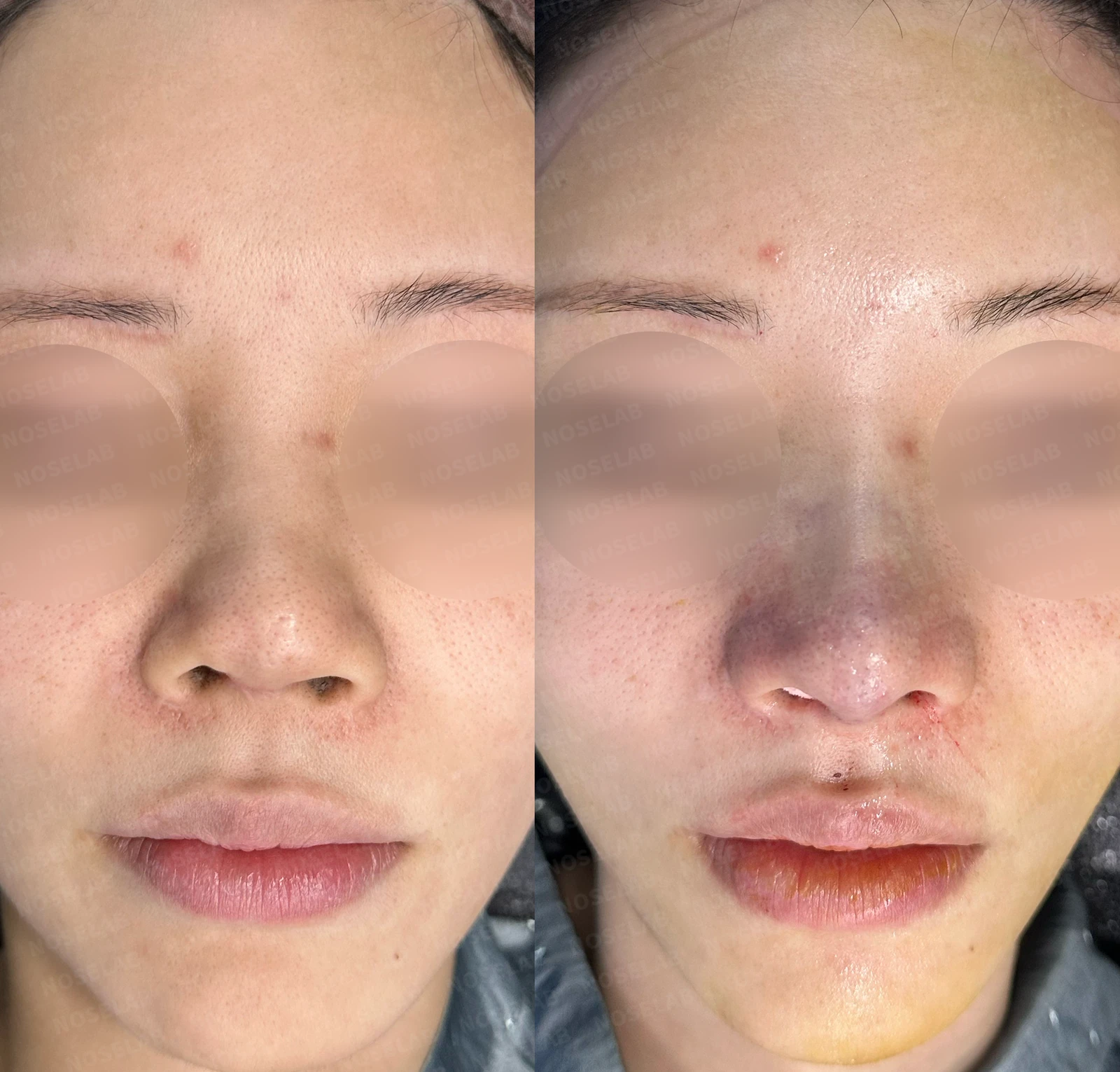
Before surgery / Day 0 immediately after surgery Photos taken immediately after surgery show swelling and may differ from the final result.
From the front, the splayed look has been corrected, and the excessive nostril show has improved. The columella has come down naturally, and the deviated appearance has also improved.
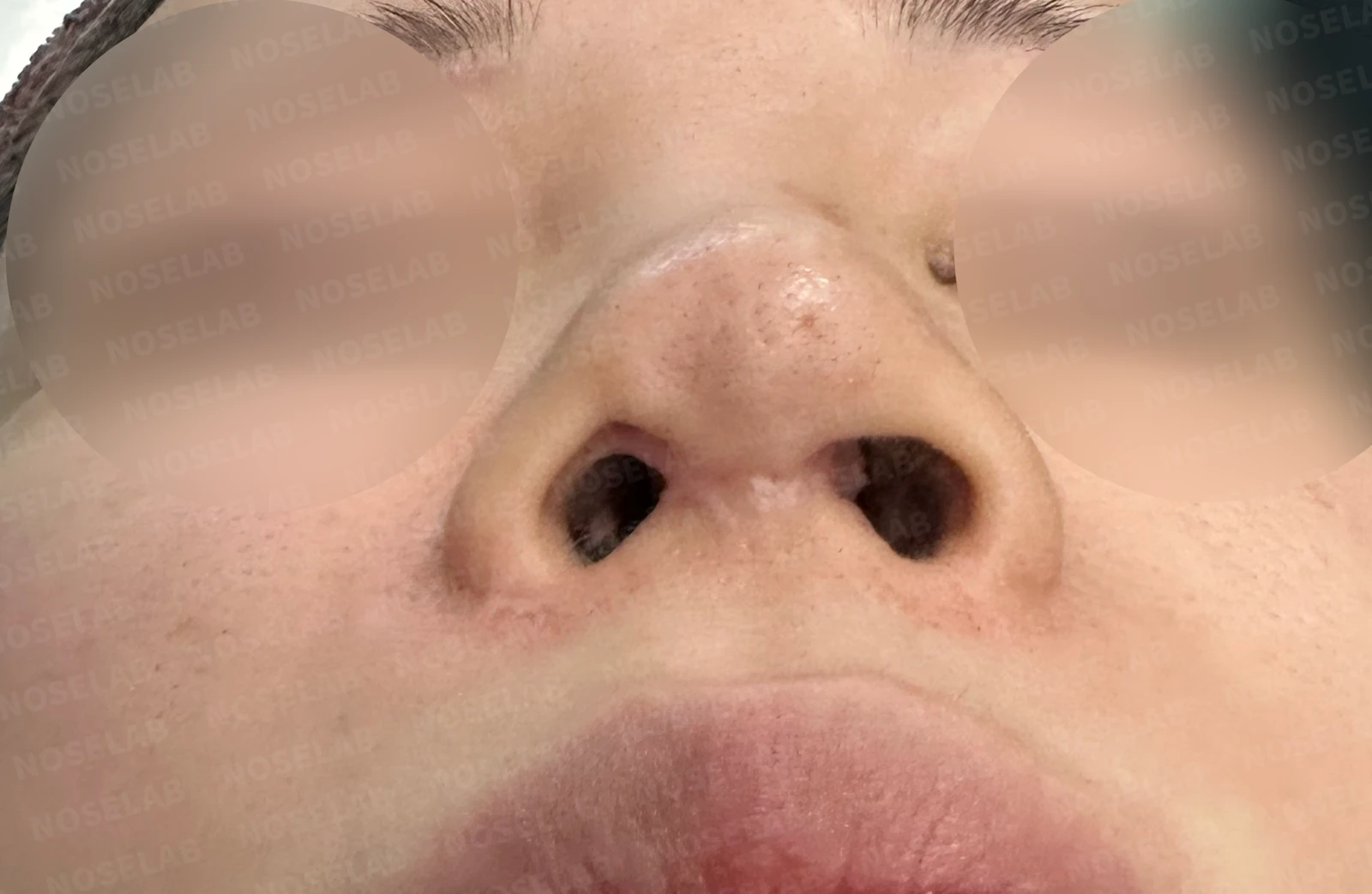
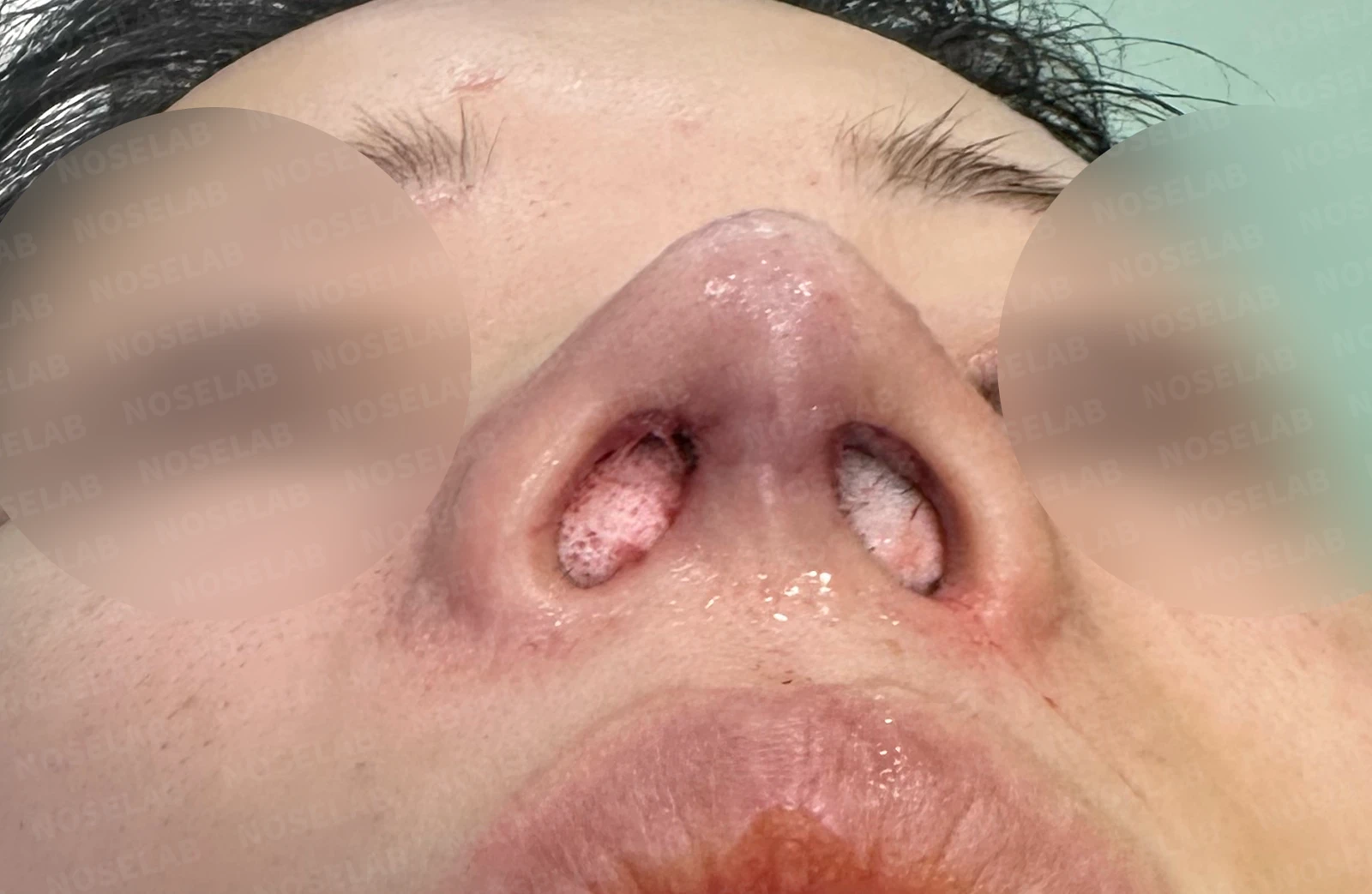
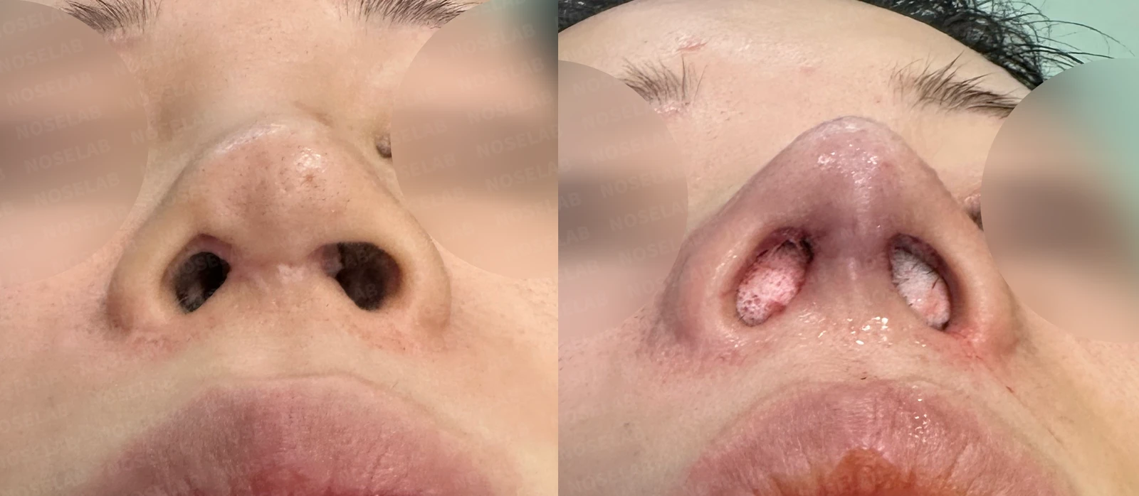
Before surgery / Day 0 immediately after surgery Photos taken immediately after surgery show swelling and may differ from the final result.
The most noticeable change is the improvement in nostril asymmetry. Scar tissue in the right nostril was removed, and by reducing the columellar thickness while shifting it to the left, the nostril asymmetry was corrected as much as possible.
Photos immediately after surgery
Conclusion
During the patient's surgery, the condition of the cartilages was worse than anticipated. Approximately 80% of the septal cartilage and the right lower lateral cartilage had been lost from the previous surgery, so these cartilages were completely reconstructed using autologous rib cartilage. Cartilage reconstruction surgery may not show a dramatic effect in the early stage, but I believe it plays a crucial role in maintaining shape and preventing deformation over time. Of course, asymmetry may temporarily appear more pronounced early on due to the reconstruction, but it is best to view this as swelling associated with the healing process.
Because this patient underwent revision surgery for contracture deformity caused by inflammation, autologous rib cartilage was used for both the bridge and the tip, and our focus must remain on preventing postoperative infection and supporting safe healing. Therefore, we plan to actively promote wound healing with antibiotic therapy, hyperbaric oxygen therapy, and laser therapy.
Today, I shared a case of contracted nose surgery. I hope this helps those who are struggling with a contracted nose.
This was Dr. Peter C. Kang. Thank you.

Limitations and risks
When autologous rib cartilage is used, scarring and pain may remain at the donor site on the chest, and pneumothorax (pleural injury) can occur rarely. Over time, the grafted cartilage may warp or undergo partial resorption.
When the septum is corrected at the same time, nasal obstruction may persist or recur, and in rare cases, septal perforation or changes in smell may occur.
Common potential risks include infection, bleeding, asymmetry, scarring, skin necrosis, and insufficient improvement; results vary depending on individual skin condition and previous surgical history.