Hello, I am Dr. Peter C. Kang.
Today, I would like to share the case of a patient who visited our clinic because they were dissatisfied with the outcome of their previous rhinoplasty. Following the prior surgery, the patient was troubled by a nasal tip that appeared bulbous, drooped, and slightly deviated to the right, along with symptoms of nasal obstruction.
🔍 Surgical history
Approximately 3 years ago: Previous rhinoplasty (silicone implant, ear cartilage, septoplasty, and concurrent rhinitis surgery)
Approximately 3 years ago: Filler injection to the forehead and glabella (not removed during the previous rhinoplasty)
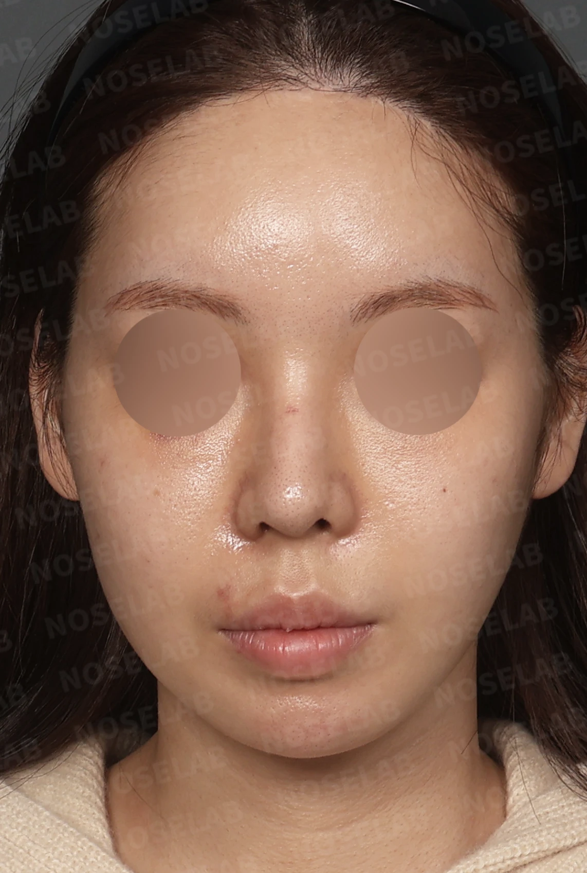
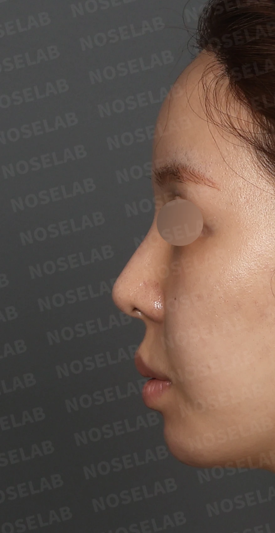
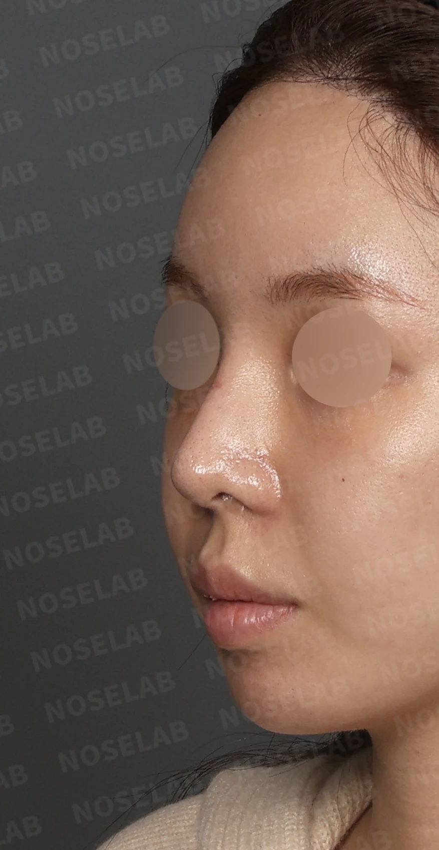
Front, left profile, and oblique views during design consultation.
💉 Patient requests
Preference for a distinct, defined nasal line
Correction of the bulbous and drooping nasal tip
Correction of the rightward deviation
Relief of nasal obstruction (accompanied by rhinitis)
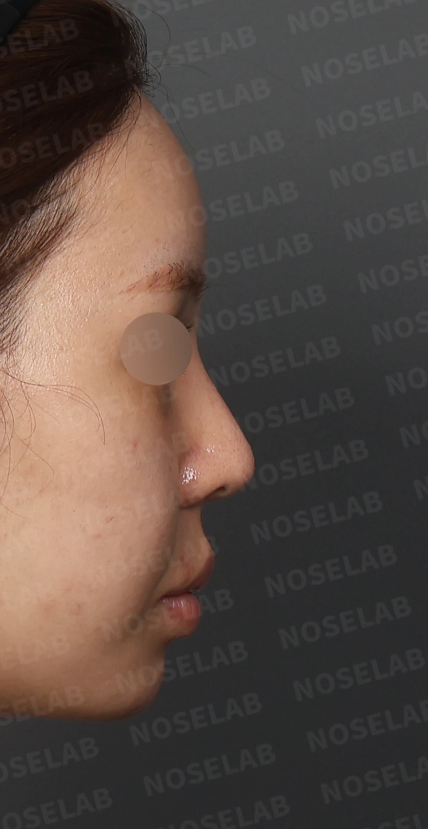
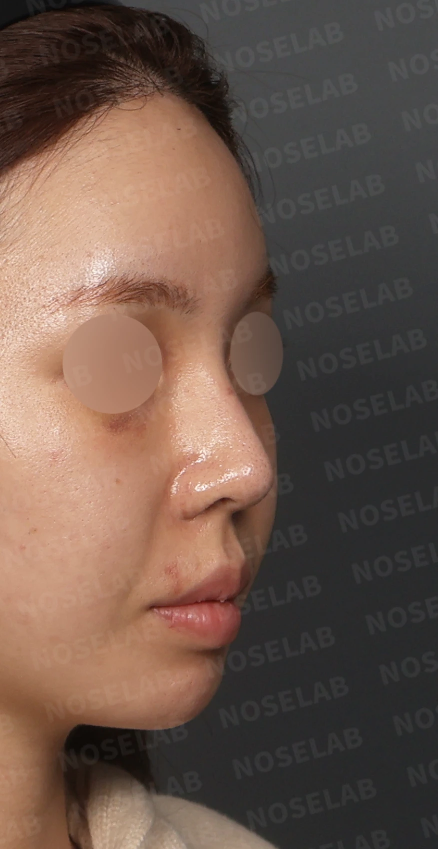
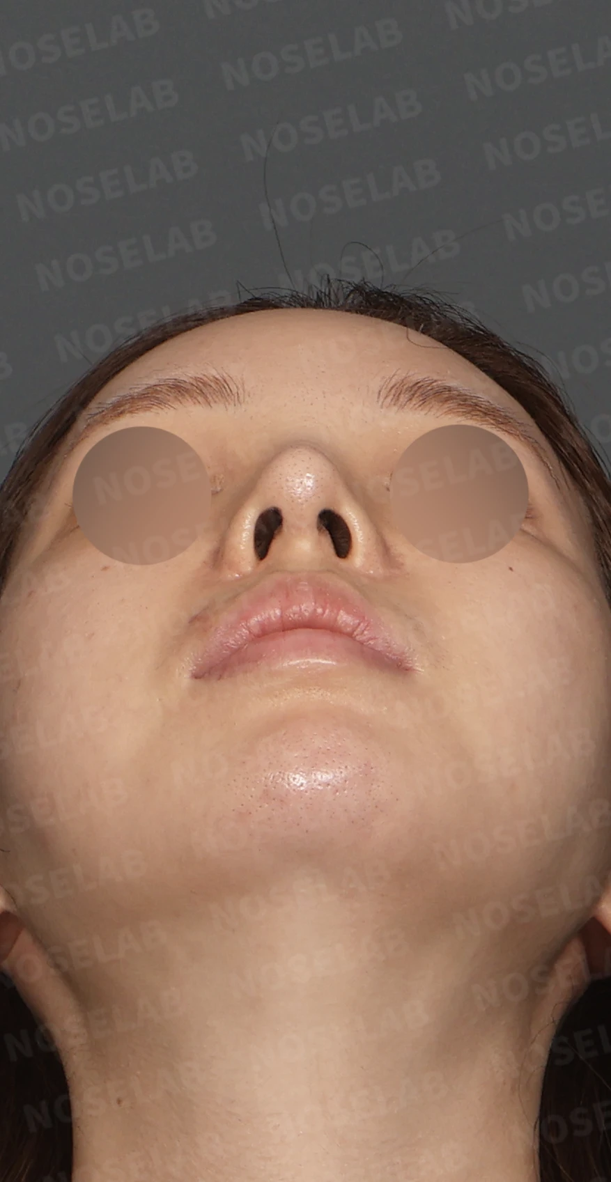
Right profile, oblique, and nostril views during design consultation.
🔧 Surgical plan and procedure
Septal reconstruction using autologous rib cartilage
Using the patient's own rib cartilage, the weakened septum was reinforced and reconstructed. A solid structural framework was established to prevent the nasal tip from drooping, resulting in a well-projected, distinct tip line.
Adjustment of the nasal bridge line using a silicone implant
The implant was precisely carved to fit the contour of the nasal bones, naturally realizing the defined bridge line the patient desired.
Septoplasty
The deviated septum was straightened to correct the appearance of rightward deviation and to improve internal nasal airflow.
Rhinitis surgery (turbinate surgery)
The hypertrophied inferior turbinates were precisely trimmed to relieve nasal obstruction and help breathing feel considerably more comfortable.
Correction of the nasolabial angle
The angle above the philtrum was refined to improve the profile line and restore balance between the nasal tip and the lips.
🌟 Surgical results
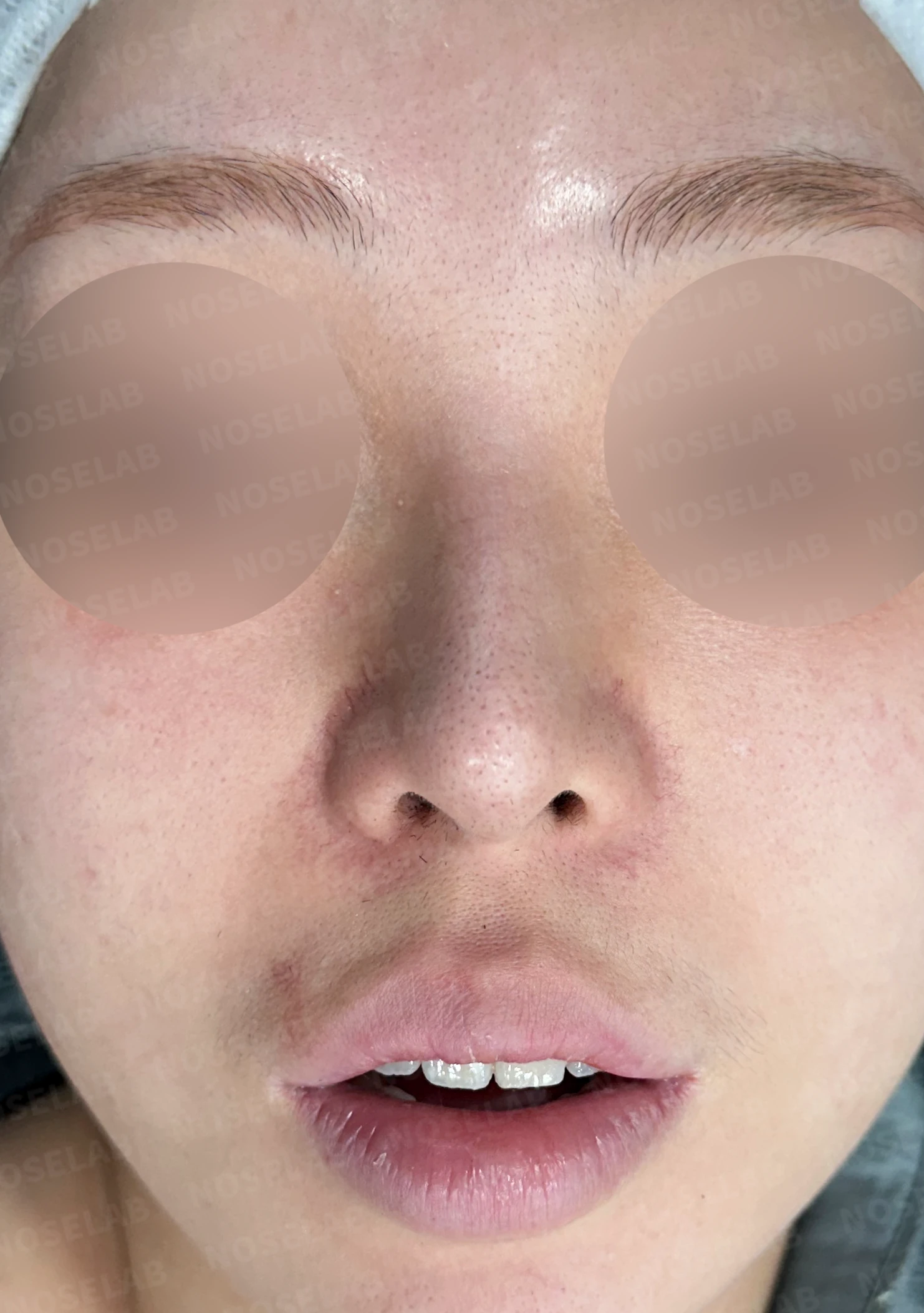
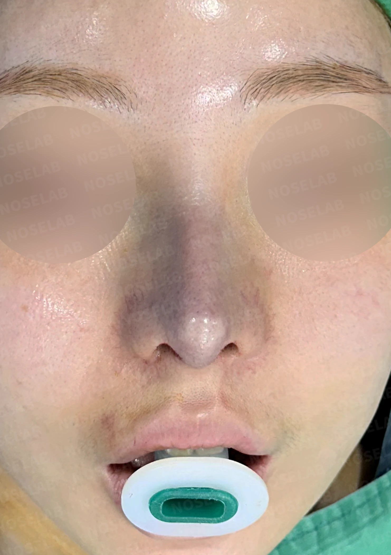
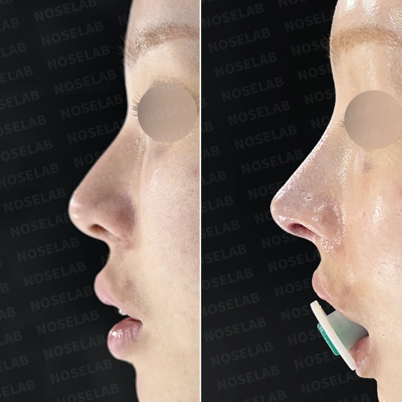
Before surgery / Day 0 immediately after surgery Photos taken immediately after surgery show swelling and may differ from the final result.
Front view changes
The bulbous nasal tip is narrowed and well-defined
The rightward deviation is corrected, establishing stable bilateral symmetry
The nasal bridge line appears clean and well-defined
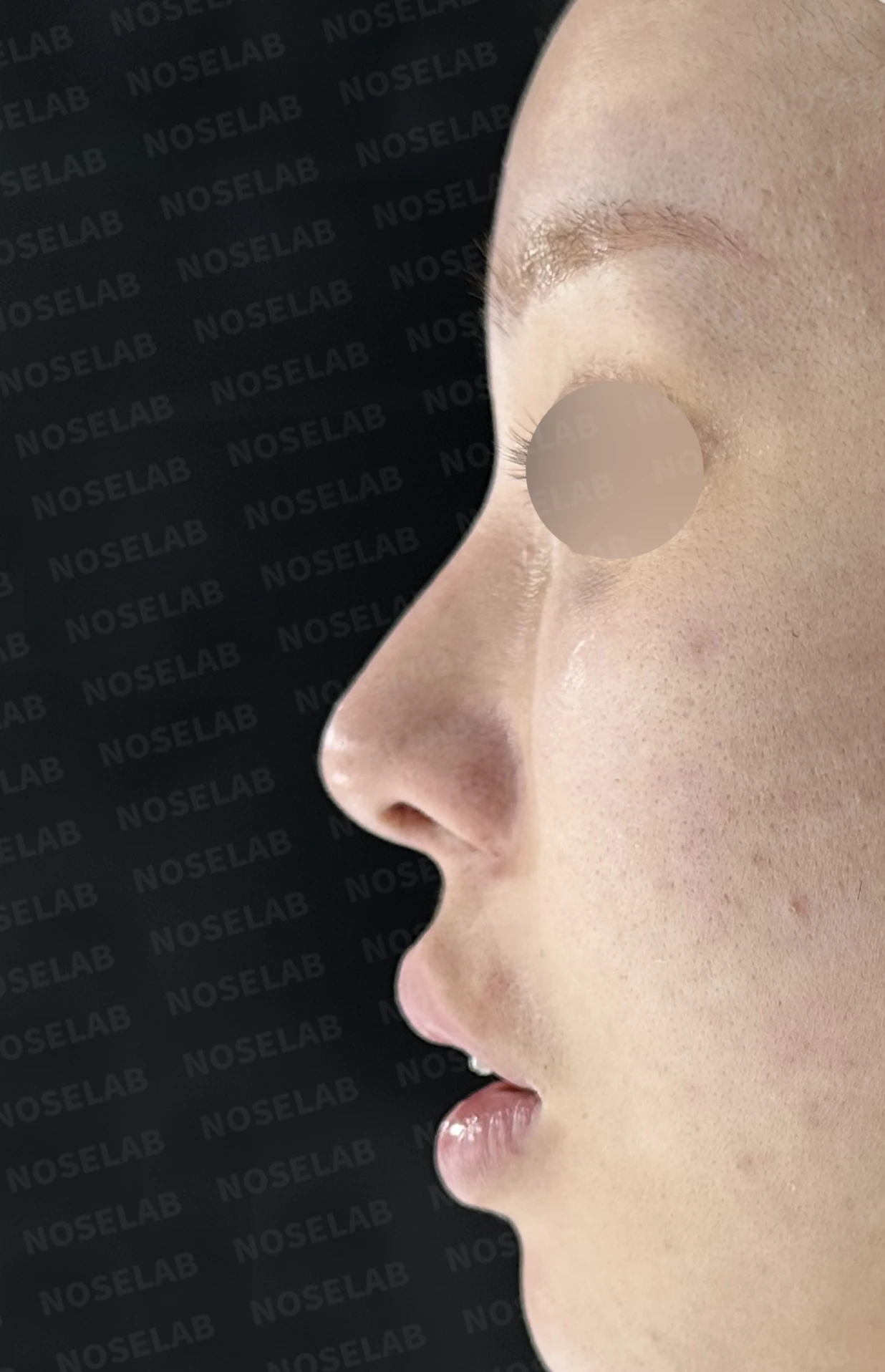
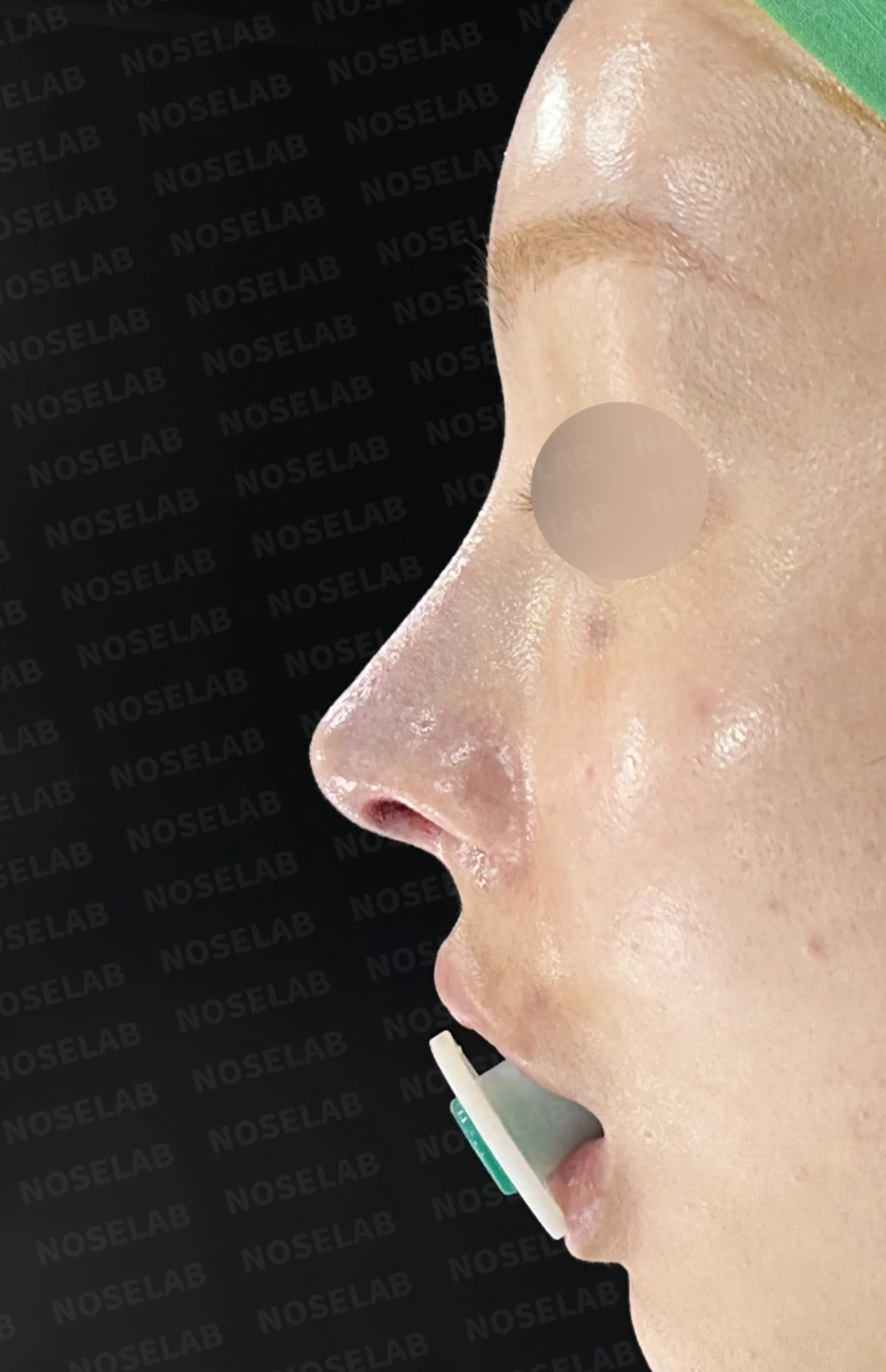
Before surgery / Day 0 immediately after surgery Photos taken immediately after surgery show swelling and may differ from the final result.
Profile view changes
The drooping nasal tip is naturally elevated
A distinct yet natural straight bridge line is formed
The nasolabial angle is refined, giving the profile a more sophisticated contour
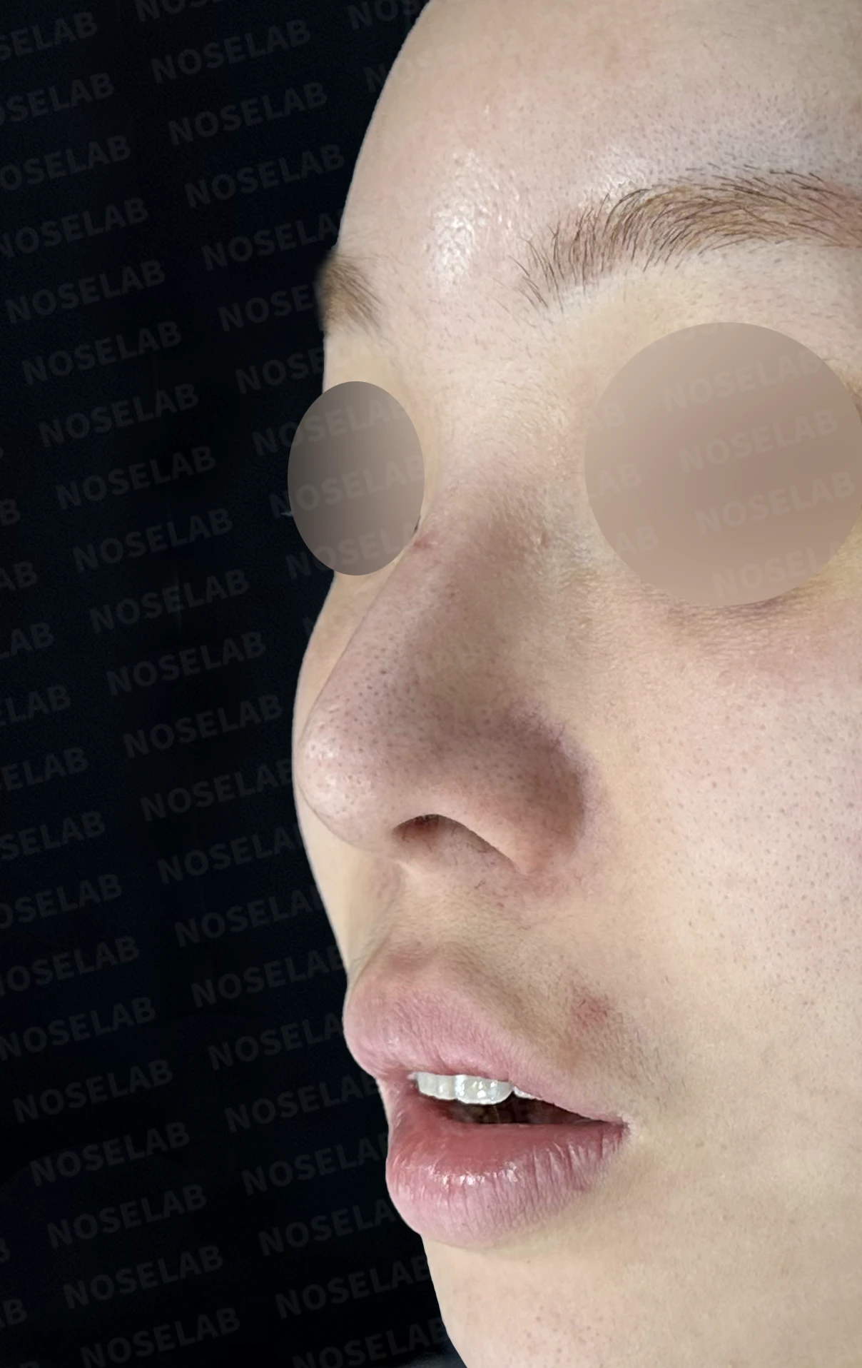
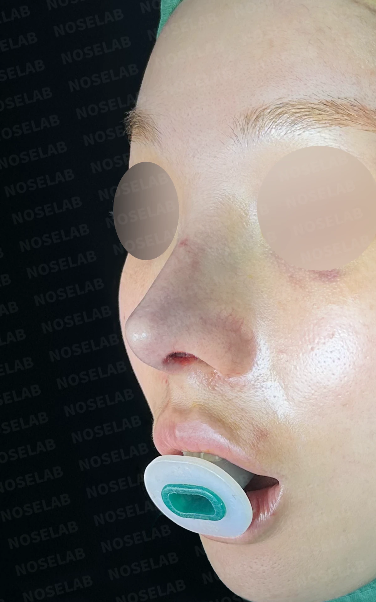
Before surgery / Day 0 immediately after surgery Photos taken immediately after surgery show swelling and may differ from the final result.
Overall change in impression
The septum is straightened, significantly relieving nasal obstruction
Rhinitis symptoms are alleviated
Breathing has become more comfortable
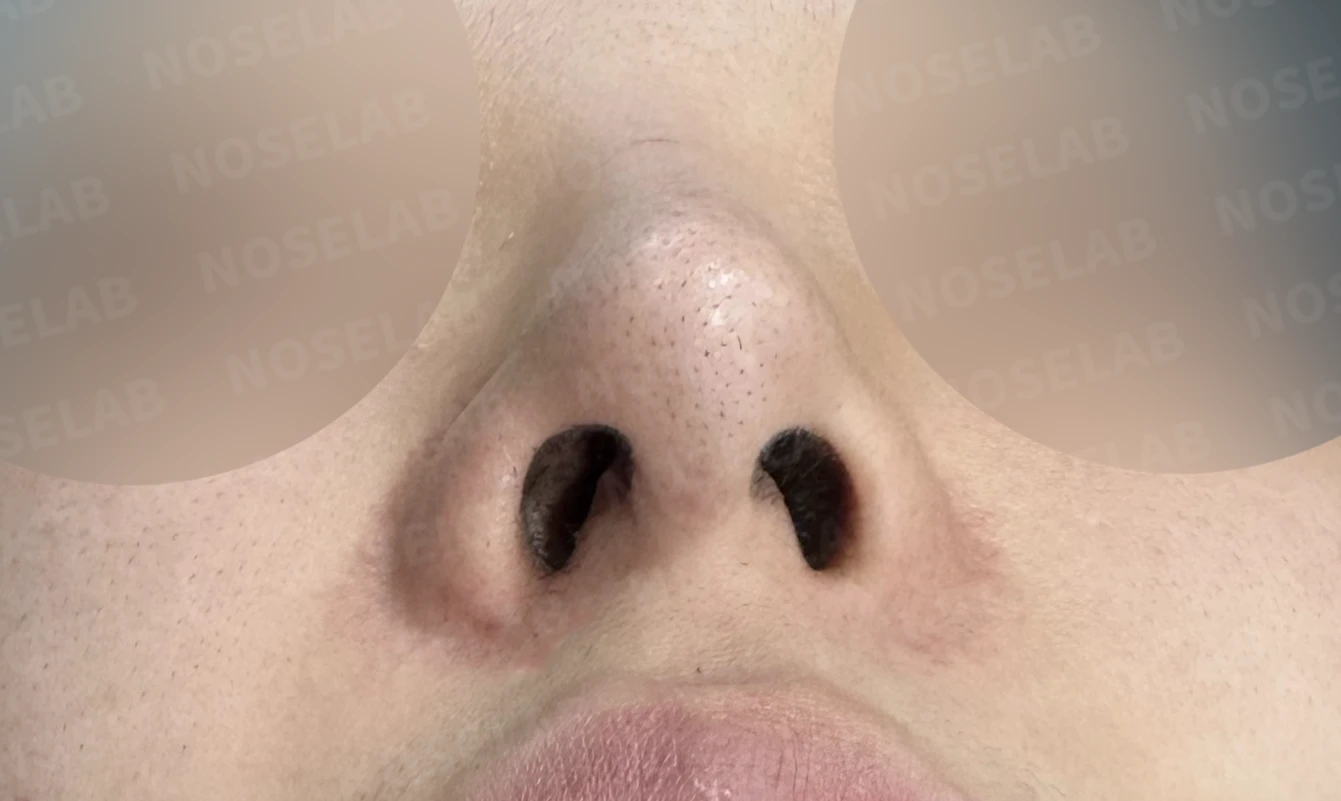
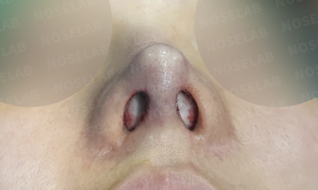
Before surgery / Day 0 immediately after surgery Photos taken immediately after surgery show swelling and may differ from the final result.
💡 Specialist opinion

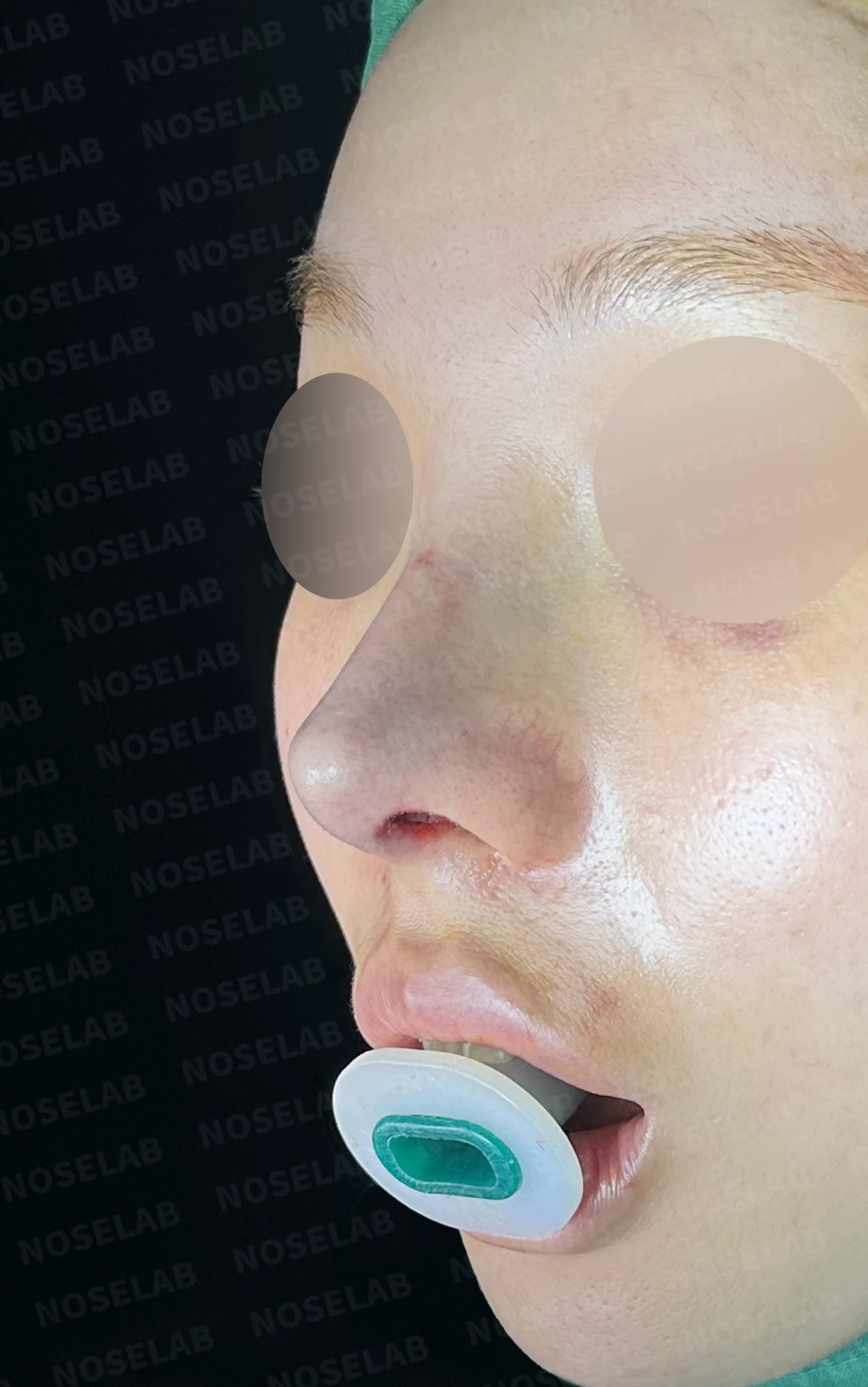
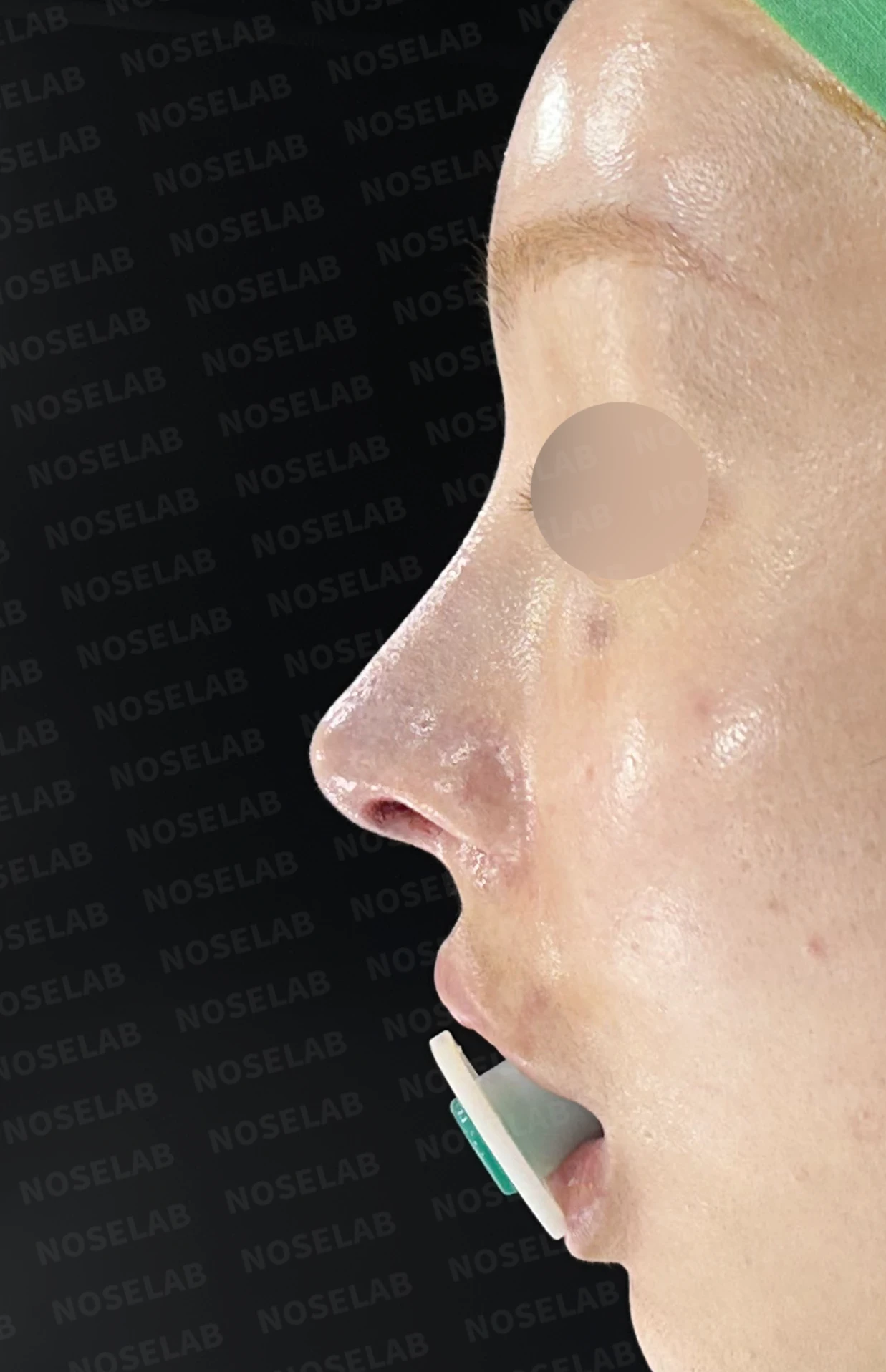
Front, oblique, and profile views on day 0 immediately after surgery. Photos taken immediately after surgery show swelling and may differ from the final result.
Revision rhinoplasty is far more complex than primary rhinoplasty. Because the septum is often weakened or thick scar tissue has formed after the first procedure, it is difficult to achieve a satisfactory outcome simply by adjusting the external shape. In this case as well, septal support was inadequate, so the foundational structure of the nose was rebuilt from the base using autologous rib cartilage.
Furthermore, for patients who also experience nasal obstruction, aesthetic correction alone cannot improve their quality of life. Pursuing both cosmetic and functional outcomes simultaneously is the core principle of closed revision rhinoplasty. In this case, septoplasty and rhinitis surgery were performed concurrently to help restore comfortable breathing.
Autologous rib cartilage carries a low risk of foreign body reaction and provides sufficient structural strength, making it a reliable material for reconstructing a weakened nasal framework. Even for patients seeking a more pronounced nasal line, utilizing autologous tissue supports a stable long-term result.
This was Dr. Peter C. Kang. Thank you.
#closedrhinoplasty #closedrevisionrhinoplasty #autologousribcartilage #septalreconstruction #revisionrhinoplasty #nasalobstructionsurgery #droopingtip #definednose

Limitations and risks
When autologous rib cartilage is used, scarring and pain may remain at the donor site on the chest, and pneumothorax (pleural injury) can occur rarely. Over time, the grafted cartilage may warp or undergo partial resorption.
When foreign materials such as injected fillers are removed and reconstructed, contracture may recur, nasal height and length may decrease, and staged surgery may be necessary depending on the skin condition.
When the septum is corrected at the same time, nasal obstruction may persist or recur, and in rare cases, septal perforation or changes in smell may occur.
Common potential risks include infection, bleeding, asymmetry, scarring, skin necrosis, and insufficient improvement; results vary depending on individual skin condition and previous surgical history.