Hello, I am Dr. Peter C. Kang.
Today, I would like to share the case of a patient who visited our clinic concerned about a severely upturned nasal tip and pinched-looking alae after the anterior portion of the septum had been completely removed during previous surgery.
🔍 Surgical history
The patient had a history of 2 previous rhinoplasty procedures.
Approximately 21 years ago: Rhinoplasty using a silicone implant and ear cartilage
Approximately 2 years ago: Revision rhinoplasty using a silicone implant and autologous rib cartilage
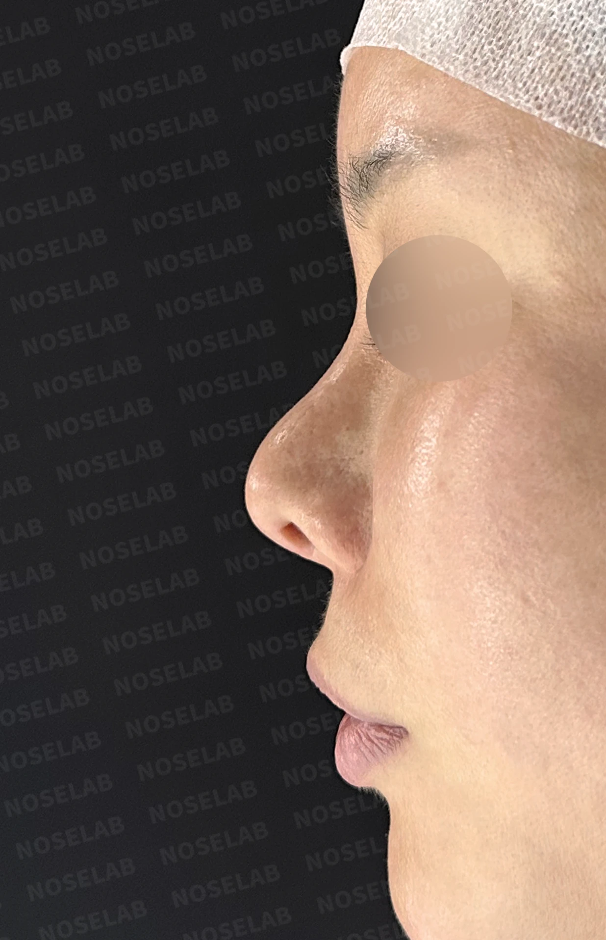
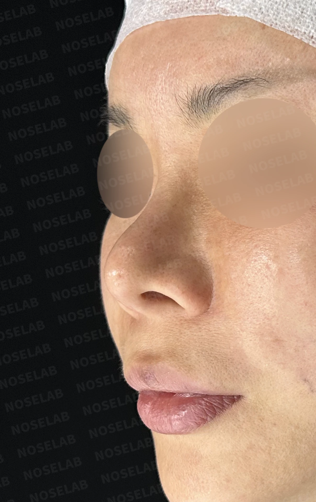
Notably, during the previous surgery, the anterior portion of the septum had been completely removed and harvested as graft material. Because the septum is the core support structure holding up the nasal tip, its loss severely weakened tip support, causing the nasal tip to become noticeably upturned. In addition, the alae appeared pinched on both sides and both sides of the nasal bridge were depressed, leaving the patient dissatisfied with the overall nasal appearance.
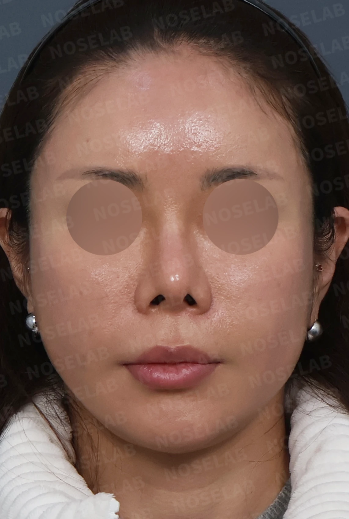
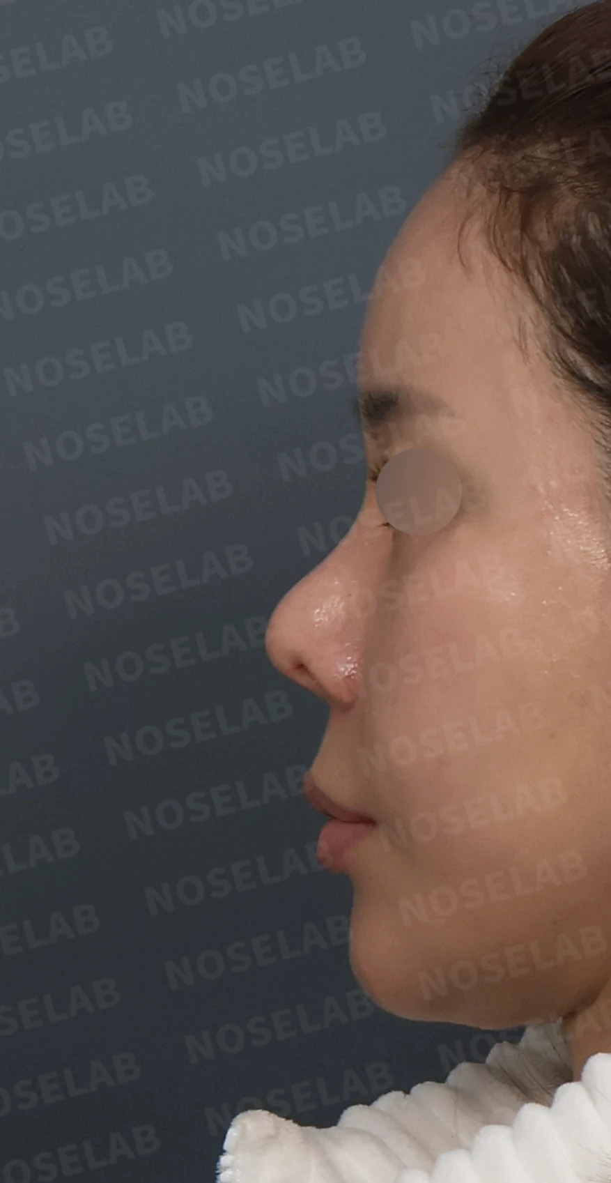
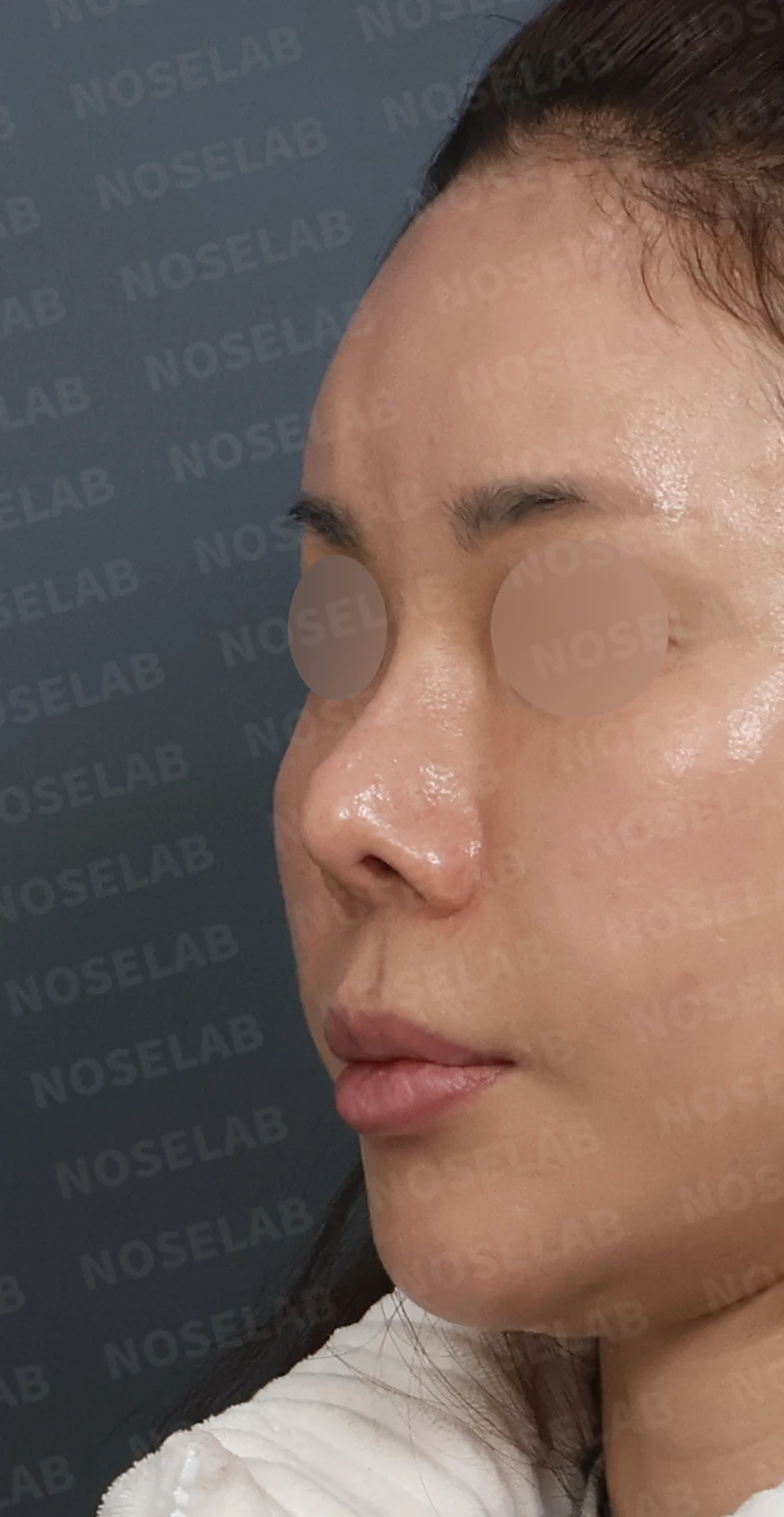
Front, left profile, and oblique views during design consultation.
💉 Patient requests
The patient's specific requests were as follows:
Correction of the upturned nasal tip
Correction of the pinched-looking alae
Desire to reduce nostril show
Overall correction of the crooked nose
Improvement of nasal obstruction
When the septum is lost, revision rhinoplasty is not merely a matter of reshaping the nose; it is closer to a reconstructive procedure that must build a new core support structure. I explained this thoroughly to the patient and designed a surgical plan centered on septal reconstruction using autologous rib cartilage.
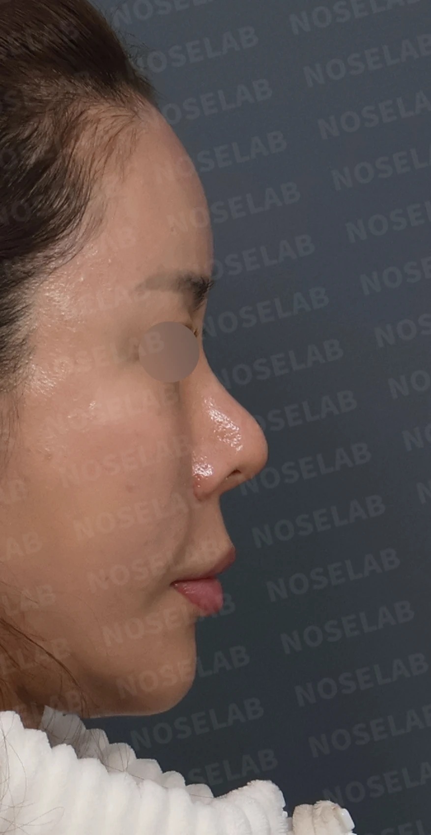
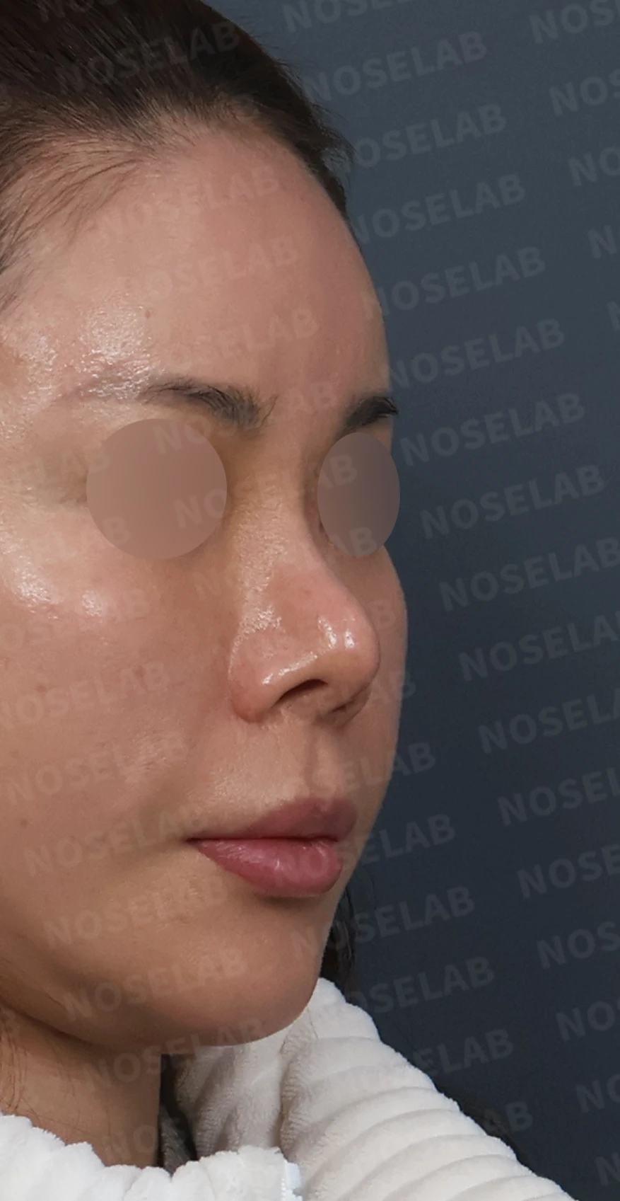
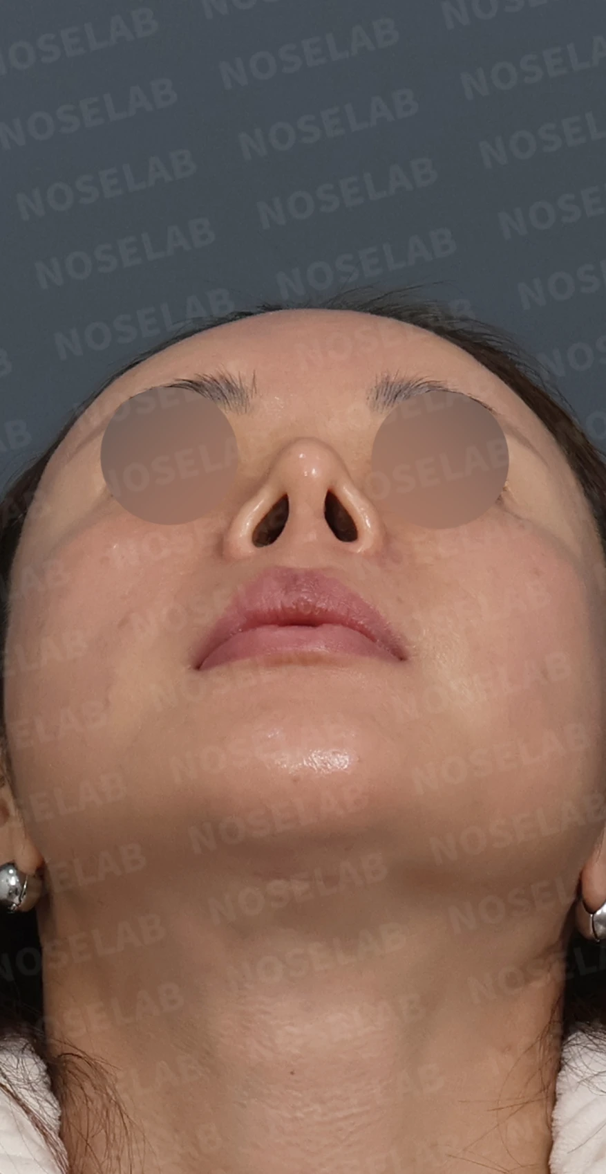
Right profile, oblique, and nostril views during design consultation.
🔧 Surgical plan and procedure
1. Septal reconstruction using autologous rib cartilage
Reconstruction of the anterior septum, which was completely removed in previous surgery, using autologous rib cartilage
Creation of a new core support framework for the nasal tip to correct the upturned tip
Securing structural stability of the septum to support a stable long-term result
2. Alar reinforcement using autologous rib cartilage
Reinforcement of the bilaterally pinched alae with autologous rib cartilage
Restoration of natural alar curvature
Reduction of nostril show to achieve a more stable frontal appearance
3. Filling depressed bridge areas with micro-particulate autologous rib cartilage
Processing autologous rib cartilage into fine micro-particles
Spreading and filling the depressed areas on both sides of the bridge to create a smooth dorsal line
Restoration of a natural bridge contour
4. Corrective surgery for deviated septum and rhinitis
Straightening the deviated septum through septoplasty
Alleviating rhinitis symptoms through inferior turbinate reduction
Breathing function normalized through relief of nasal obstruction
🌟 Surgical results
Front view changes
The previously upturned nasal tip is lowered to a natural position, creating a stable shape
The pinched-looking alae are reinforced with natural curvature
Reduced nostril show provides a more relaxed, balanced frontal impression
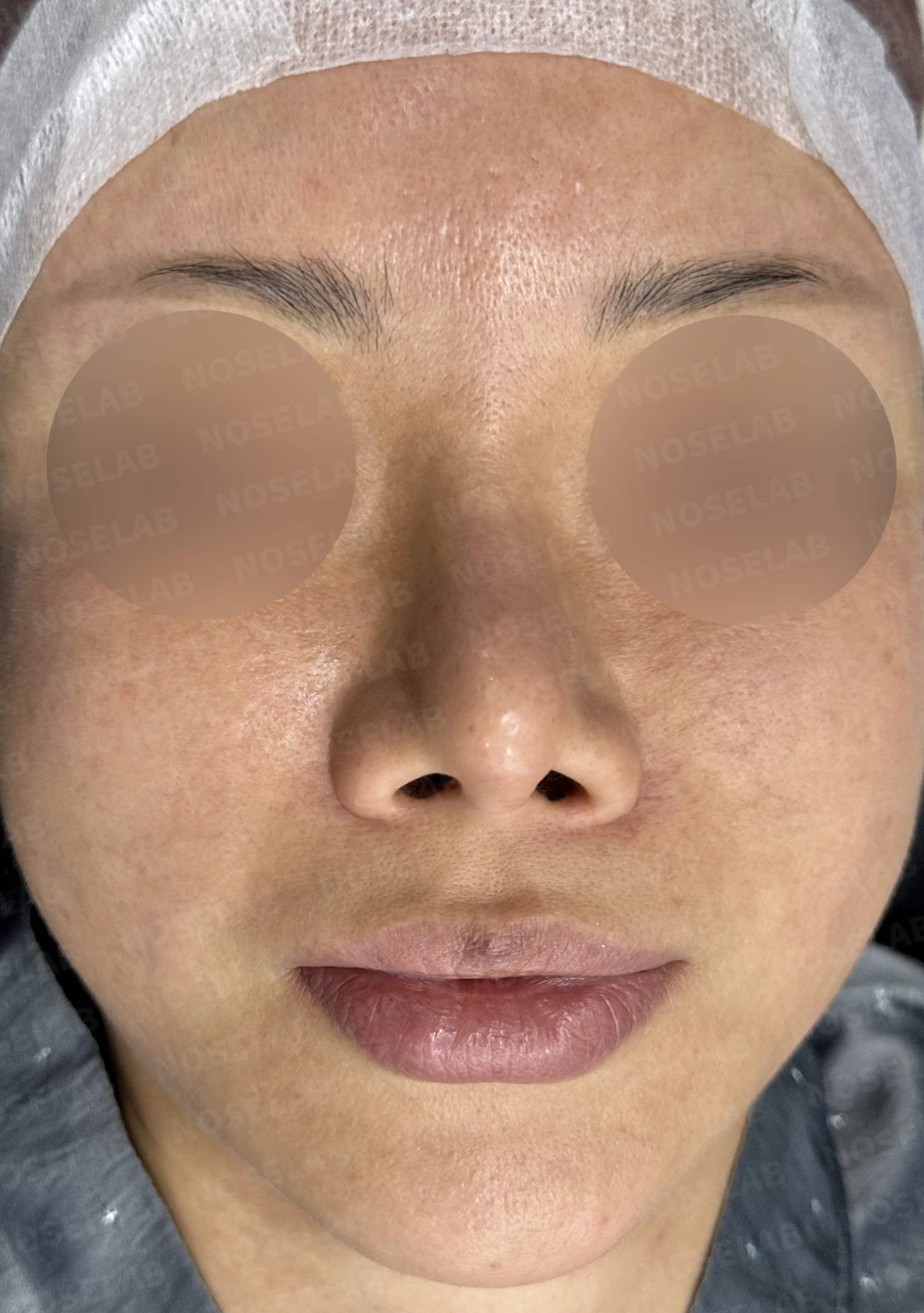
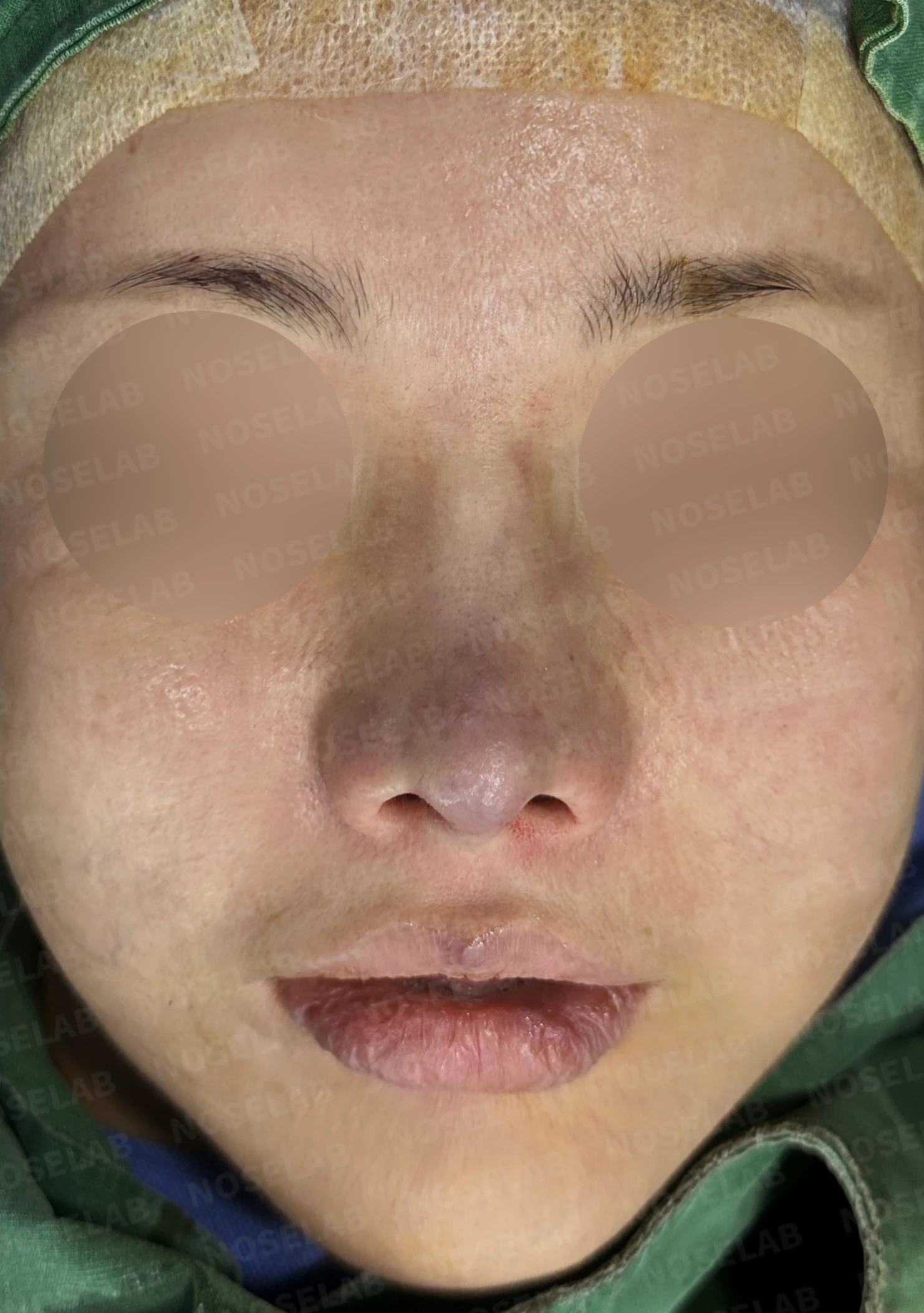
Before surgery / Day 0 immediately after surgery Photos taken immediately after surgery show swelling and may differ from the final result.
Profile view changes
Lowering the upturned tip establishes a natural nasal profile line
Septal reconstruction secures a stable support structure for the nasal tip
An overall balanced lateral silhouette
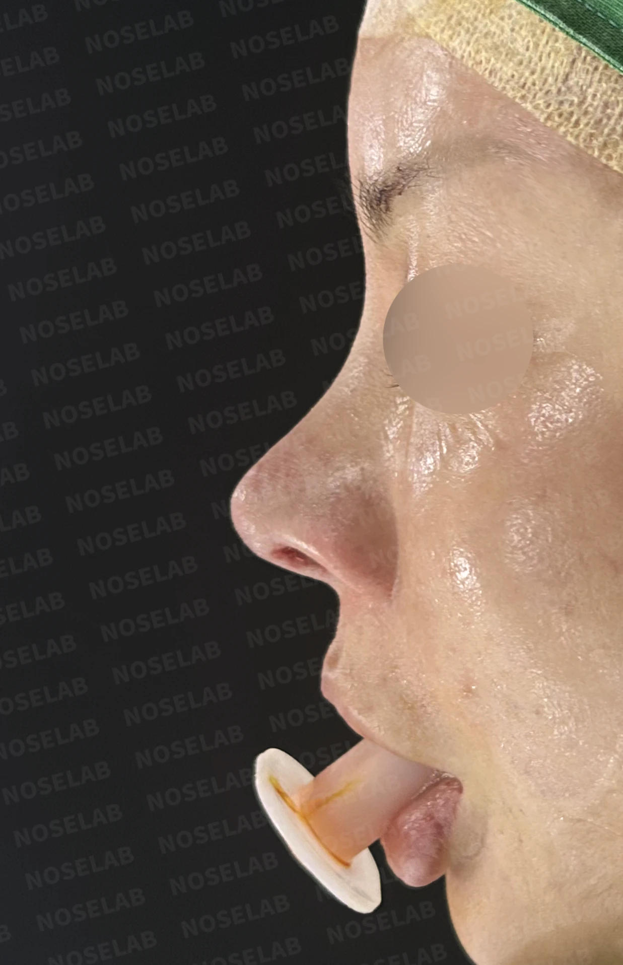
Before surgery / Day 0 immediately after surgery Photos taken immediately after surgery show swelling and may differ from the final result.
Changes on the oblique view
Natural transition between the nasal bridge and nasal tip from the 45-degree angle
Depressed areas on both sides of the bridge are smoothly filled
An overall soft and natural nasal contour
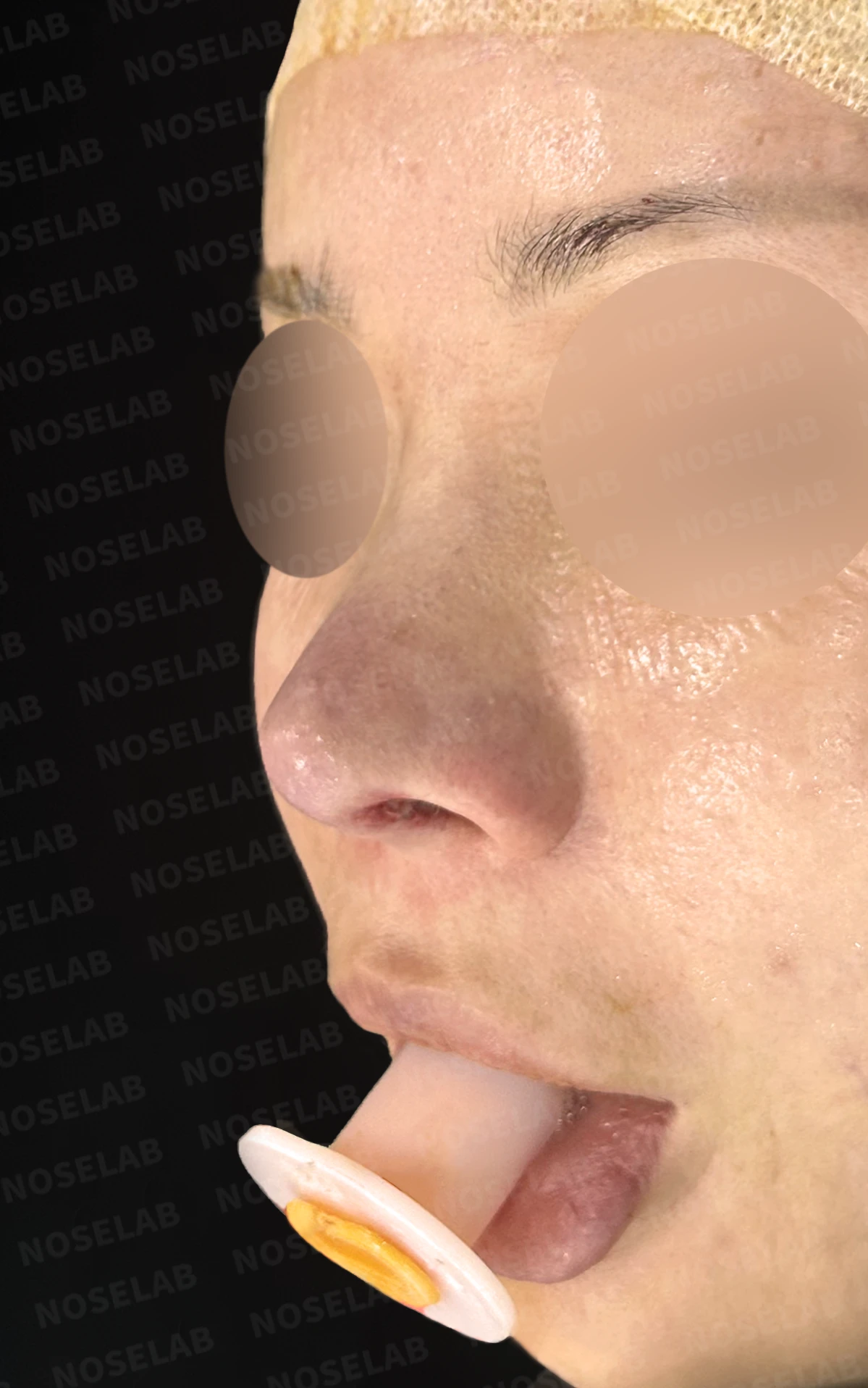
Before surgery / Day 0 immediately after surgery Photos taken immediately after surgery show swelling and may differ from the final result.
Changes in nostril shape
The pinched alae are reinforced, restoring a natural nostril shape
Excessive nostril show improves as the nasal tip is brought down
Balanced, symmetric nostril shape
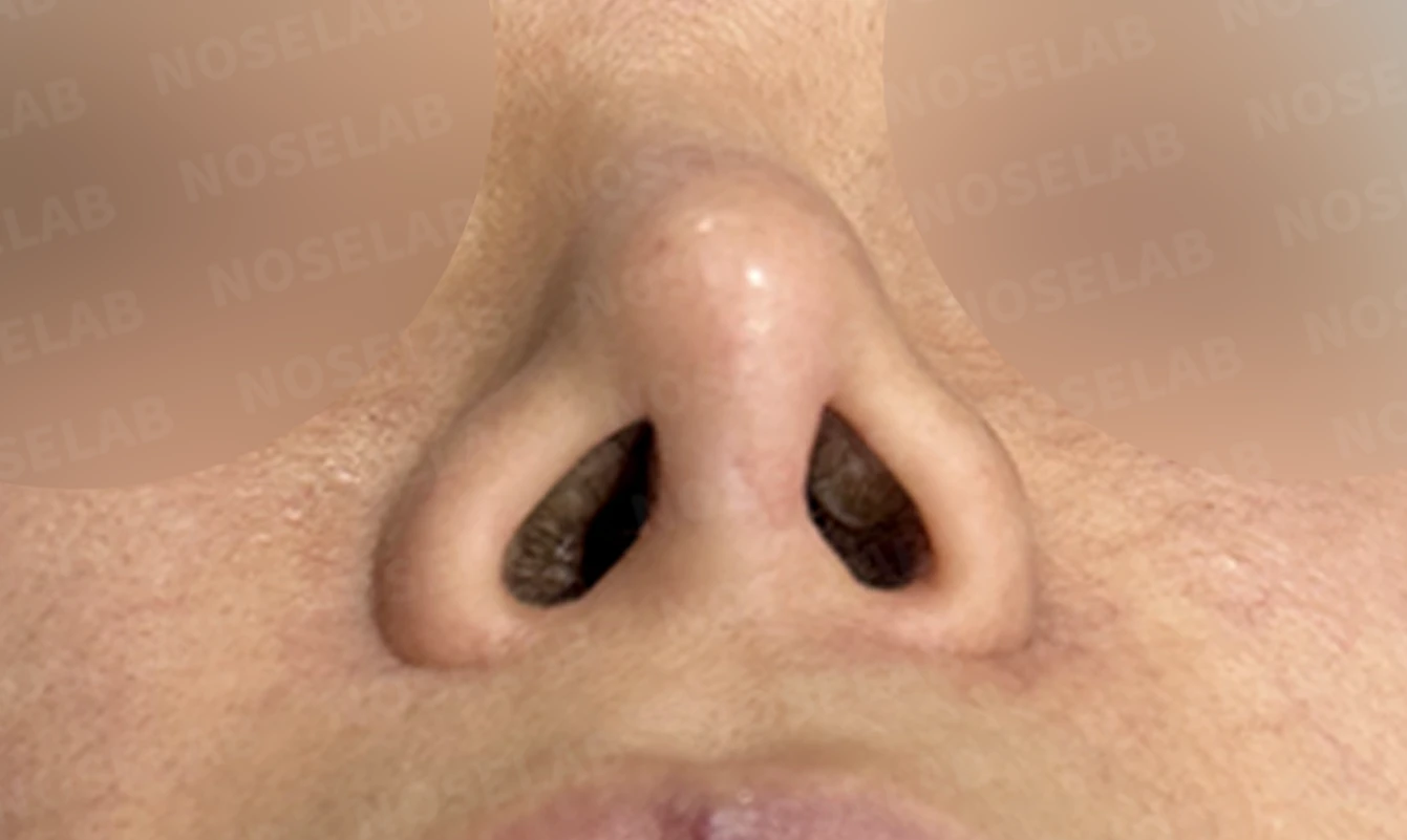
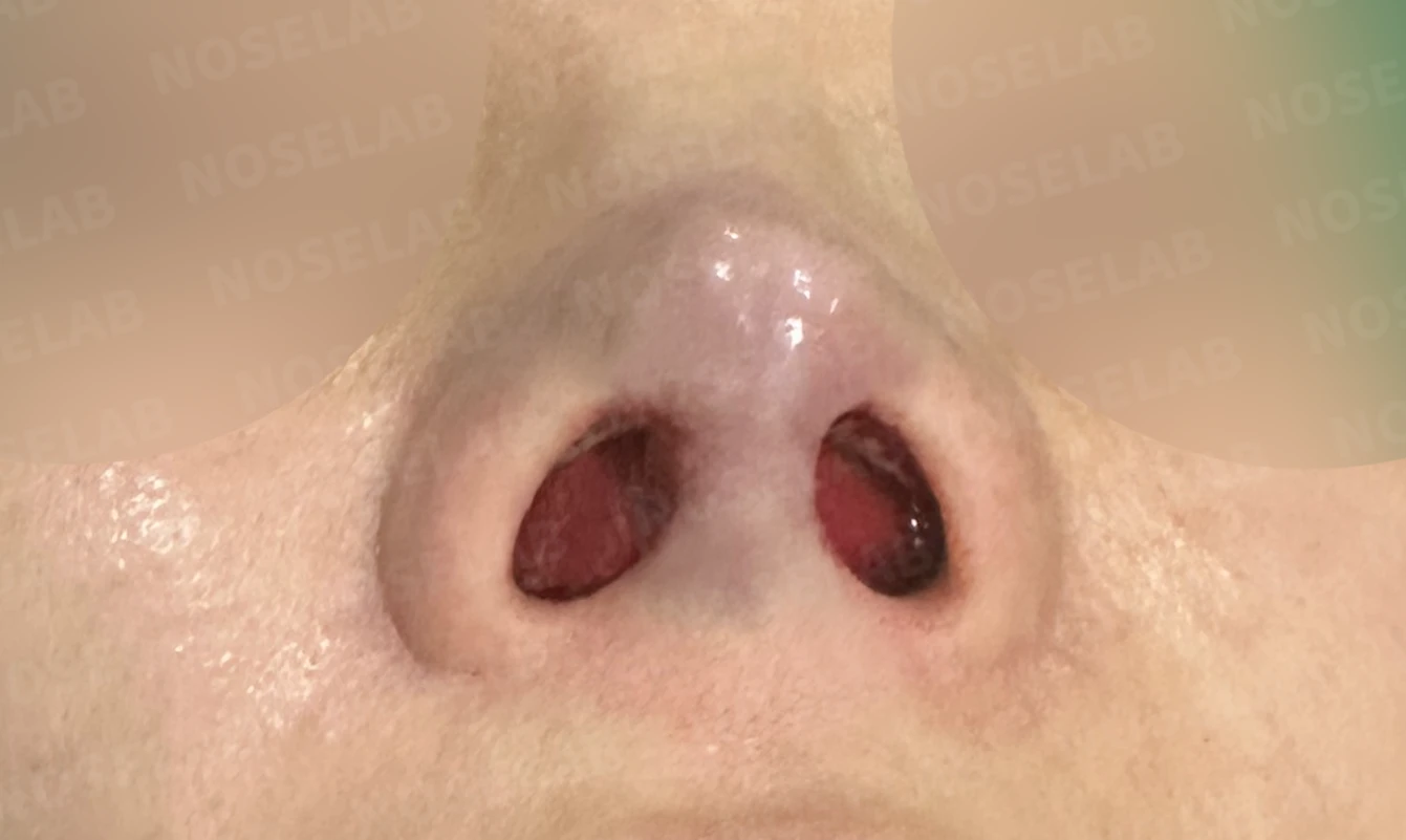
Before surgery / Day 0 immediately after surgery Photos taken immediately after surgery show swelling and may differ from the final result.
💡 Specialist opinion
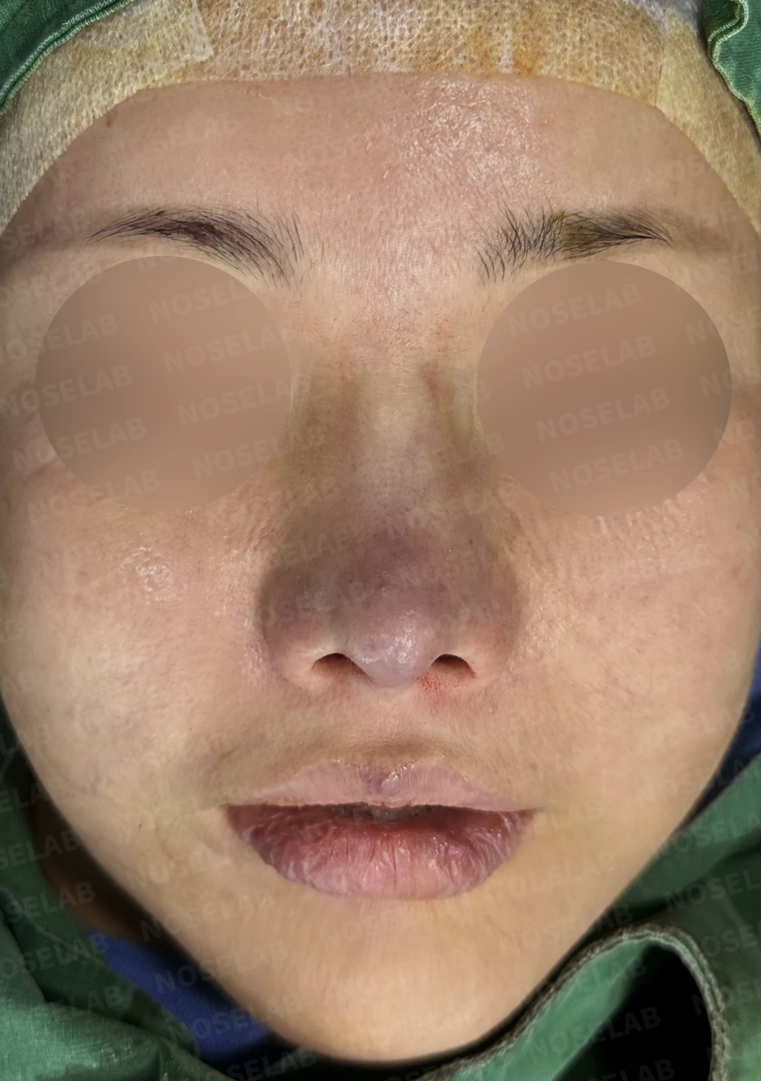
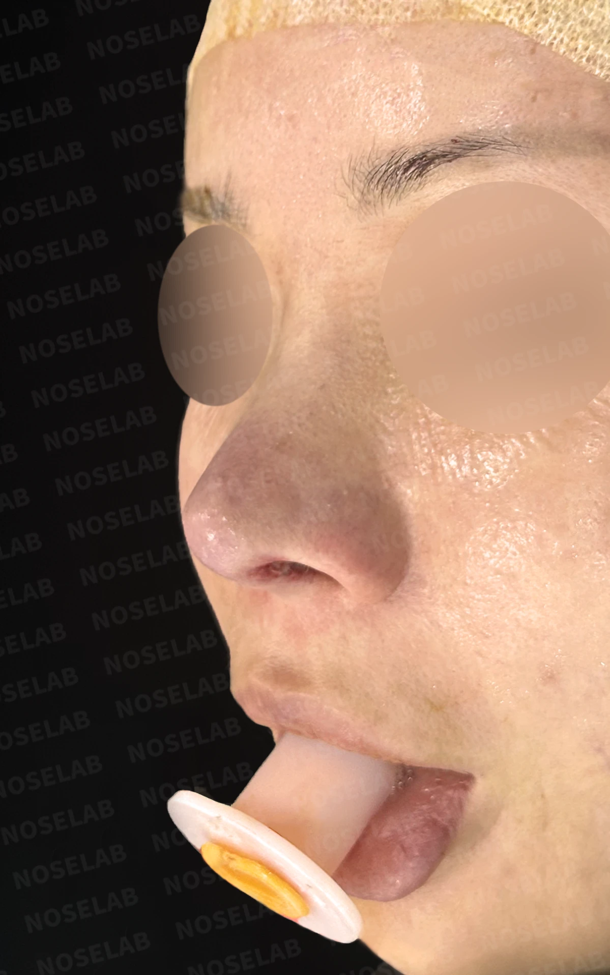
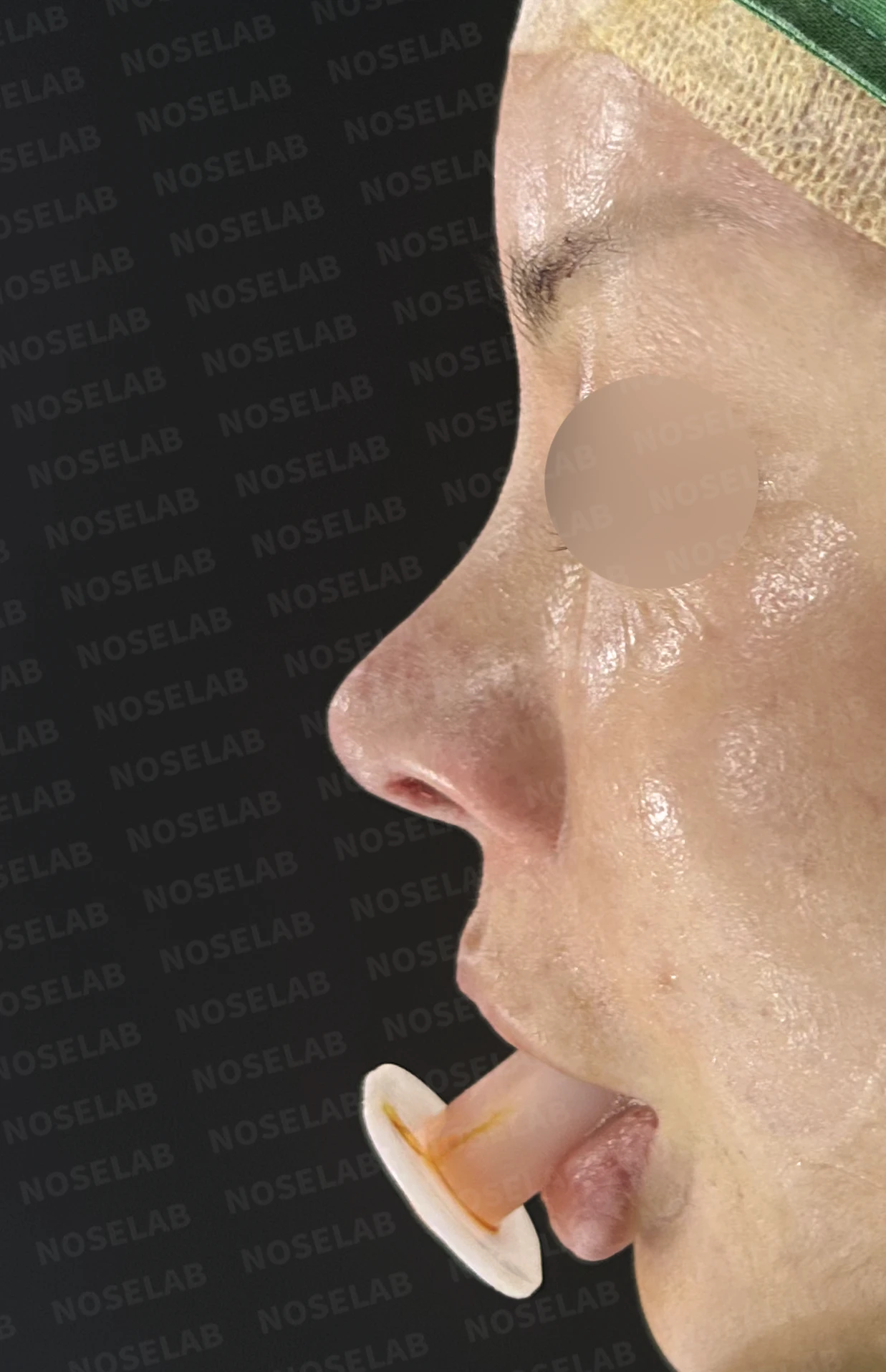
Front, oblique, and profile views on day 0 immediately after surgery. Photos taken immediately after surgery show swelling and may differ from the final result.
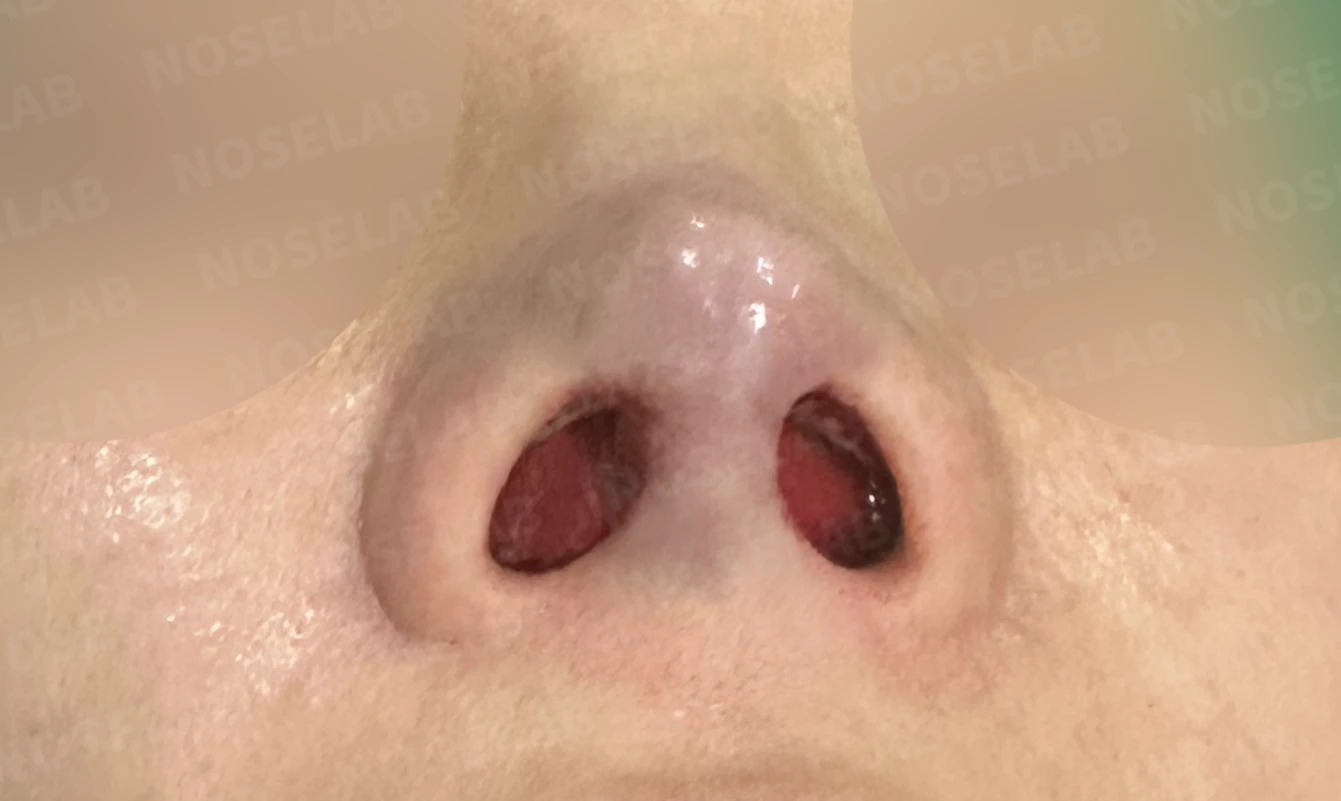
Nostril view on day 0 immediately after surgery. Photos taken immediately after surgery show swelling and may differ from the final result.
This was a highly challenging revision rhinoplasty case where the anterior septum had been completely excised in previous surgery. The septum is the foundational pillar supporting the nasal tip; when this structure is lost, tip support drops sharply, resulting in problems such as an upturned or collapsed tip.
In cases of septal loss like this, simply refining the external shape of the tip cannot provide a fundamental solution. Only by reconstructing the septum, the nose's core support pillar, can we prevent the tip from turning upward or deforming again. In this surgery, we precisely reconstructed the missing septal segments with autologous rib cartilage, establishing a stable structural foundation for the nasal tip.
Furthermore, both alae appeared pinched as a consequence of previous surgery. We reinforced the alar structures with autologous rib cartilage to restore natural curvature. For the depressions on both sides of the bridge, we finely diced autologous rib cartilage and spread it evenly to build a smooth dorsal line.
Revision rhinoplasty after septal loss requires far greater surgical skill and experience than standard revision cases. This is because a substantial amount of structural framework must be rebuilt under compromised conditions, including preexisting scar tissue and decreased vascularity. Even in such complex reconstructive cases, NOSELAB thoroughly analyzes each patient's structural status to deliver the intended result.
This was Dr. Peter C. Kang. Thank you.
#closedrhinoplasty #closedrevisionrhinoplasty #revisionrhinoplasty #septalreconstruction #autologousribcartilage #upturnednosecorrection #pinchednose #alarreinforcement #bridgedepressioncorrection #nasalobstructionsurgery

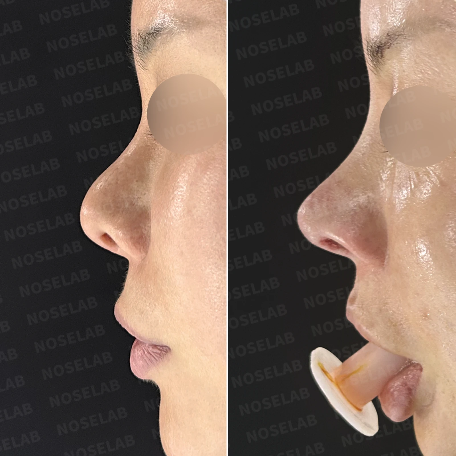
Limitations and risks
When autologous rib cartilage is used, scarring and pain may remain at the donor site on the chest, and pneumothorax (pleural injury) can occur rarely. Over time, the grafted cartilage may warp or undergo partial resorption.
When the septum is corrected at the same time, nasal obstruction may persist or recur, and in rare cases, septal perforation or changes in smell may occur.
Common potential risks include infection, bleeding, asymmetry, scarring, skin necrosis, and insufficient improvement; results vary depending on individual skin condition and previous surgical history.