Hello, I am Dr. Peter C. Kang.
Today, I would like to share a revision rhinoplasty case of a male patient who had concerns about severe left-sided nasal obstruction and a noticeably drooping tip.
🔍 Surgical history
In this case, the patient had undergone two prior nasal surgeries.
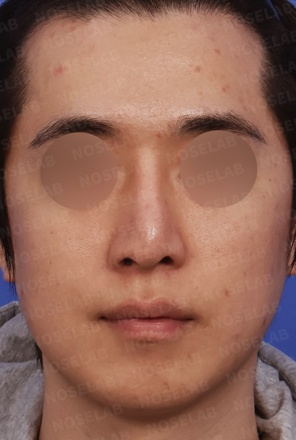
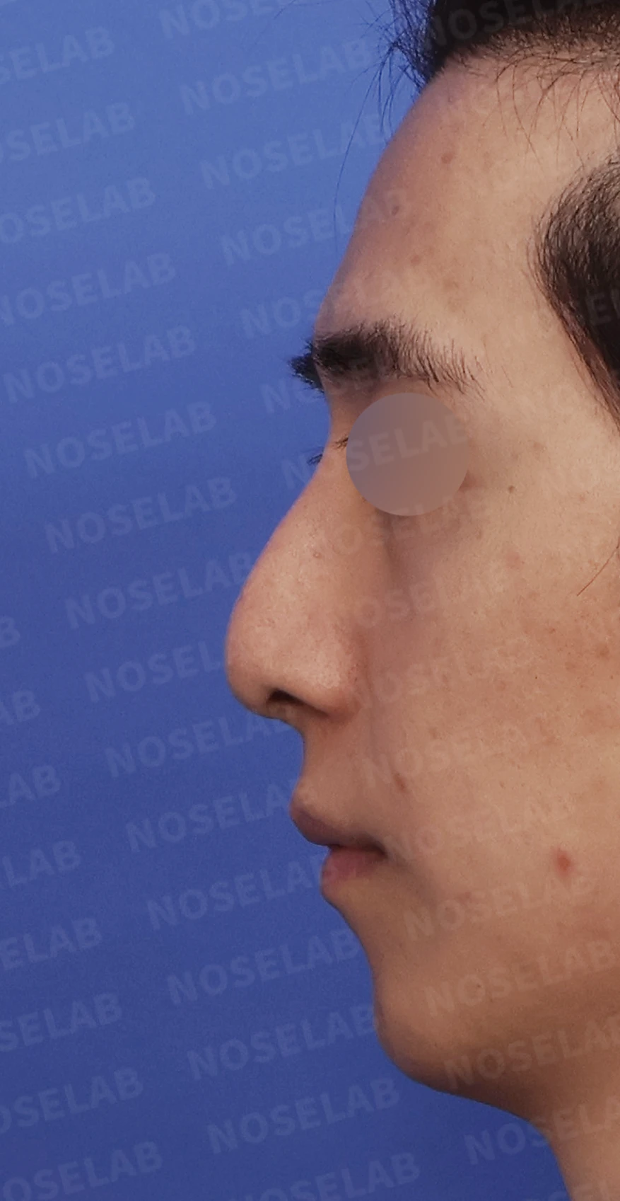
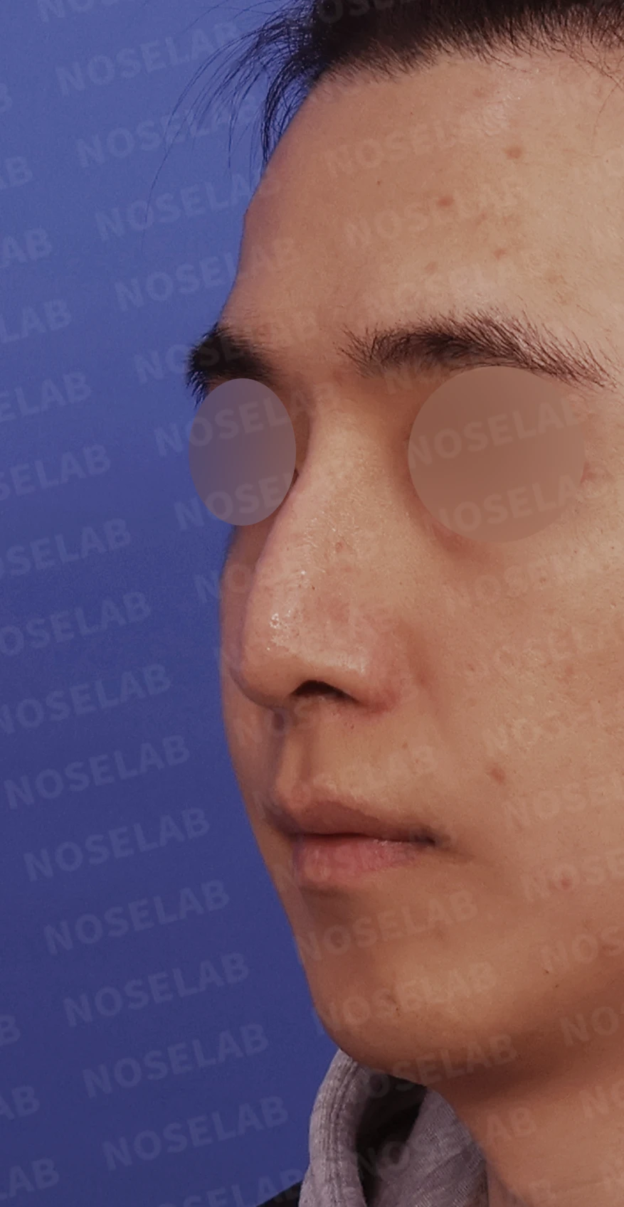
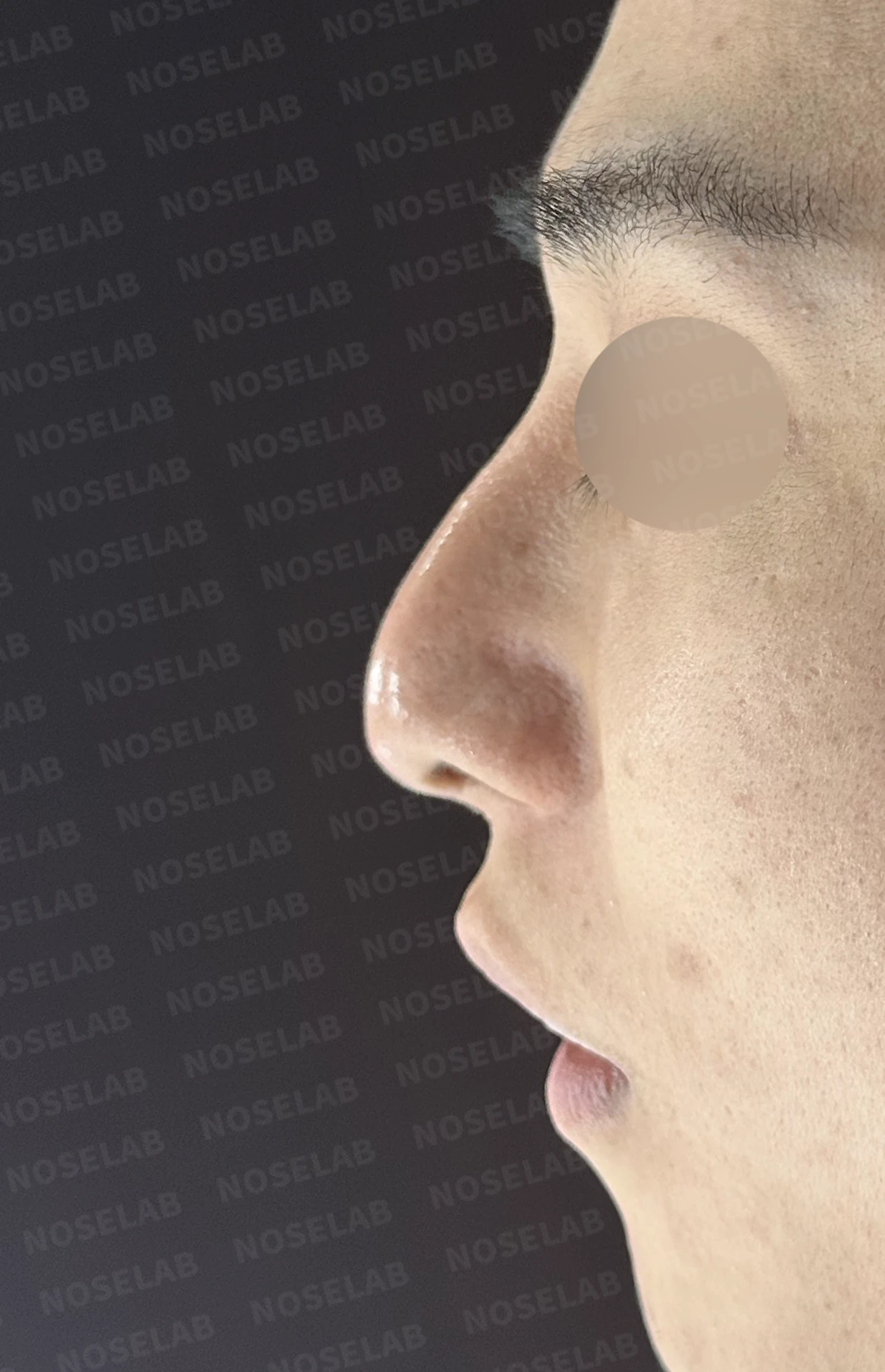
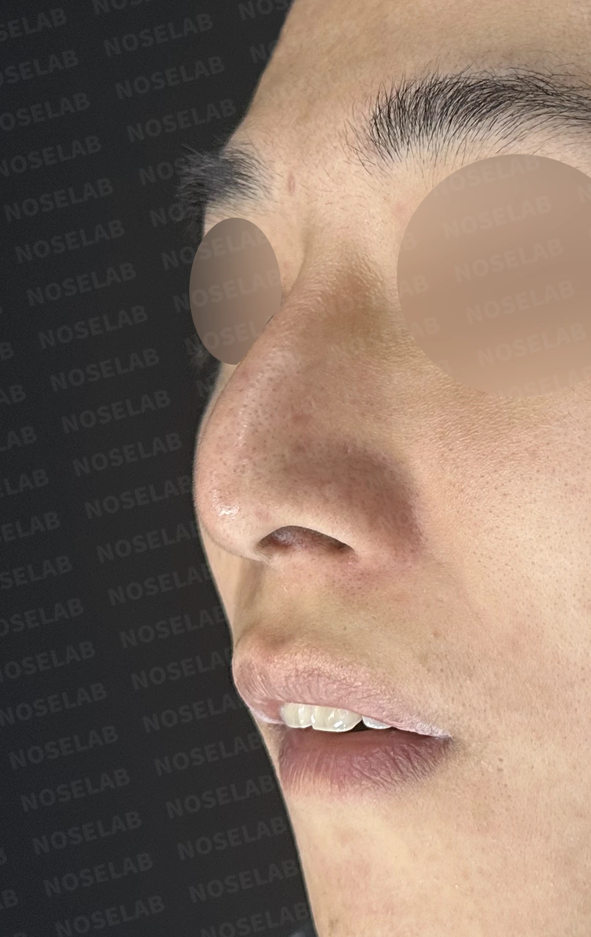
Front, left profile, and oblique views during design consultation.
Approximately 17 years ago: Reduction for a nasal bone fracture (at a university hospital)
Approximately 10 years ago: Septal surgery and rhinoplasty with a silicone implant
However, from previous procedures, the septum had completely collapsed to the left, leaving almost no airway space in the left nasal cavity and causing severe nasal obstruction.
💉 Patient requests
When the patient visited our clinic for consultation, the requests were as follows:
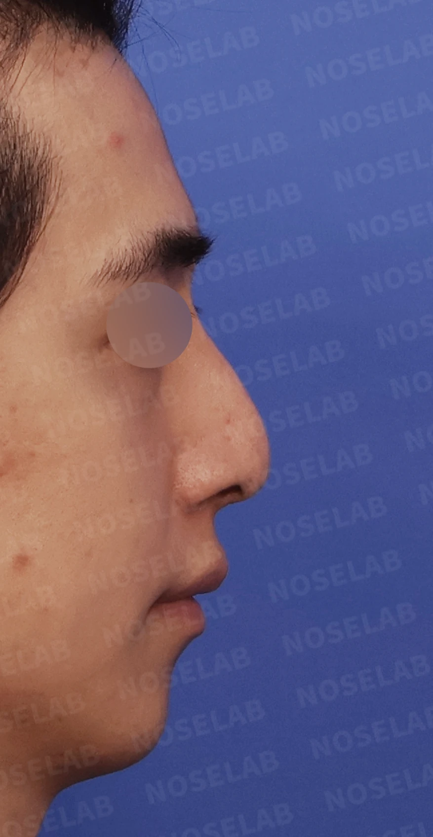
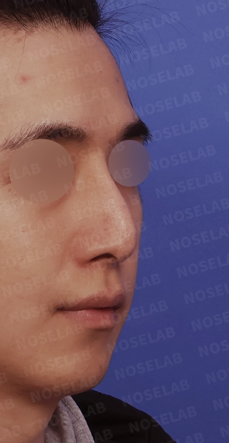
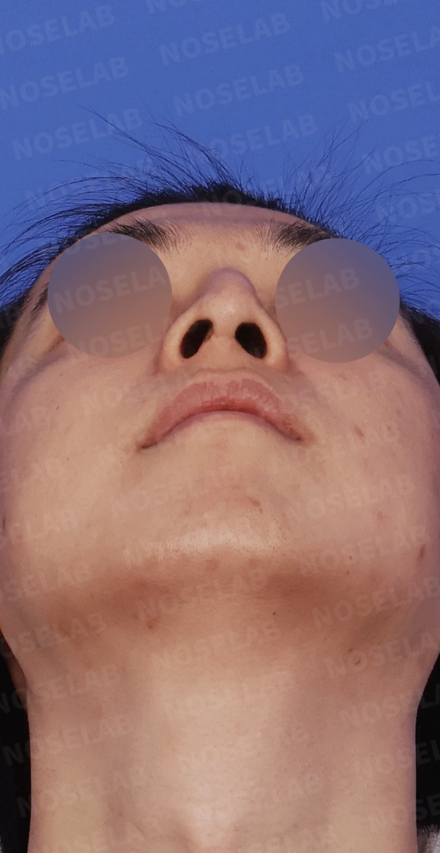
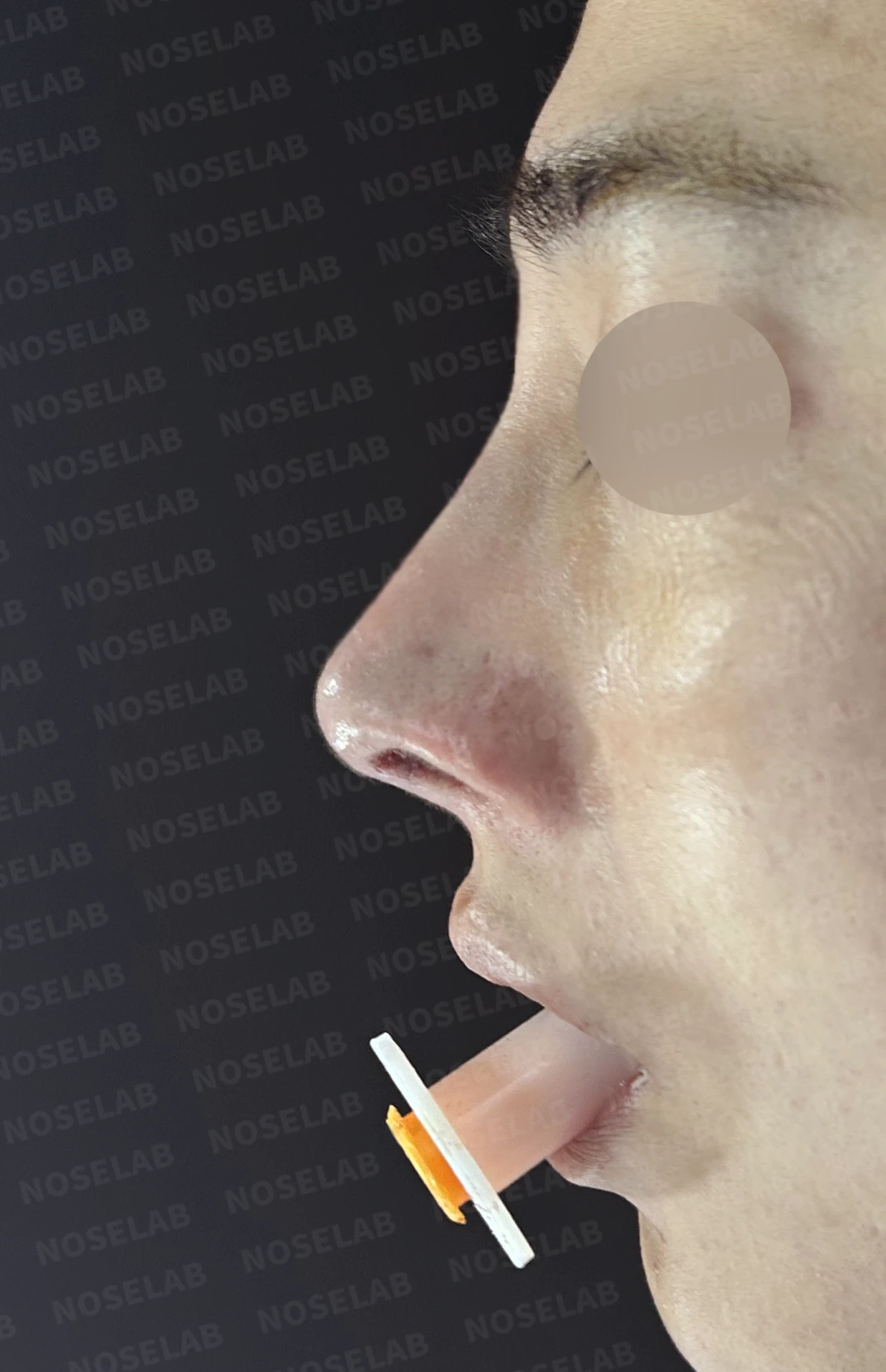
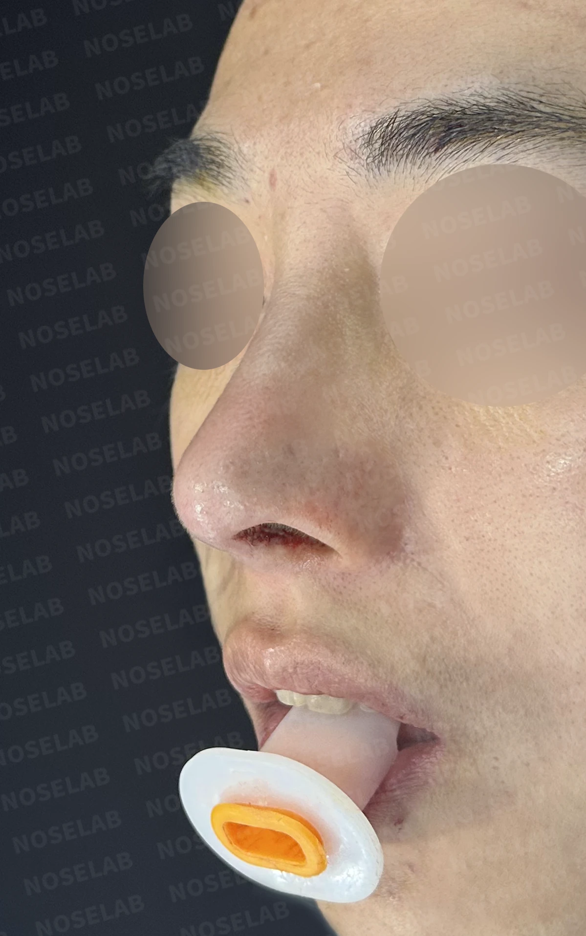
Right profile, oblique, and nostril views during design consultation.
Relief of left nasal obstruction (primary objective)
Correction of crooked nose
Improvement of a drooping tip
Preference for autologous rib cartilage
Improvement of scar tissue inside the nose
The left nasal obstruction in particular caused significant discomfort in daily life, so functional improvement was the patient's highest priority.
🔧 Surgical plan and procedure
Preoperative examination revealed that the septum had completely collapsed to the left, leaving virtually no left nasal cavity space. In addition, the nasal tip was noticeably drooping, and the underlying cartilage and implant were visible through the skin.
1. Septal reconstruction using autologous rib cartilage
Reconstruction of the completely collapsed septum using autologous rib cartilage
Stable fixation by drilling holes in the maxilla for cartilage anchoring
Structural restoration to secure space in the left nasal cavity
2. Septoplasty
Straightening the deviated septum
Root cause of nasal obstruction addressed
Securing balanced airflow in both nasal passages
3. Septal extension
Extending the nasal tip based on the reconstructed septum
Naturally elevating the drooping tip
Covering the cartilage and implant that showed through
4. Correction of nasolabial angle
Adjustment of the angle between the nose and the lips
Maximizing correction of the drooping tip
Achieving a masculine yet refined profile line
5. Rhinitis surgery
Reduction of hypertrophic inferior turbinates
Resolution of nasal obstruction symptoms
Restoring comfortable breathing function
🌟 Surgical results
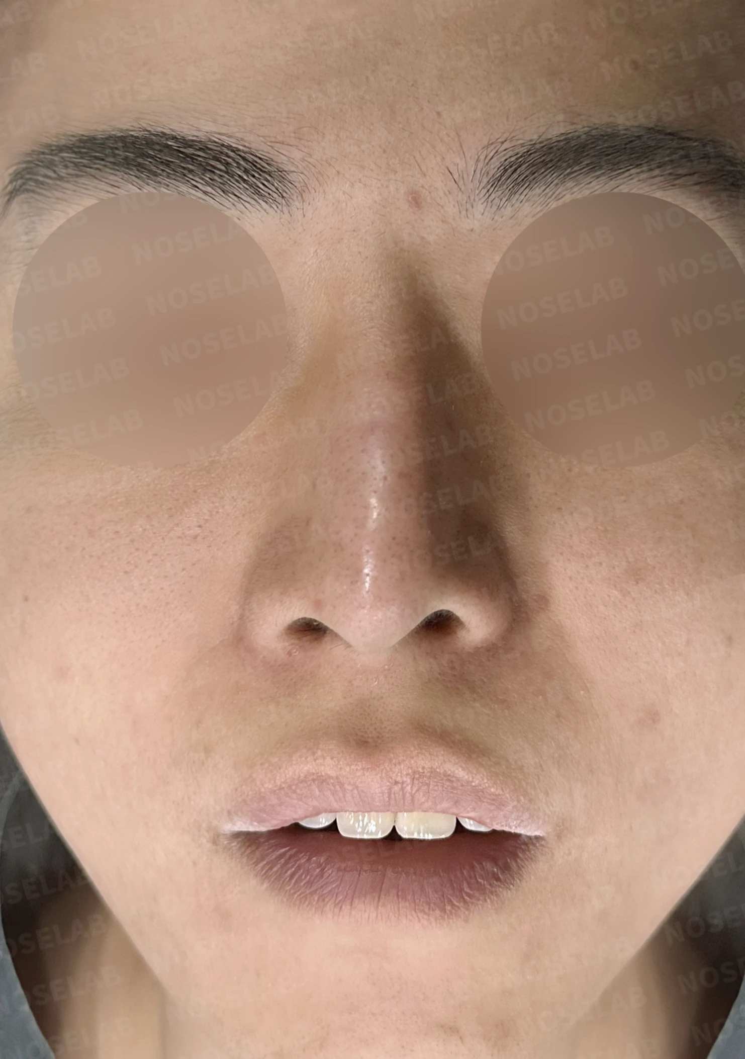
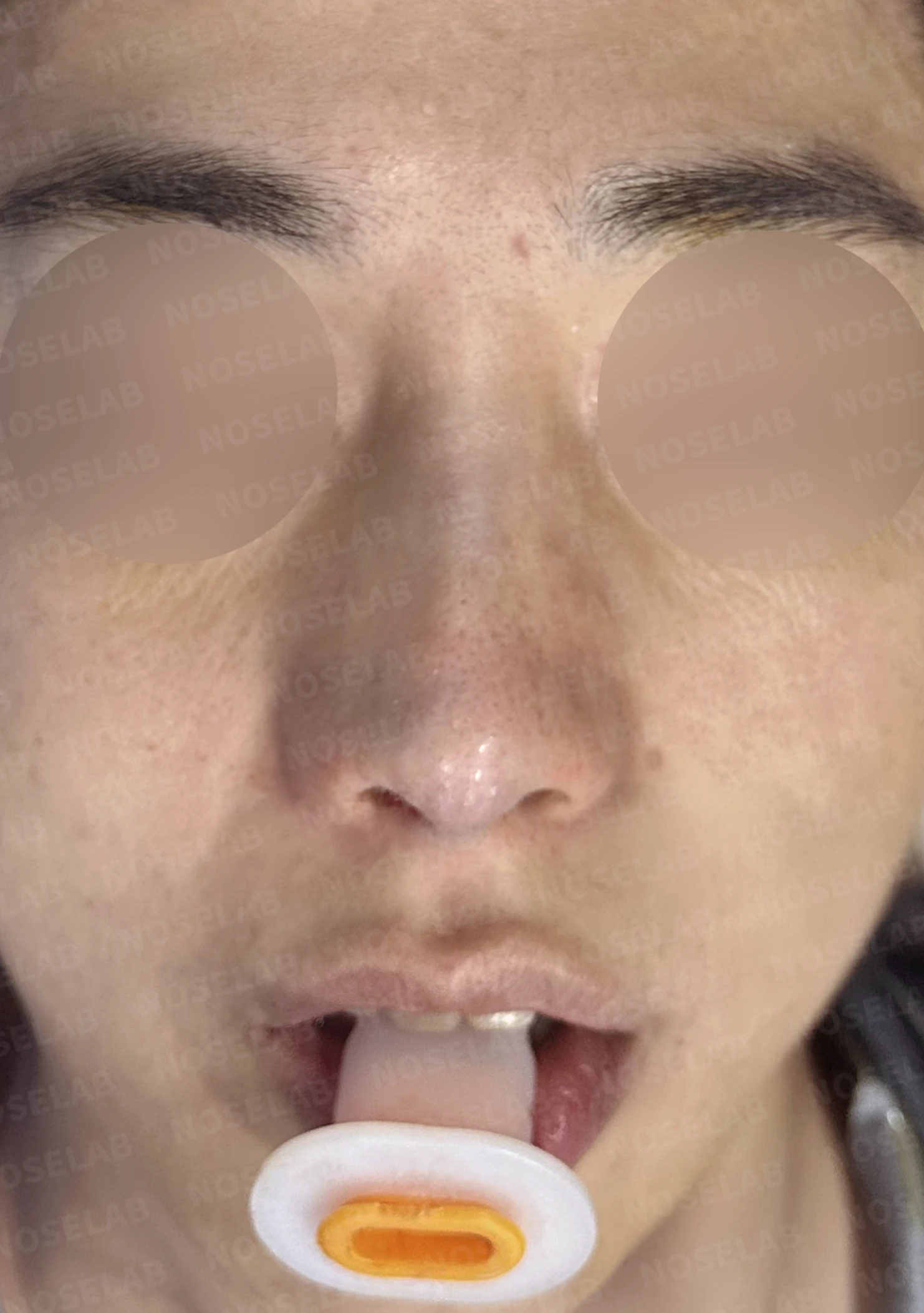
Before surgery / Day 0 immediately after surgery Photos taken immediately after surgery show swelling and may differ from the final result.
1. Frontal changes
The crooked nose was straightened
Overall neat and refined impression
Achieving bilateral symmetry
Before surgery / Day 0 immediately after surgery Photos taken immediately after surgery show swelling and may differ from the final result.
2. Profile changes
The drooping tip was naturally elevated
Cartilage and implant show-through were corrected
A masculine and natural nasal bridge line
Before surgery / Day 0 immediately after surgery Photos taken immediately after surgery show swelling and may differ from the final result.
3. Functional changes
Nasal obstruction relieved by securing left nasal cavity space
Long-term functional maintenance supported by septal reconstruction
Overall breathing improved through rhinitis surgery
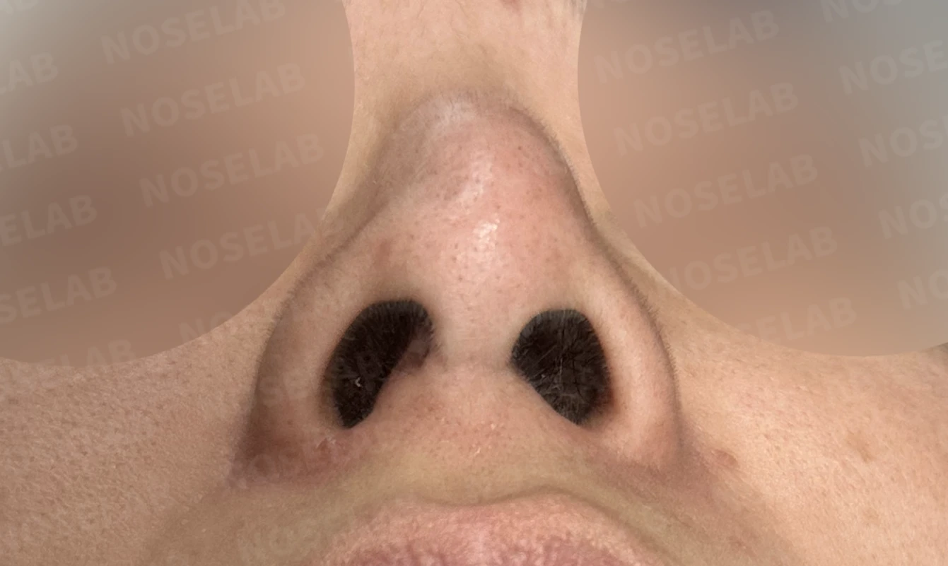
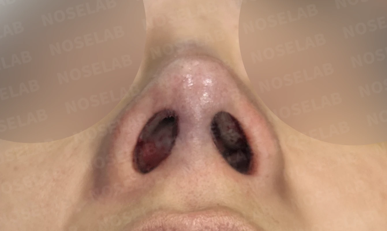
Before surgery / Day 0 immediately after surgery Photos taken immediately after surgery show swelling and may differ from the final result.
💡 Specialist opinion
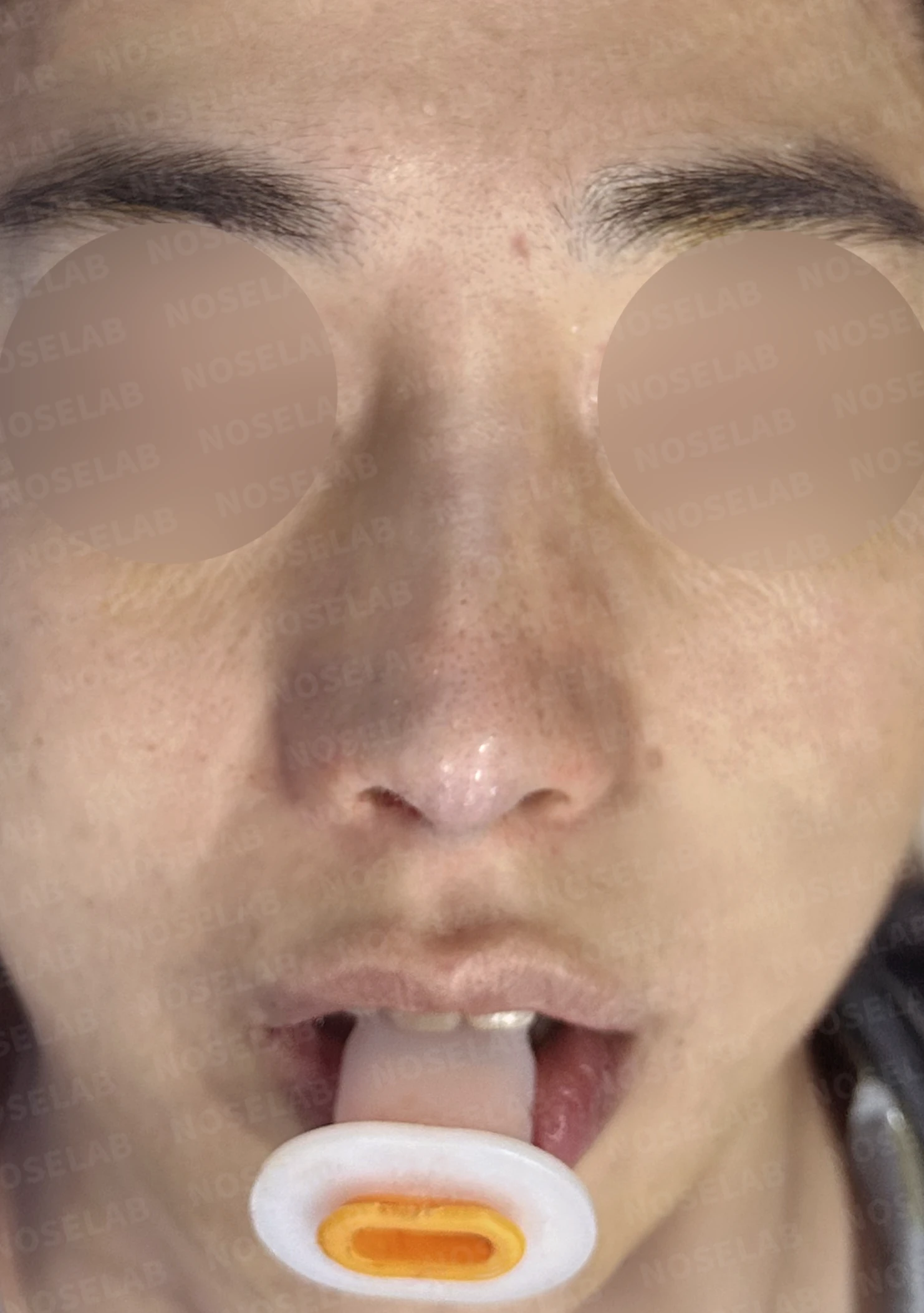
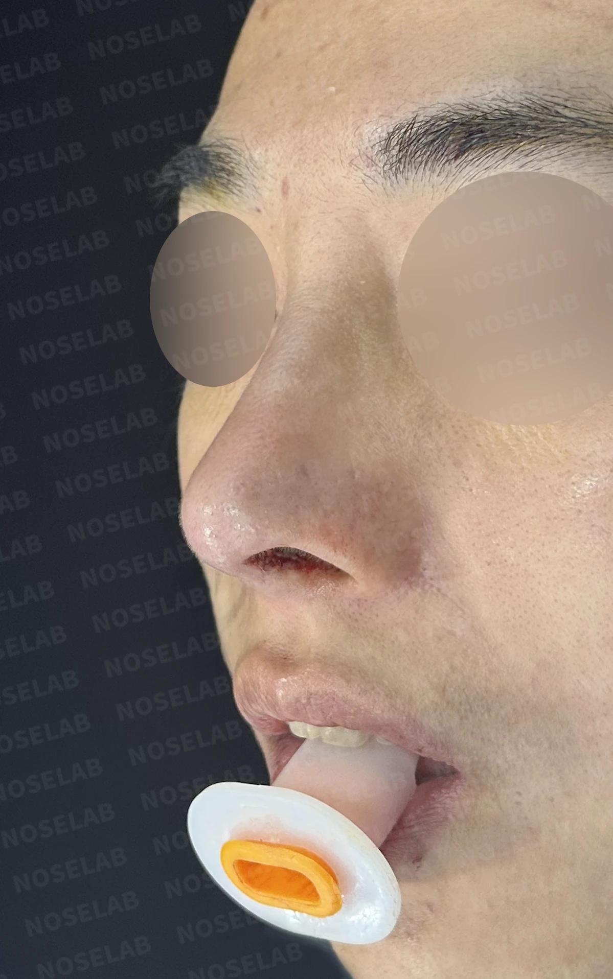
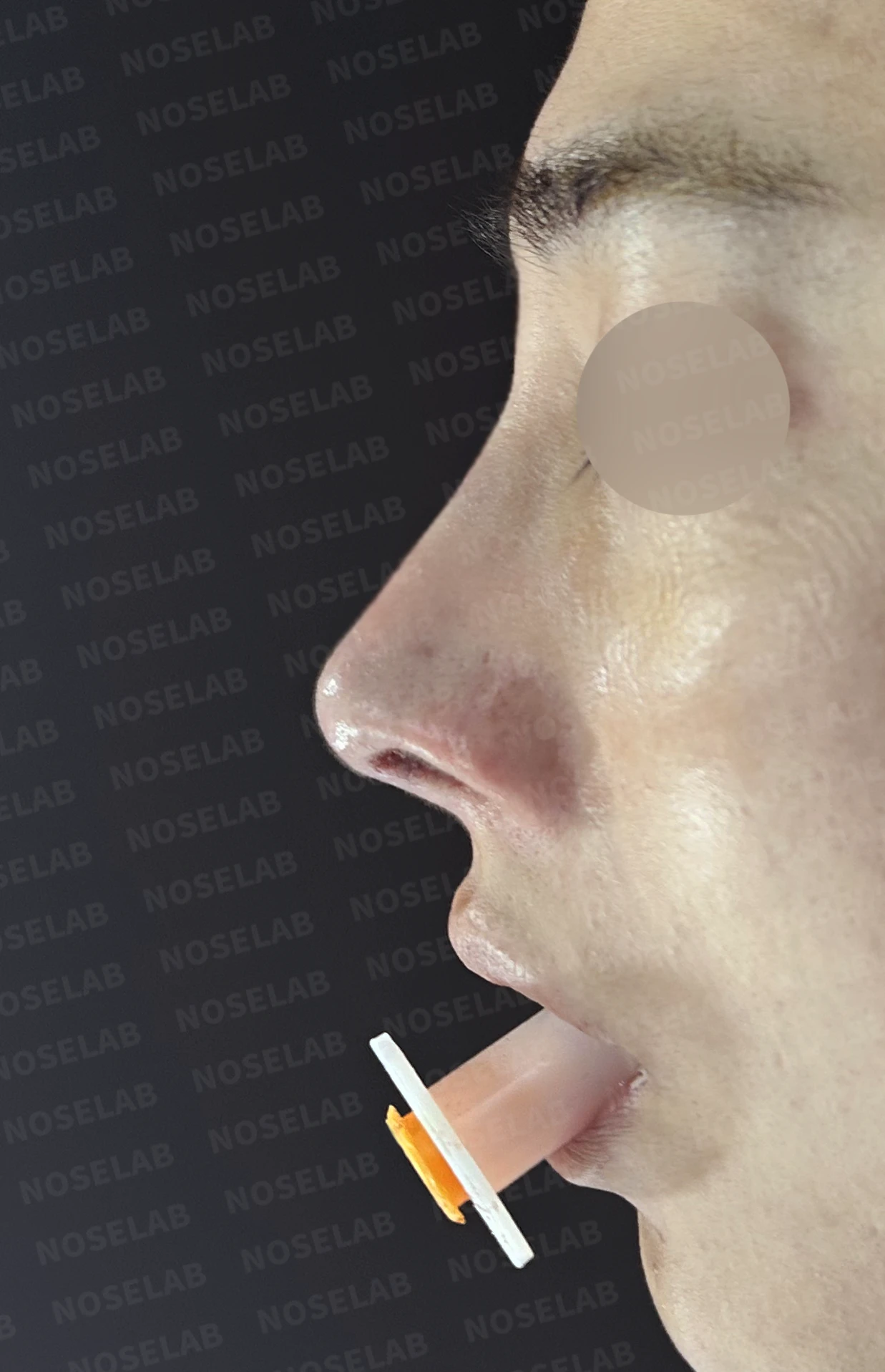
Front, oblique, and profile views on day 0 immediately after surgery. Photos taken immediately after surgery show swelling and may differ from the final result.
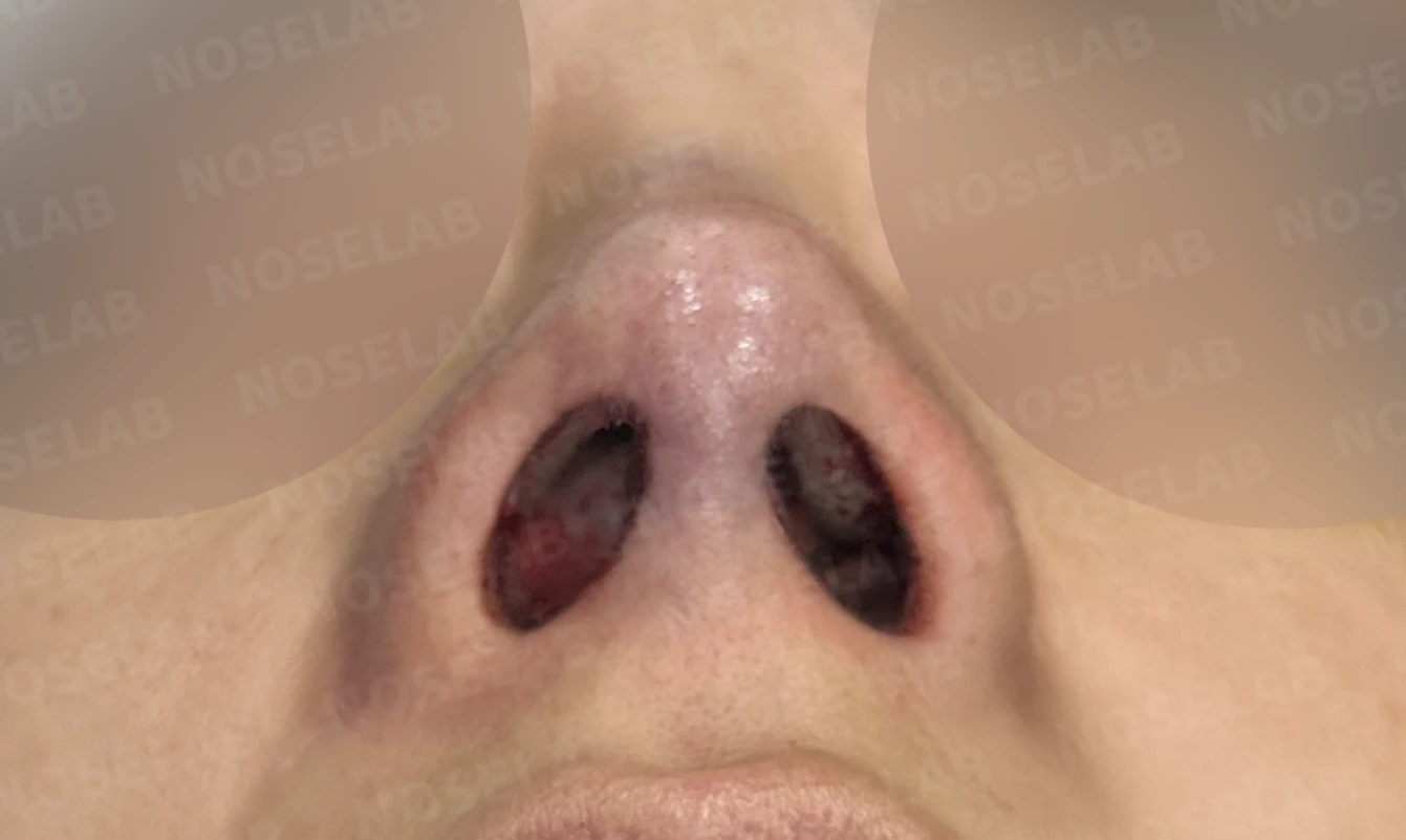
Nostril view on day 0 immediately after surgery. Photos taken immediately after surgery show swelling and may differ from the final result.
When the septum has completely collapsed and the nasal cavity space is nearly gone, conventional septal correction alone has limitations. In such cases, the septum itself must be reconstructed using autologous rib cartilage.
In septal reconstruction, there is a process of drilling holes into the maxilla to secure the cartilage. In most cases, 1 to 3 or 4 fixation points are created. Bleeding can occur during this process, and particularly in patients who bleed more heavily, bleeding can sometimes arise from within the bone even after thorough hemostasis, occasionally leading to a septal hematoma. In this case, there was also a somewhat higher amount of bleeding during surgery, so I managed the recovery process with extra care, taking into account the potential for swelling, bruising, and hematoma formation.
Even in these circumstances, patients recover smoothly with appropriate postoperative management, so there is no need for excessive concern. The key is to closely monitor progress after surgery and take timely measures whenever needed.
I will continue to prioritize safety and recovery to provide the intended surgical outcomes. I hope this case provides helpful information to those with similar concerns. This has been Medical Director Dr. Peter C. Kang. Thank you.
#closedrhinoplasty #closedrevisionrhinoplasty #septalreconstruction #autologousribrhinoplasty #nasalobstructionsurgery #septoplasty #rhinitissurgery #malerhinoplasty #droopingtipcorrection

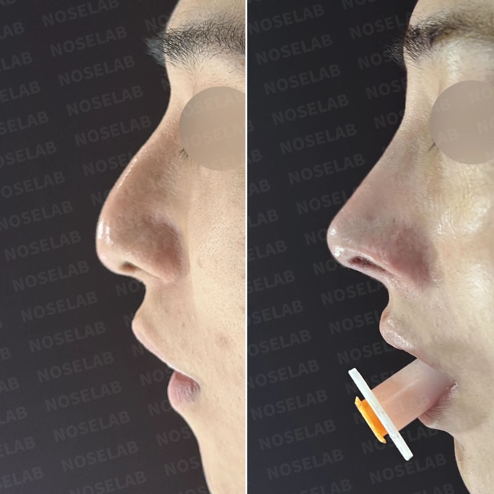
Limitations and risks
When autologous rib cartilage is used, scarring and pain may remain at the donor site on the chest, and pneumothorax (pleural injury) can occur rarely. Over time, the grafted cartilage may warp or undergo partial resorption.
When the septum is corrected at the same time, nasal obstruction may persist or recur, and in rare cases, septal perforation or changes in smell may occur.
Common potential risks include infection, bleeding, asymmetry, scarring, skin necrosis, and insufficient improvement; results vary depending on individual skin condition and previous surgical history.