Hello, I am Dr. Peter C. Kang.
Today, I would like to share the case of a patient who came to us with concerns about inflammation and contracture following multiple rhinoplasties.
🔍 Surgical history
2007: L-shaped silicone implant
August 2021: Silicone implant + ear cartilage → Inflammation occurred
November 2022: Silicone implant + mesh + alar base / premaxillary augmentation (cat surgery) → Upturned nose developed
November 2023: Silicone implant + autologous rib cartilage + dermis + periosteum
10 days later: Treatment for inflammation (irrigation) and 16 sessions of PRP
💉 Primary symptoms
Contracture deformity (deformity caused by chronic inflammation)
Abnormal edema (possible inflammation caused by residual foreign material)
Nasal tip depression (silicone implant extending all the way down to the tip)
Tenderness around the right philtrum (the area of paranasal augmentation)
Persistent pulling sensation and discomfort in the left septal mucosa (caused by mesh remaining in the septum)
Left-sided nasal obstruction (deviated septum)
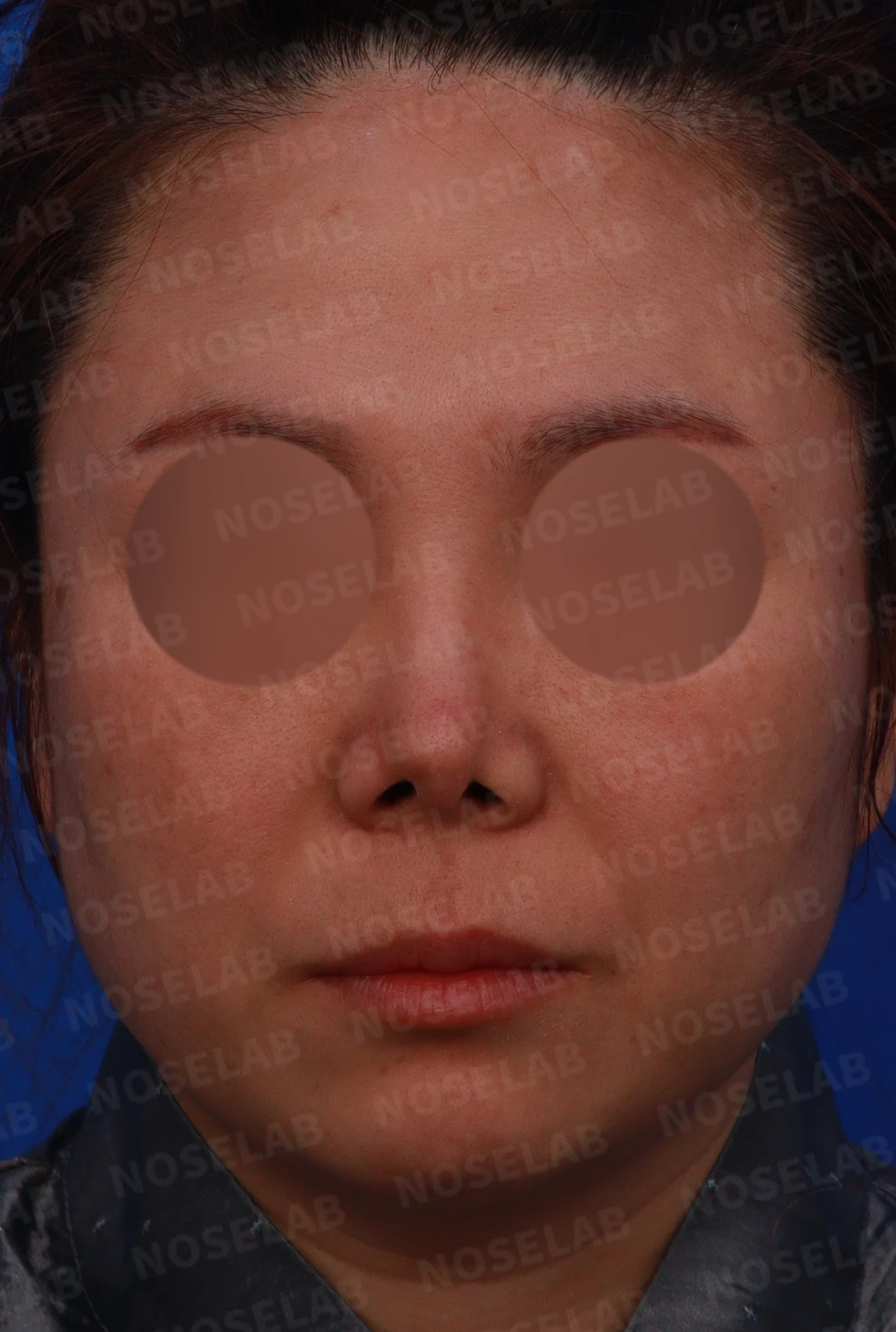
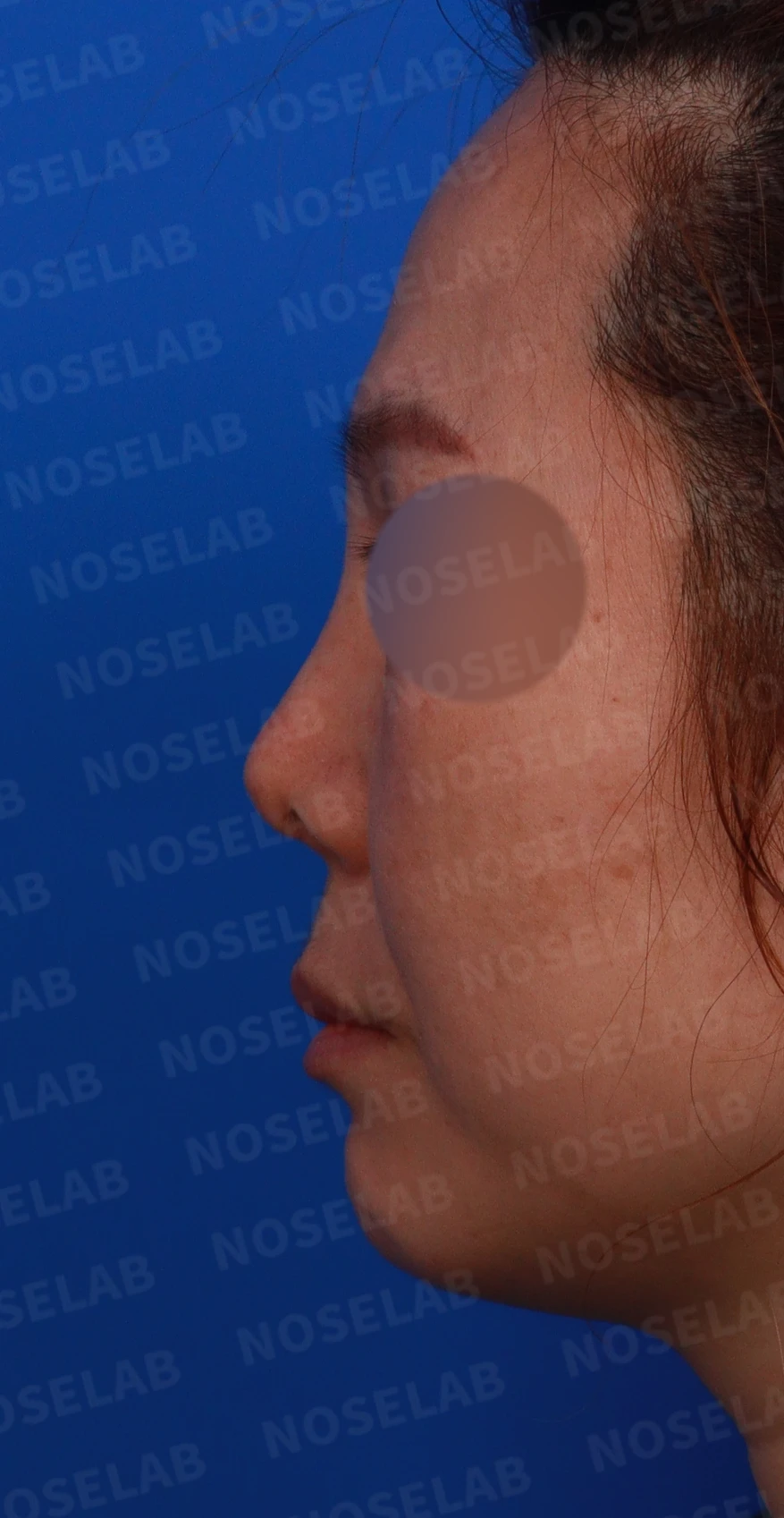
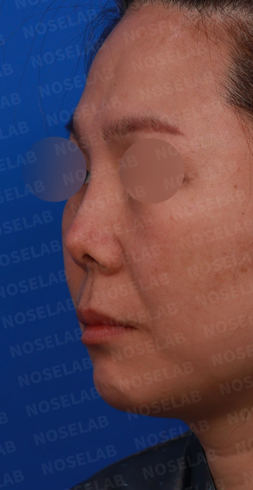
Front, left profile, and oblique views during design consultation.
🔧 Surgical plan
Reconstruction of the lower lateral cartilage and septum using autologous rib cartilage
Alar lowering
Correction of the nasolabial angle
Stem cell therapy
Functional improvement (septal and rhinitis surgery)
Intravenous antibiotic injections
Septal reconstruction, foreign material removal, and skin grafting as needed
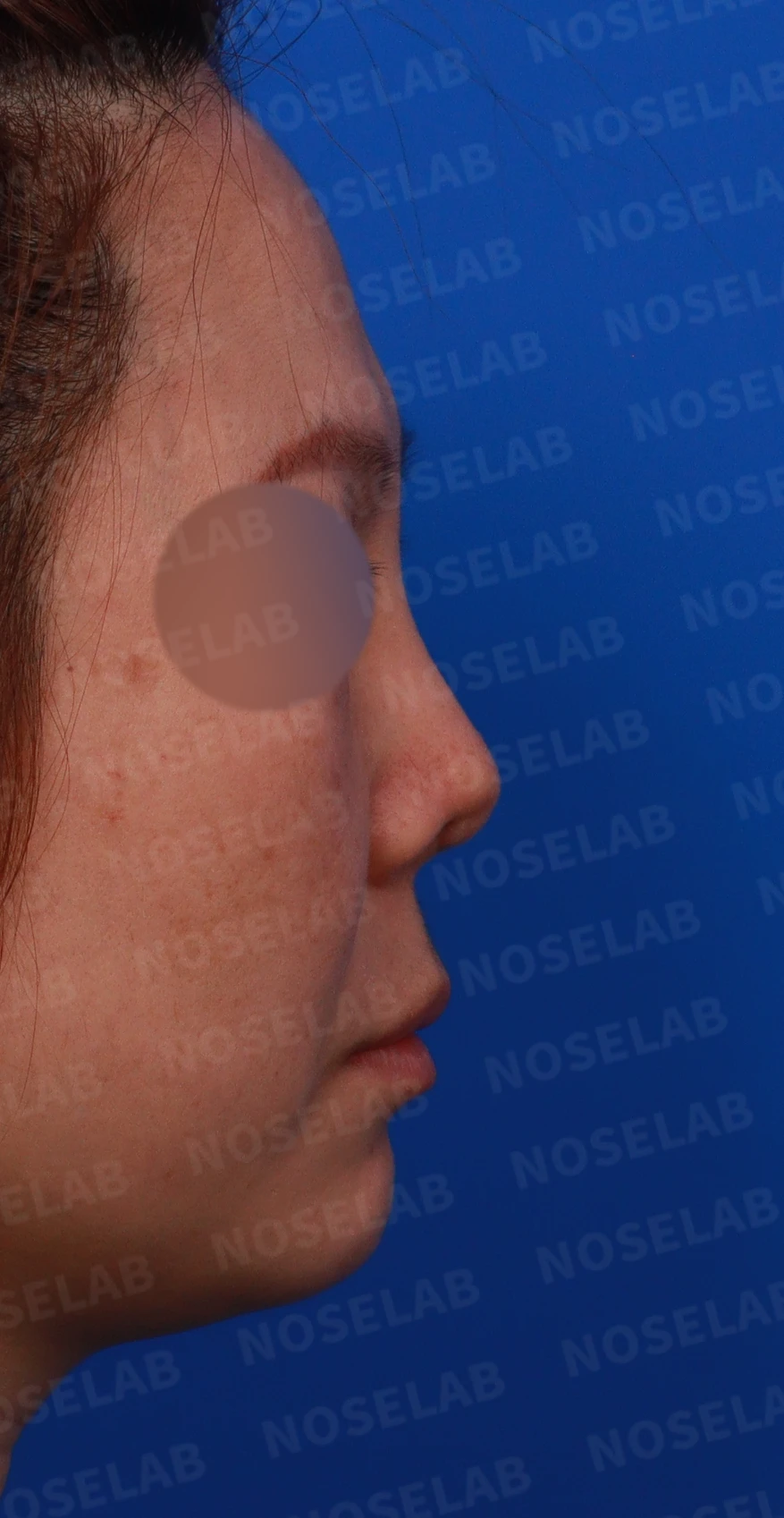
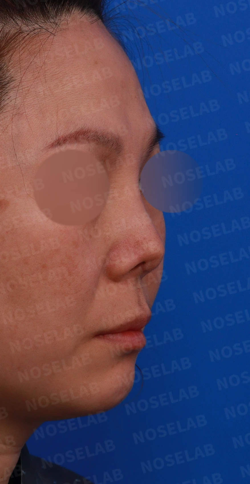
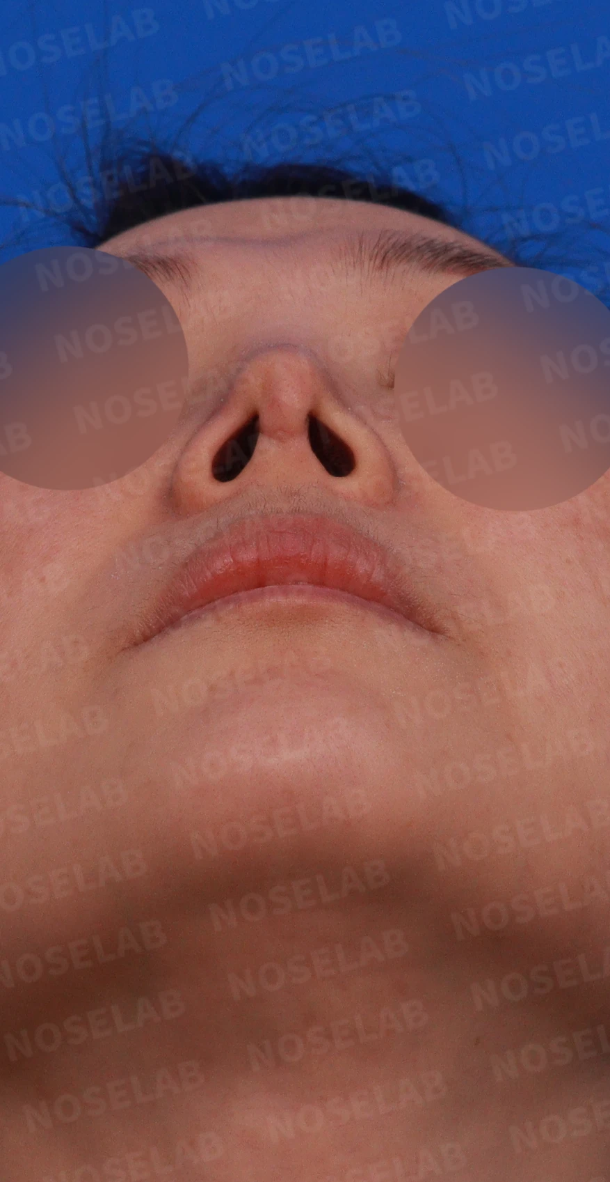
Right profile, oblique, and nostril views during design consultation.
🔧 Intraoperative findings and procedures
During the meticulous surgical procedure, the following issues were identified:
The caudal end of the silicone implant was located immediately beneath the depressed area of the nasal tip.
Residual mesh was identified in the left septal mucosa of the tip and removed.
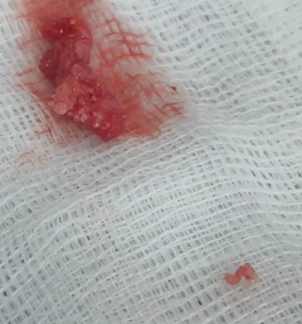
Fragments of mesh removed during surgery
Removal of an autologous rib cartilage fragment deviated to the right at the paranasal augmentation site
Chronic foreign body reaction and inflammatory response caused by residual foreign material appeared to have been present.
-> Completely removed and thoroughly irrigated
Most of the septal cartilage had been lost, and the remaining septum was deviated to the left.
-> Septal reconstruction was performed using autologous rib cartilage.
Because there was insufficient skin while correcting the shortened nose, retroauricular skin was grafted to both sides of the columella.
💉 Preoperative analysis
1. Front view
Retracted nostrils
Pinched alae
Asymmetry
Deviated nasal bridge
Nasal bone asymmetry
2. Profile view
High radix starting point
Short and collapsed nasal tip
Obtuse nasolabial angle
Upturned nose
3. Nostrils
Left-right asymmetry
Columella tilted to the left
Pinched nostrils
🌟 Surgical results
1. Frontal view improvement
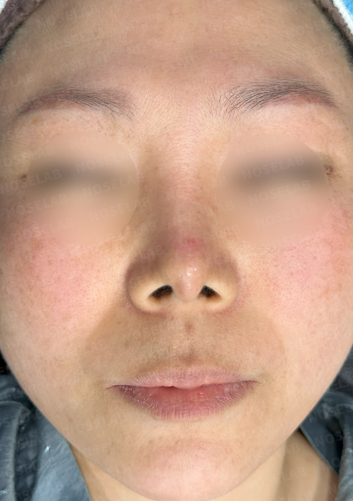
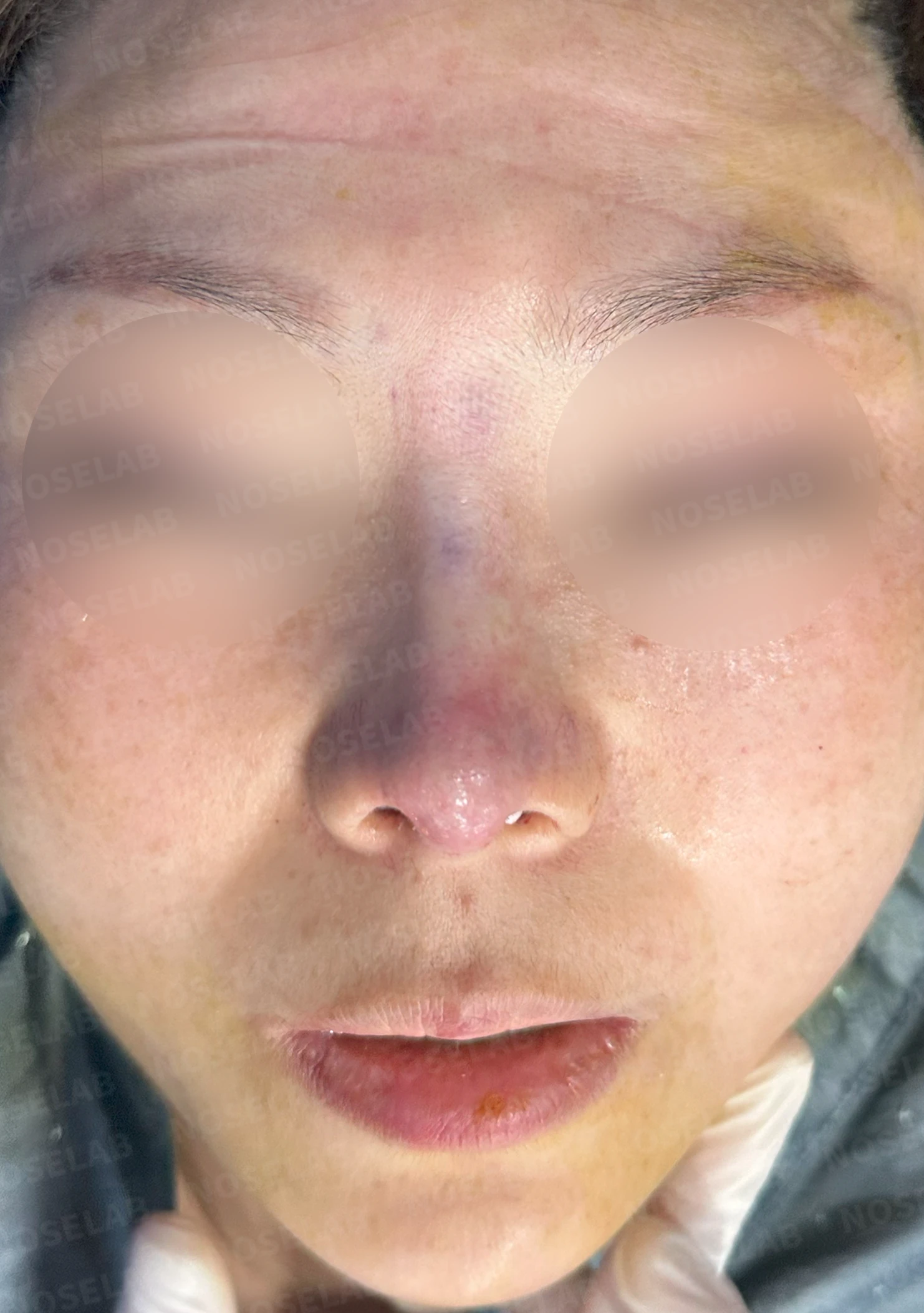
Before surgery / Day 0 immediately after surgery Photos taken immediately after surgery show swelling and may differ from the final result.
Correction of retracted nostrils
Improvement of pinched alae
Improved asymmetry
Straight bridge line
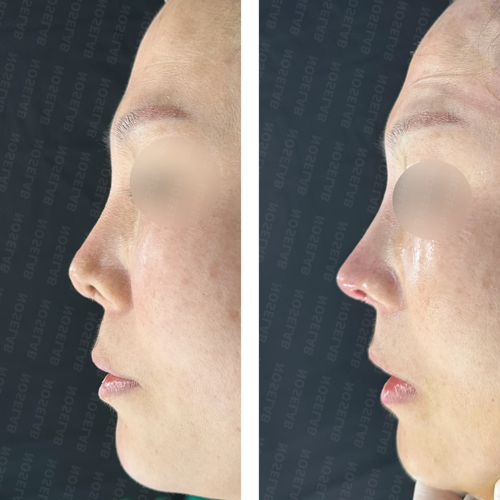
2. Profile view improvement
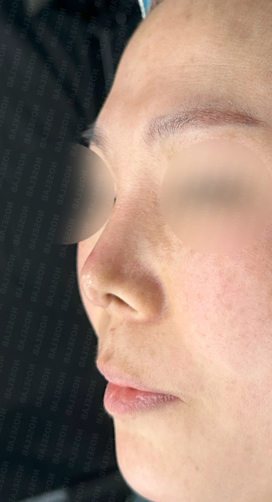
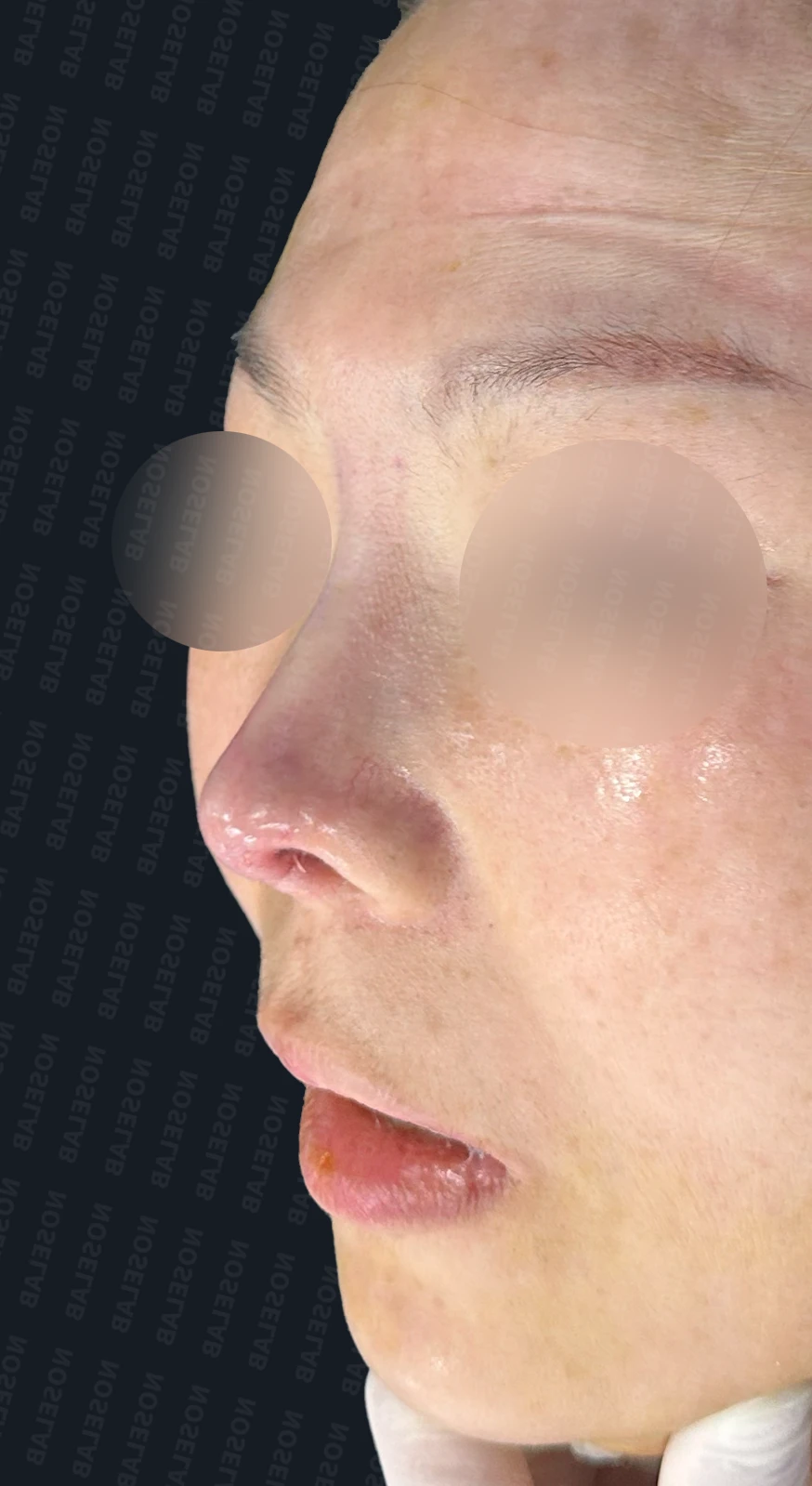
Before surgery / Day 0 immediately after surgery Photos taken immediately after surgery show swelling and may differ from the final result.
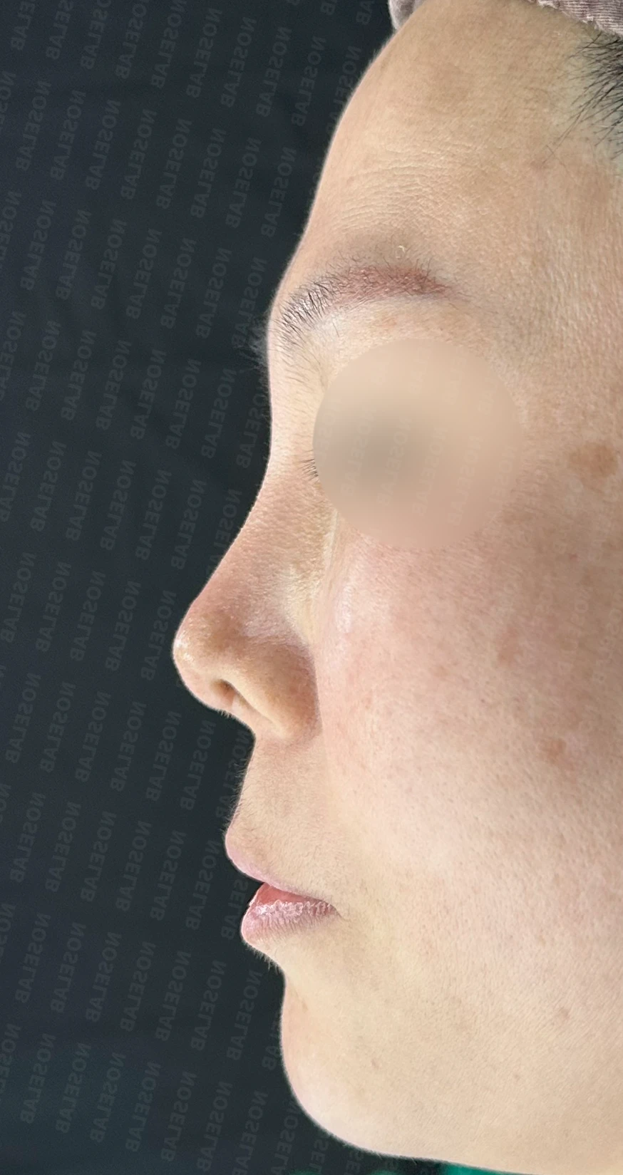
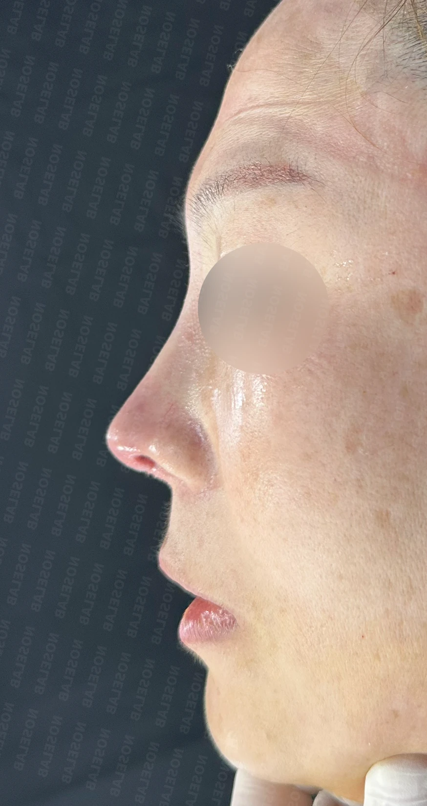
Before surgery / Day 0 immediately after surgery Photos taken immediately after surgery show swelling and may differ from the final result.
Natural radix starting point
Stable tip shape
Appropriate nasolabial angle
3. Nostril improvement
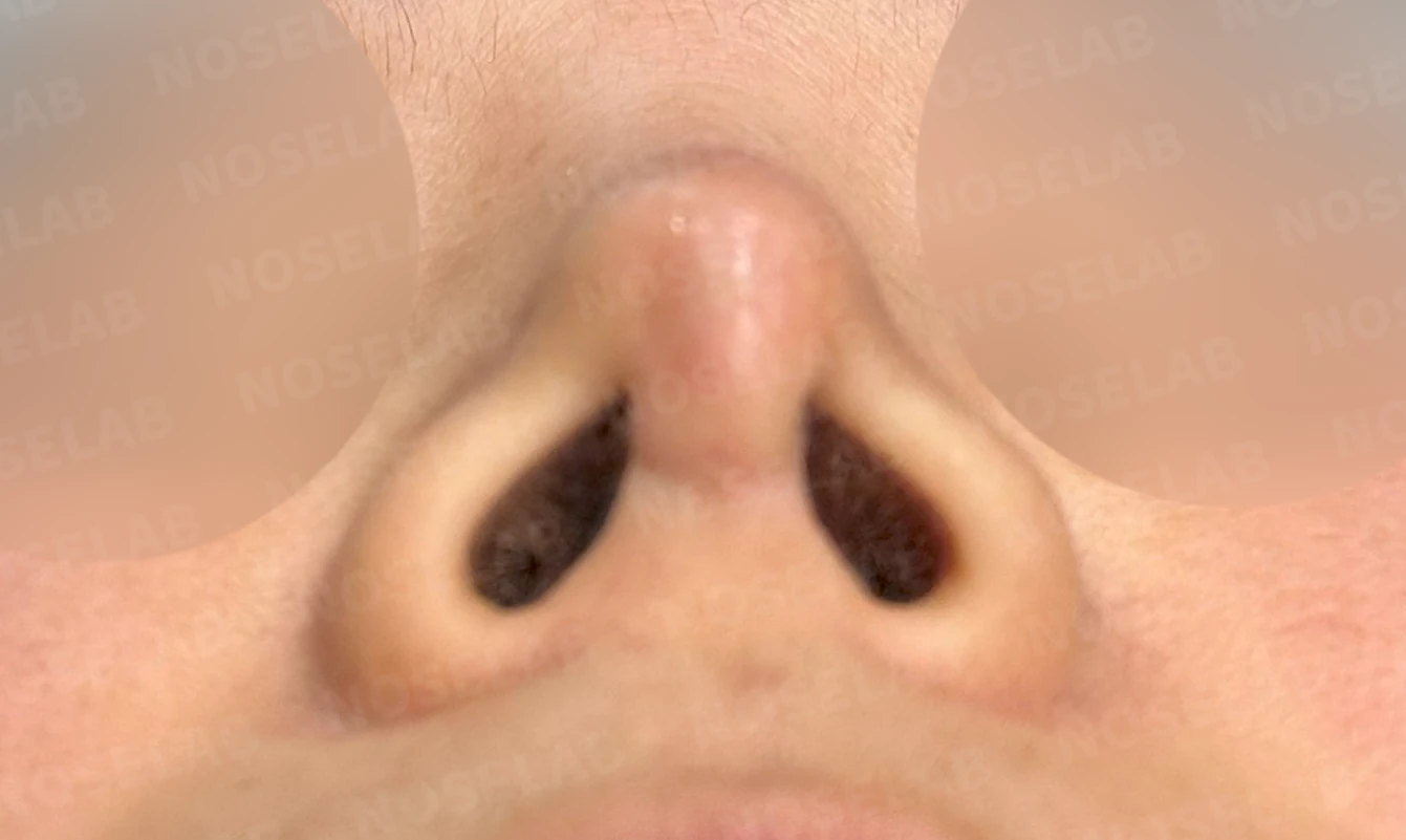
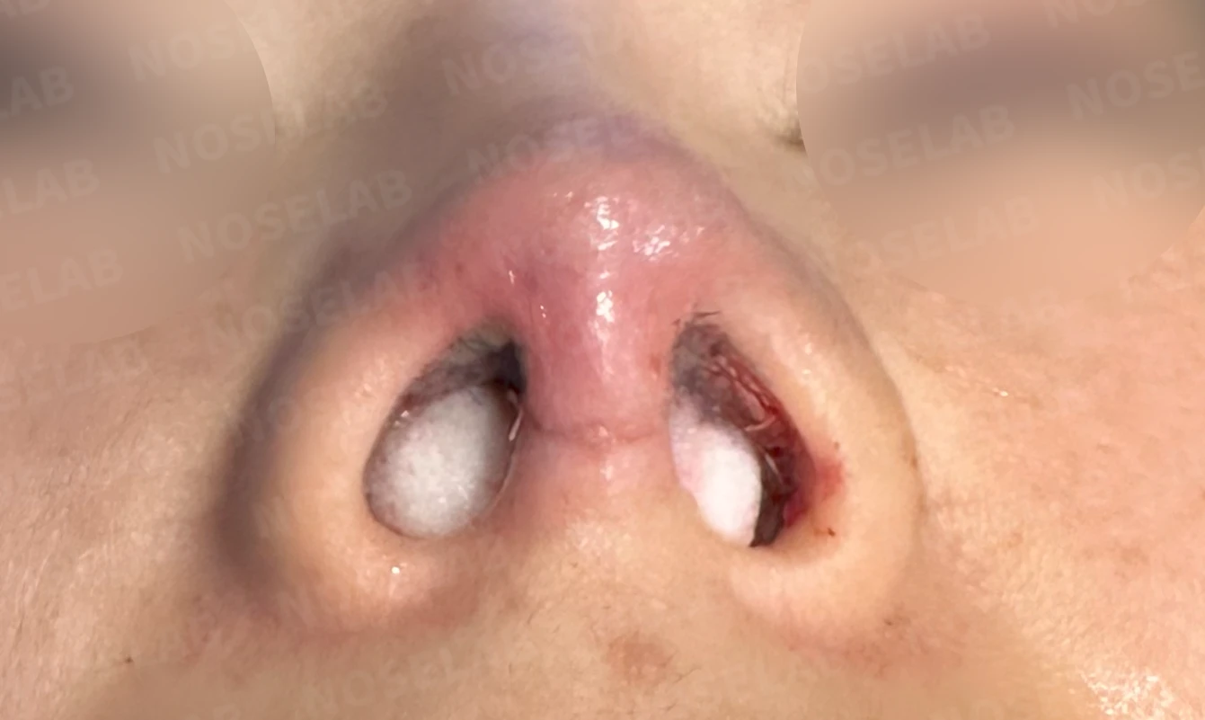
Before surgery / Day 0 immediately after surgery Photos taken immediately after surgery show swelling and may differ from the final result.
Improved symmetry
Centrally positioned columella
Natural nostril shape
💡 Special notes
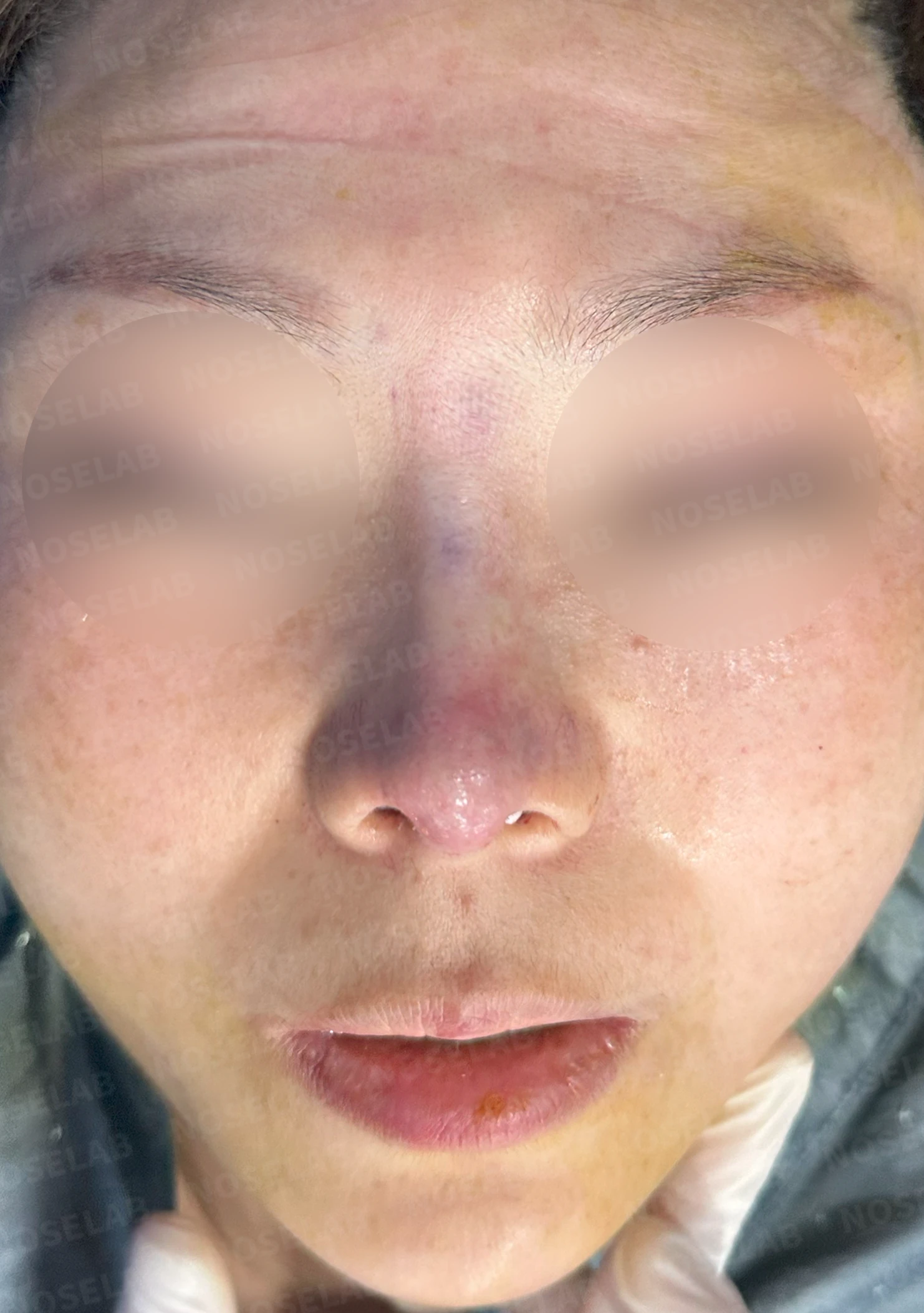
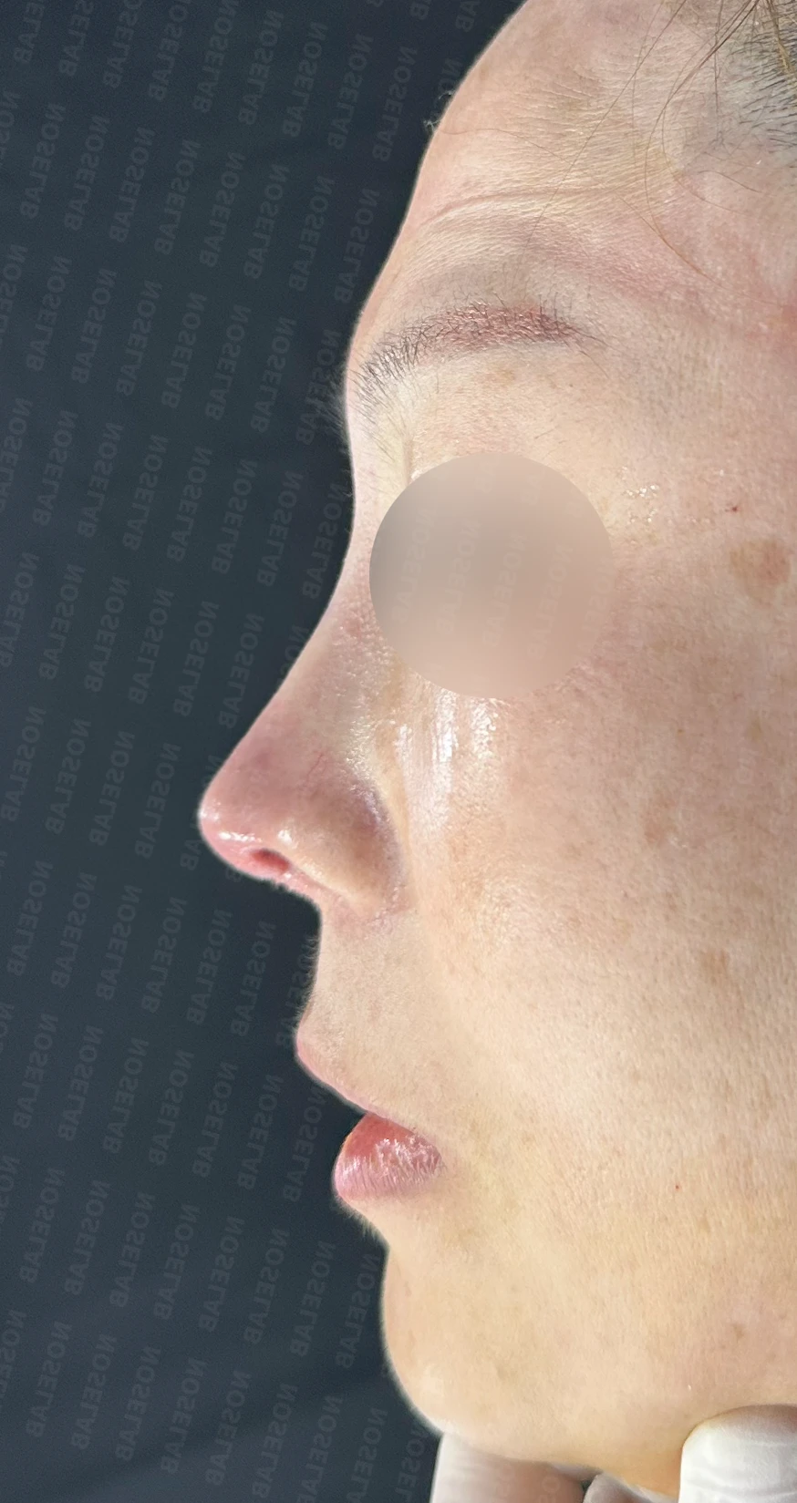
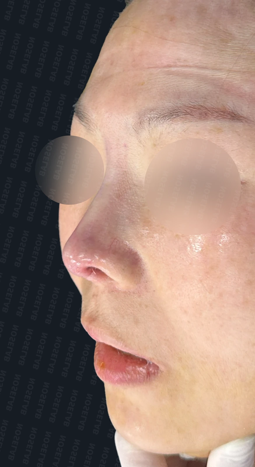
Immediately after surgery (Day 0) front, profile, and oblique viewsImmediate postoperative photos show swelling and may differ from the final result.
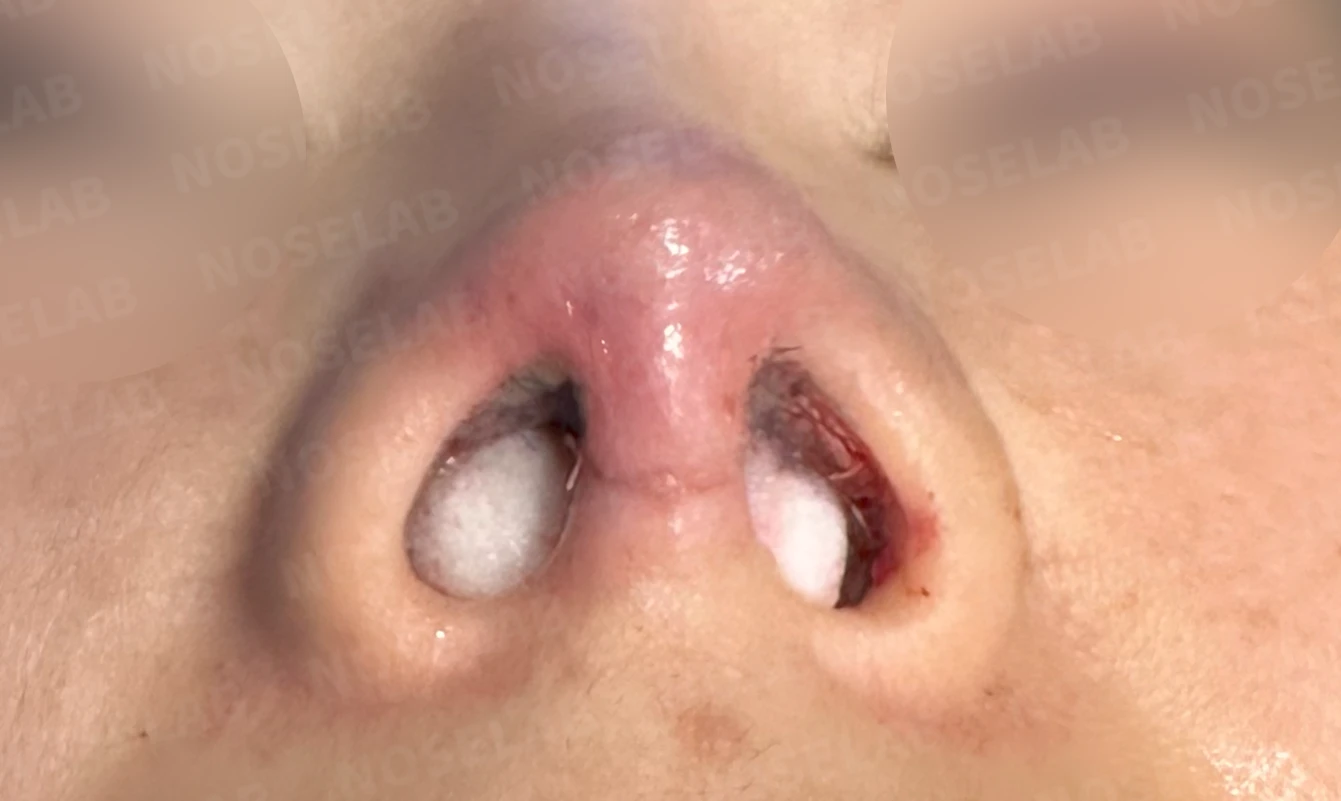
This was a case of a contracted nose resulting from inflammation caused by previous failure to completely remove mesh material.
Notably, the implant placed along the bridge extended down to the tip, typical skin depression was observed where it pressed against the nasal tip, and normal cartilage tissue was severely damaged by chronic inflammation.
All inflamed tissue was removed, and every area of damaged normal tissue was reconstructed, which is expected to naturally improve the nasal obstruction that had developed as a result. Furthermore, the contracture deformity—where the tip had twisted and turned upward—was successfully improved through stem cell therapy and reconstruction using autologous rib cartilage.
By combining active intravenous antibiotic therapy with stem cell treatments during the recovery period, I expect the patient to recover smoothly.
I hope this case helps those who are experiencing similar concerns.
This was Dr. Peter C. Kang. Thank you.
Thank you.
#closedrevisionrhinoplasty #closedcontractedrhinoplasty #closedautologousribrhinoplasty #revisionrhinoplasty #contracturecorrection #postinflammatoryreconstruction #autologousribcartilage #nasalobstructionimprovement #customrhinoplasty

Limitations and risks
When autologous rib cartilage is used, scarring and pain may remain at the donor site on the chest, and pneumothorax (pleural injury) can occur rarely. Over time, the grafted cartilage may warp or undergo partial resorption.
When foreign materials such as injected fillers are removed and reconstructed, contracture may recur, nasal height and length may decrease, and staged surgery may be necessary depending on the skin condition.
When the septum is corrected at the same time, nasal obstruction may persist or recur, and in rare cases, septal perforation or changes in smell may occur.
Common potential risks include infection, bleeding, asymmetry, scarring, skin necrosis, and insufficient improvement; results vary depending on individual skin condition and previous surgical history.