Hello, I am Dr. Peter C. Kang.
Today, I would like to introduce the case of a patient who visited our clinic with the primary concerns of nasal obstruction and nostril asymmetry.
While nasal obstruction can have many causes, in this case, mucosal synechiae formed inside the nasal cavity from previous surgeries, severely blocking both sides of the nasal airway.
What were the surgical history and patient requests?
Surgical and procedure history:
2021: Ear cartilage + filler
2022: Septal cartilage + autologous rib cartilage + Gore-Tex
October 2022 philtrum reduction
2023: Paranasal augmentation (noble surgery)
Requests:
Bilateral nasal obstruction
Improved left-right asymmetry
Reduce nostril show
Natural yet refined and defined
What did the preoperative design consultation and facial analysis show?
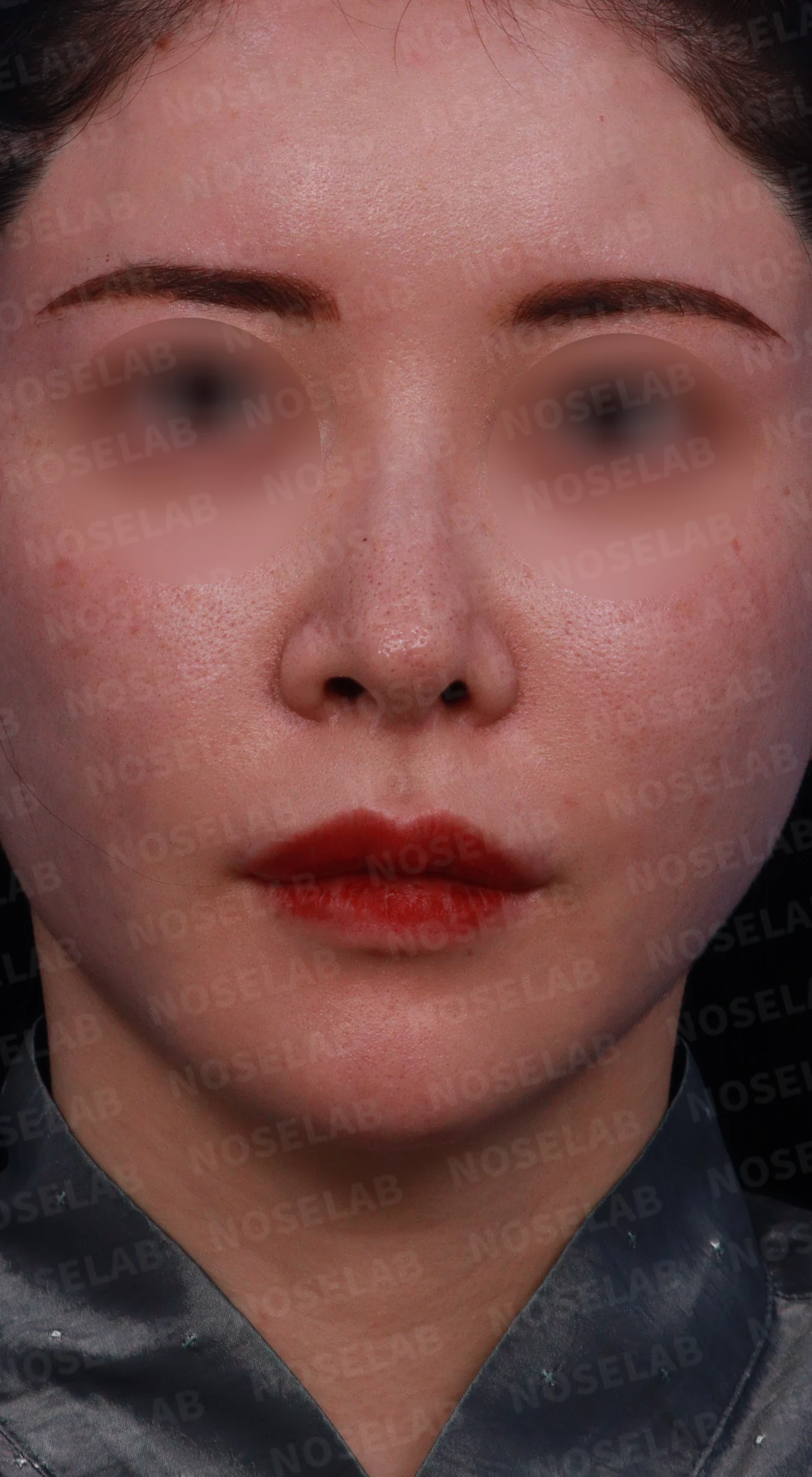
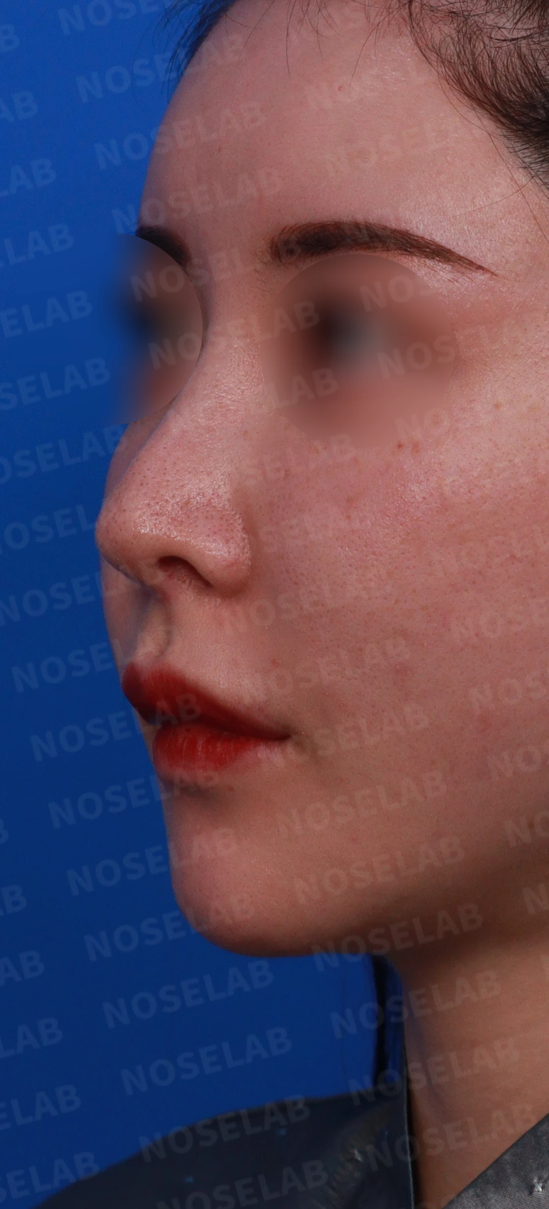
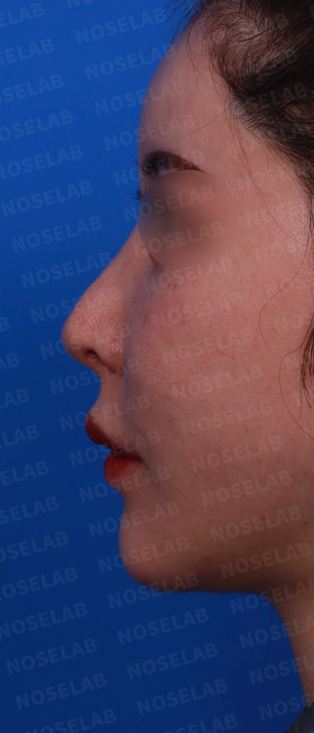
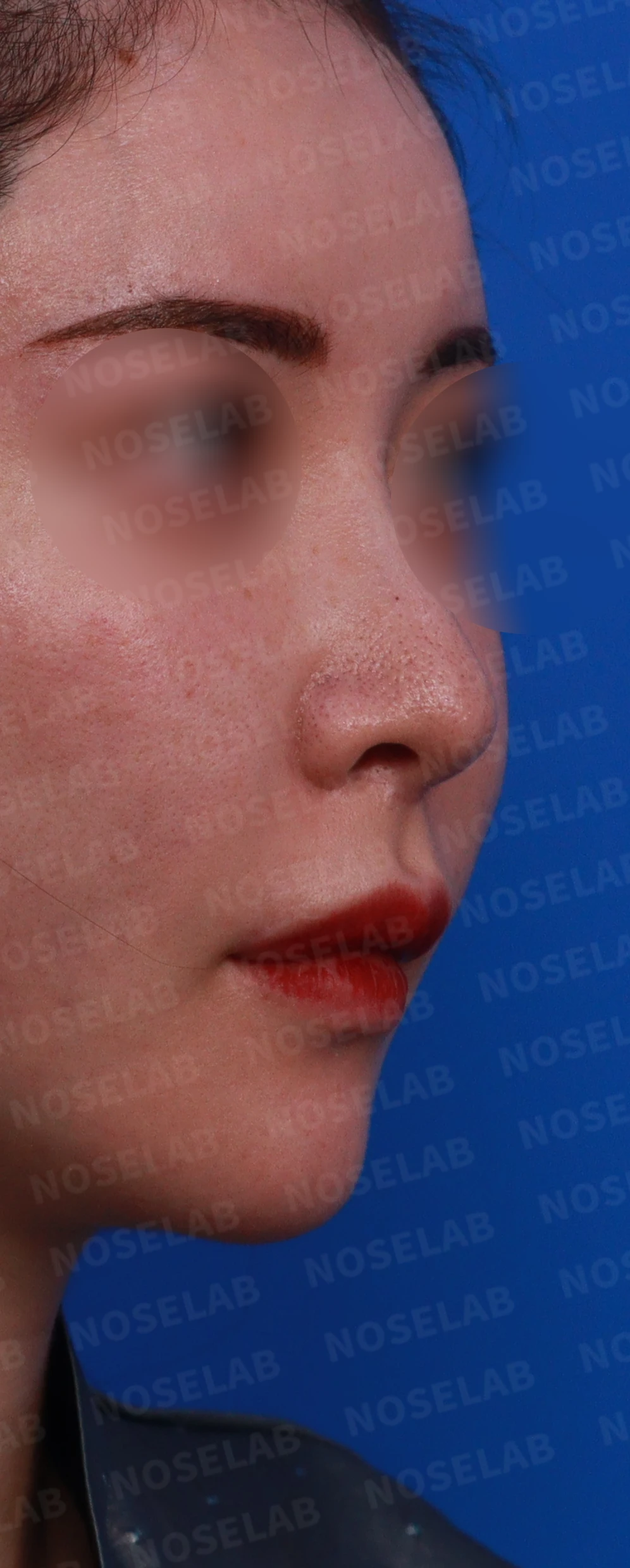
Preoperative design: front view, left oblique view, profile view
On the front view, the nasal tip appeared deviated to the left, with excessive nostril show and evident nostril asymmetry. On the profile view, the tip was drooping with a dorsal hump, the radix started high, and the tip appeared upturned. The nasolabial angle appeared greater than 95 degrees.
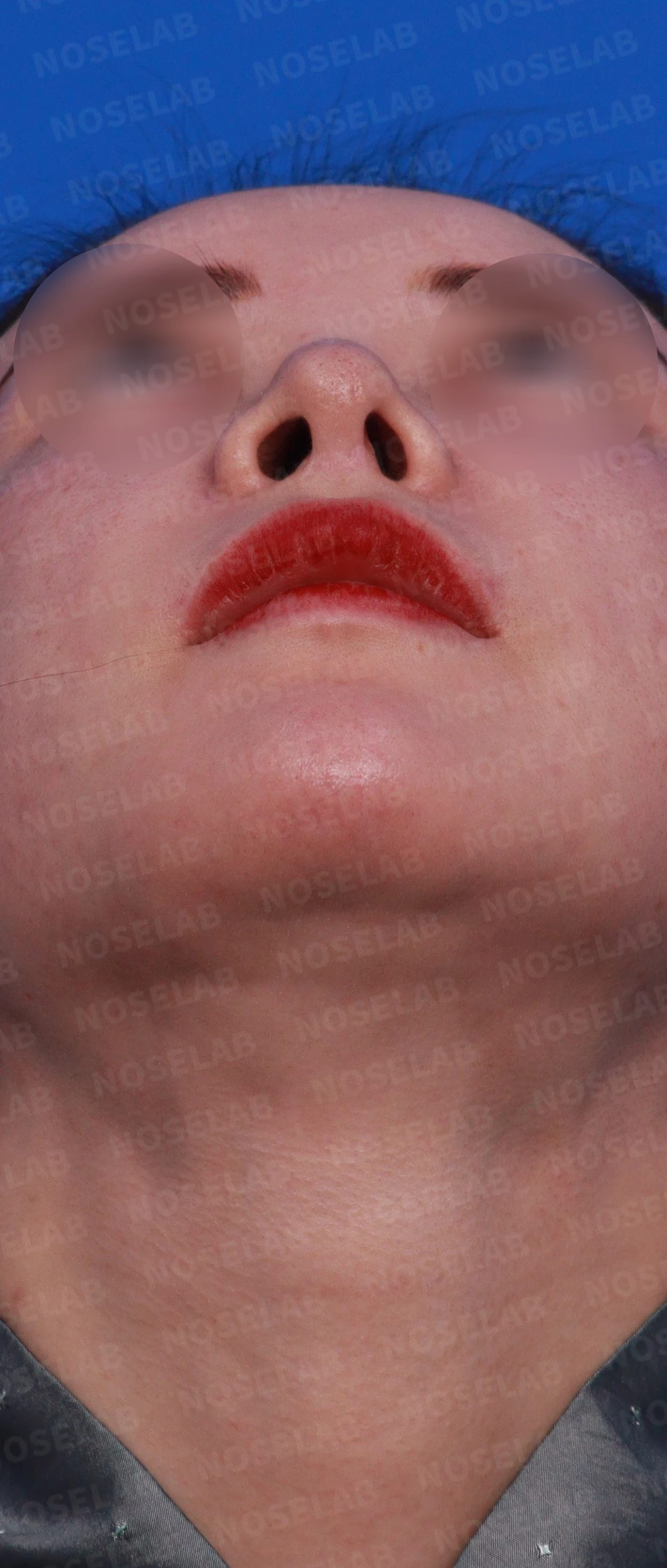
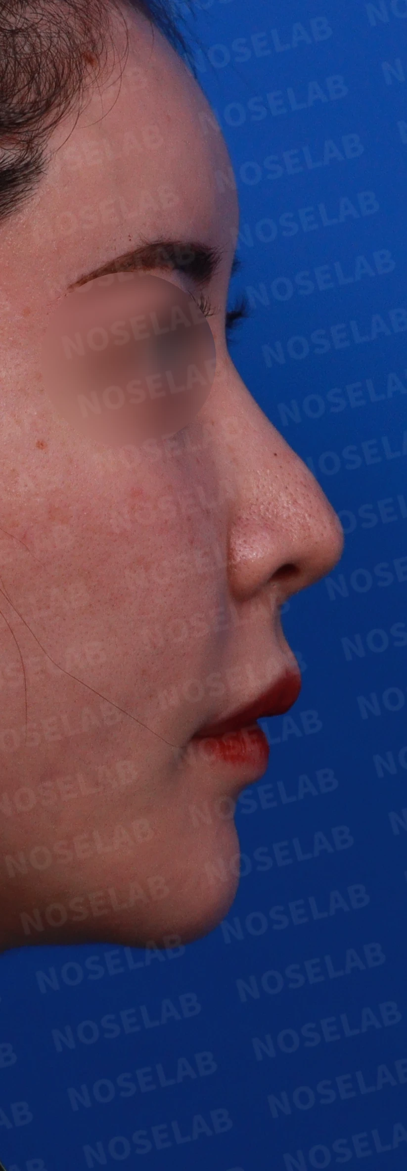
Preoperative design: basal view, right oblique view, profile view
Looking at the basal view, the columella was tilted slightly to the right, and the left nostril appeared smaller and more elongated than the right. Nostril asymmetry was clearly visible.
Were diagnostic evaluations for nasal obstruction performed?
A nasal CT scan taken at the initial visit revealed a deviated septum and intranasal mucosal synechiae, which were confirmed by nasal endoscopy.
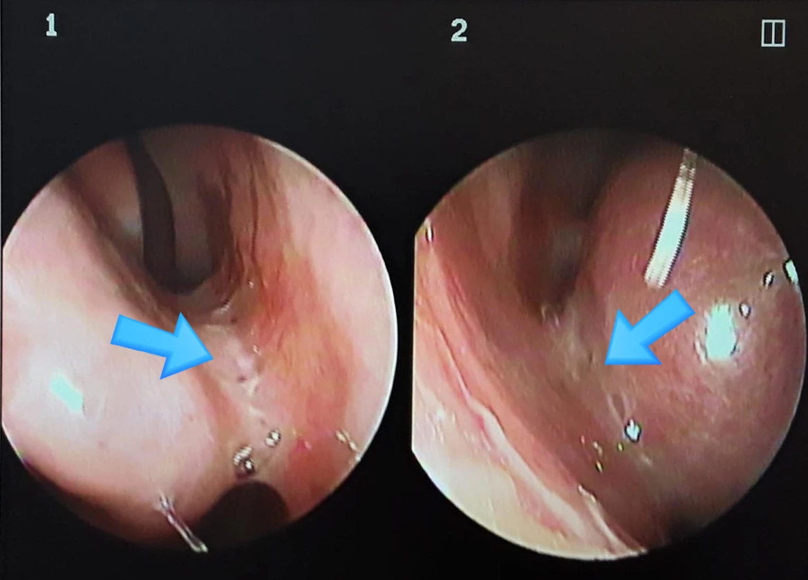
Mucosal synechia between the nasal septum and the inferior turbinate (arrow)
Surgical plan
Septal extension graft (autologous rib cartilage)
Septal reconstruction (autologous rib cartilage)—to reposition the septal cartilage to the right and correct the deviated septum
Alar rim extension (alar cartilage repositioning and cartilage grafting)—to correct nostril asymmetry and reduce nostril show
Removal of Gore-Tex and replacement with a silicone implant to lower the radix
Alar cartilage repositioning and suturing—to refine the tip shape
Nasolabial angle correction (autologous rib cartilage)
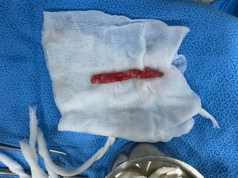
Removed Gore-Tex implant
Our core principle is to reconstruct or repair cartilage damaged in previous surgeries
using autologous rib cartilage as much as possible, restoring normal anatomical structure before creating a new contour.
When harvesting autologous rib cartilage, we identify the rib with the least calcification on chest X-rays to ensure a secure harvest. In this patient, extensive calcification was present in both costal cartilages, so we harvested a partial segment of the right 8th costal cartilage.
Additionally, when addressing nasal obstruction caused by intranasal mucosal synechiae, although the improvement can be noticeable, the risk of recurrence is high. Therefore, meticulous care is essential during surgery to prevent recurrent adhesions.
How did the nose change between before surgery and immediately after surgery?
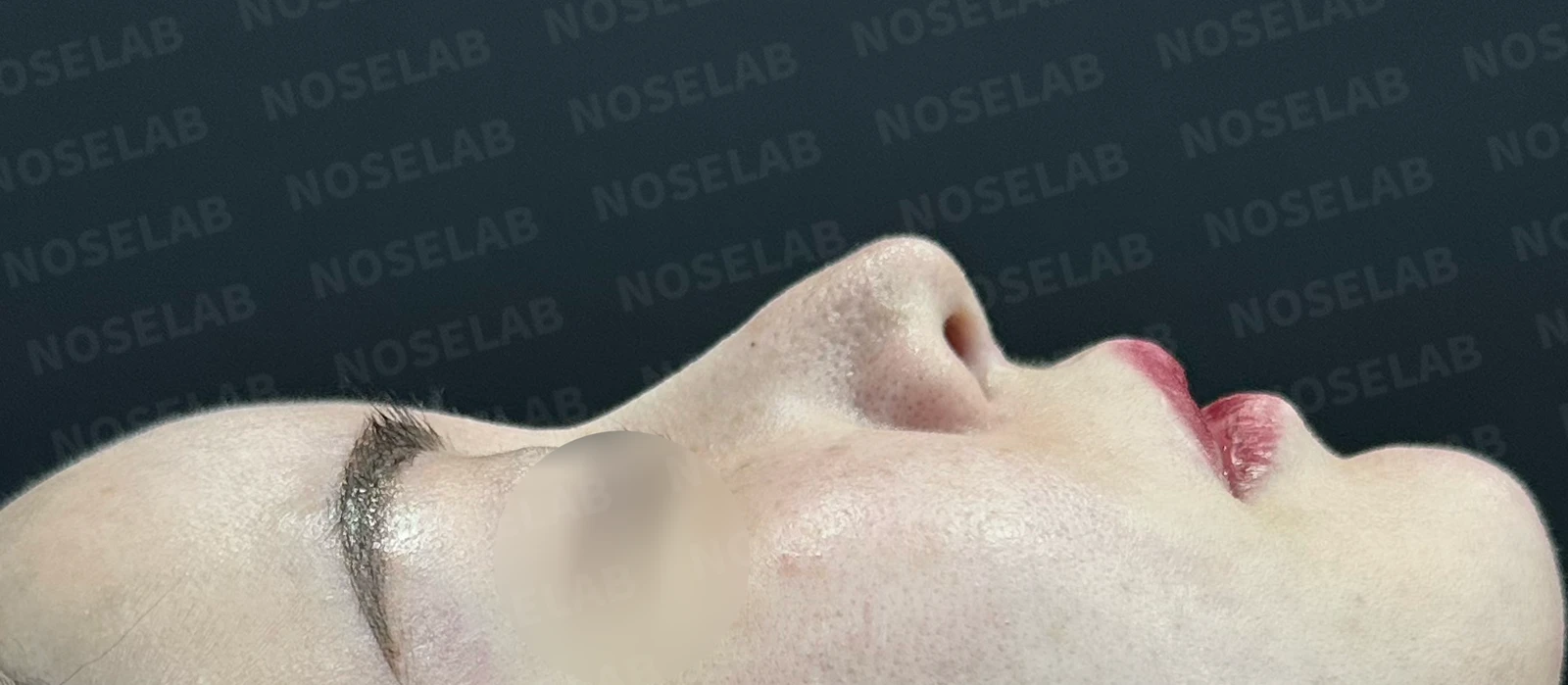
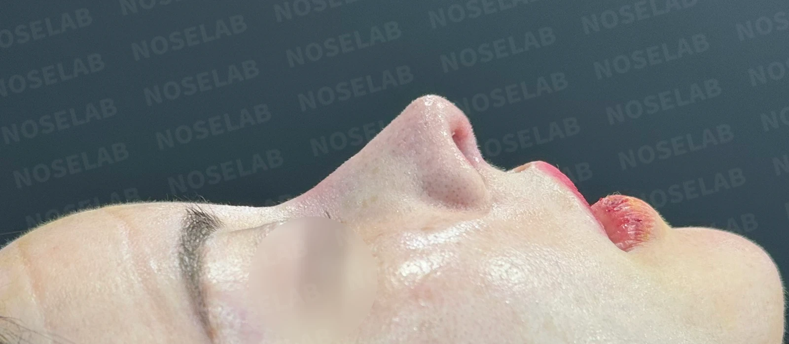
Before surgery / Day 0 immediately after surgery Photos taken immediately after surgery show swelling and may differ from the final result.
Comparing the profile views, the drooping appearance of the tip has been fully resolved, the dorsal hump has been smoothed out, and the nasolabial angle has been corrected naturally.
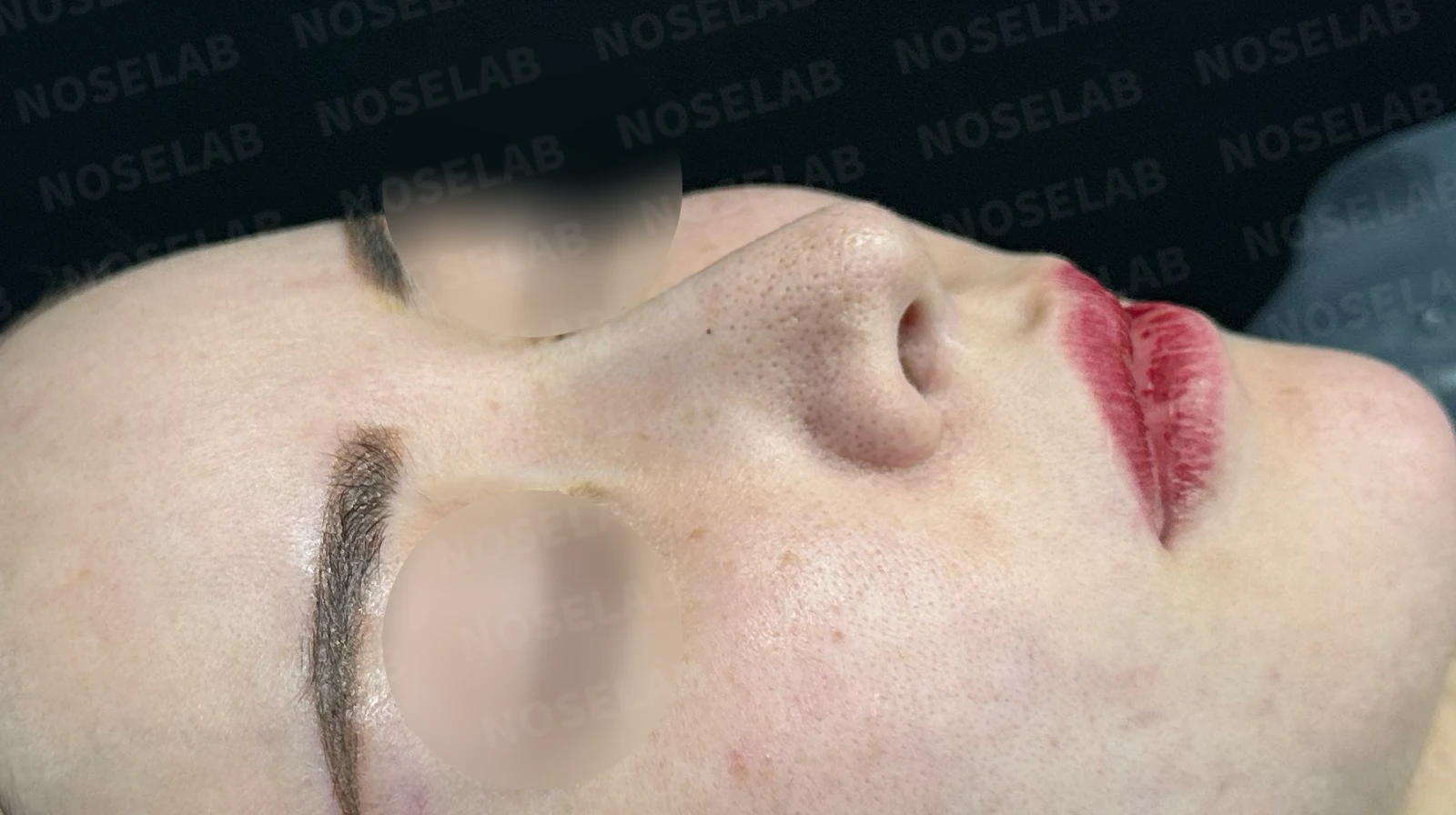
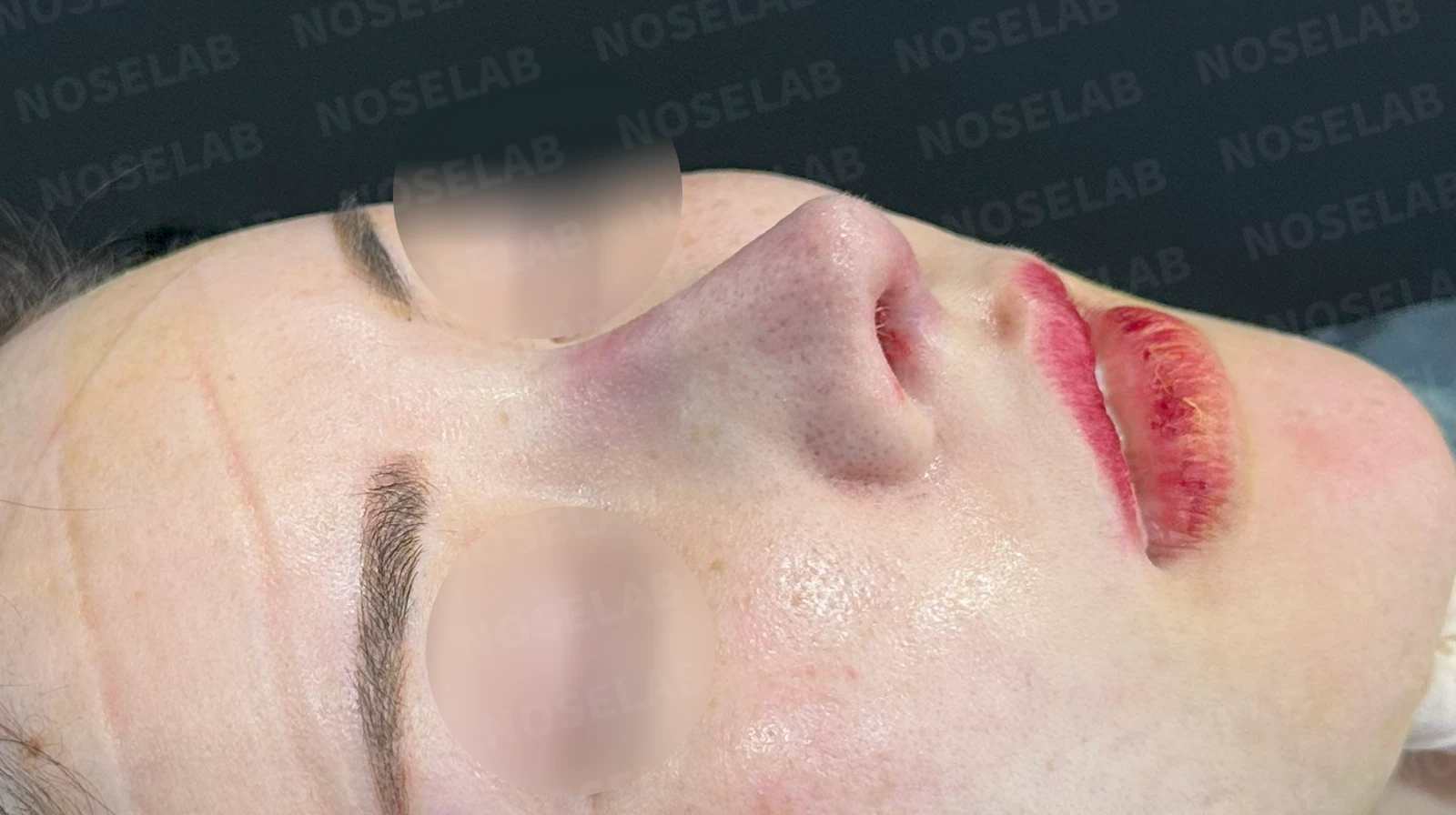
Before surgery / Day 0 immediately after surgery Photos taken immediately after surgery show swelling and may differ from the final result.
On the oblique view, it is notable that the radix now begins naturally at the pupil line.
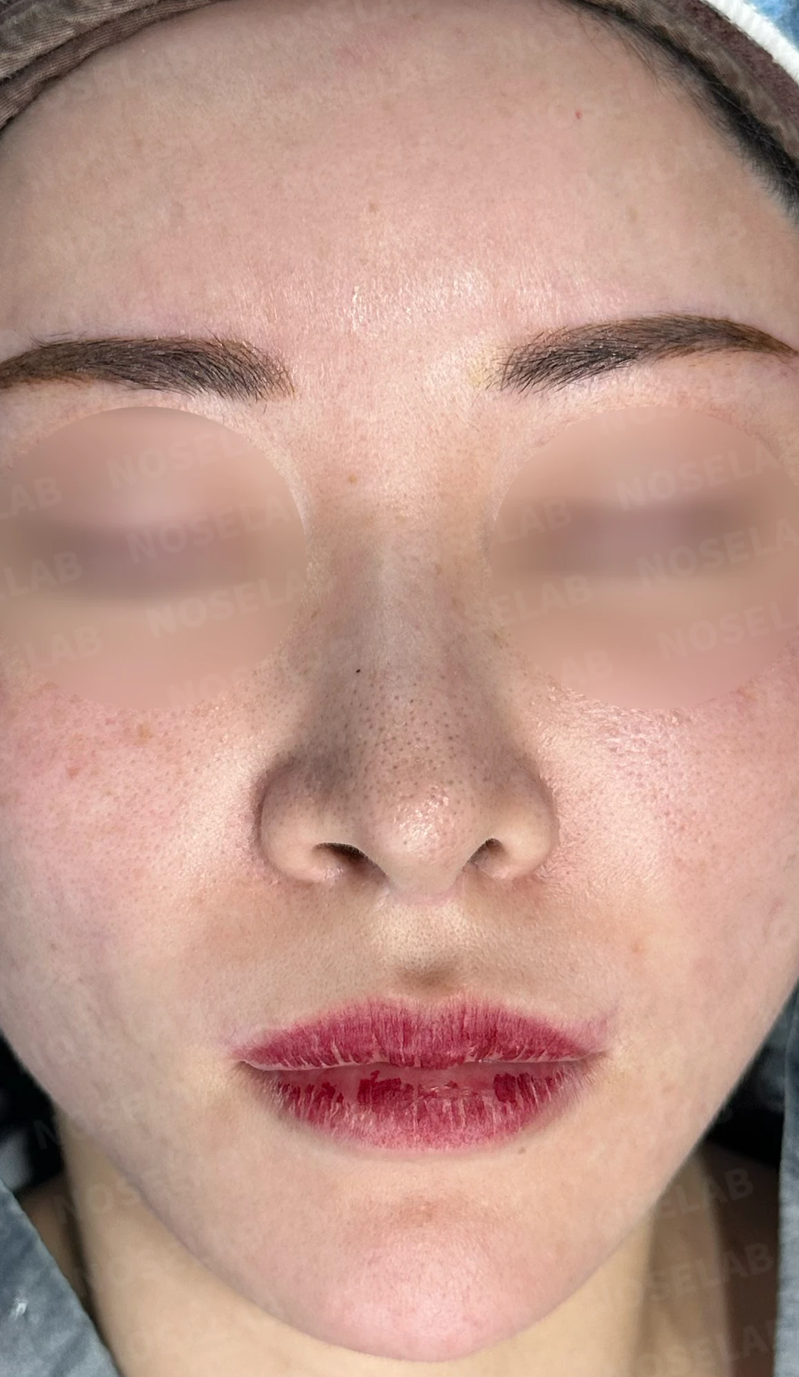
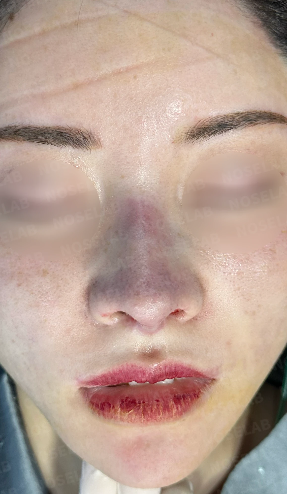
Before surgery / Day 0 immediately after surgery Photos taken immediately after surgery show swelling and may differ from the final result.
The frontal view shows the most marked change. Nostril show has been reduced by more than half, and the asymmetry has noticeably improved. Furthermore, the overall deviation of the nose has been substantially corrected.
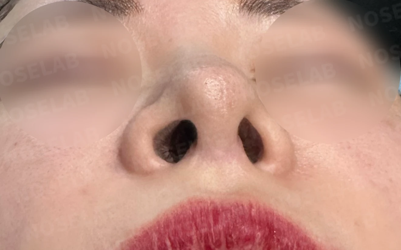
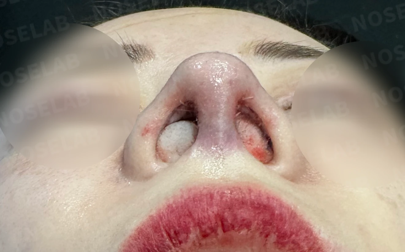
Before surgery / Day 0 immediately after surgery Photos taken immediately after surgery show swelling and may differ from the final result.
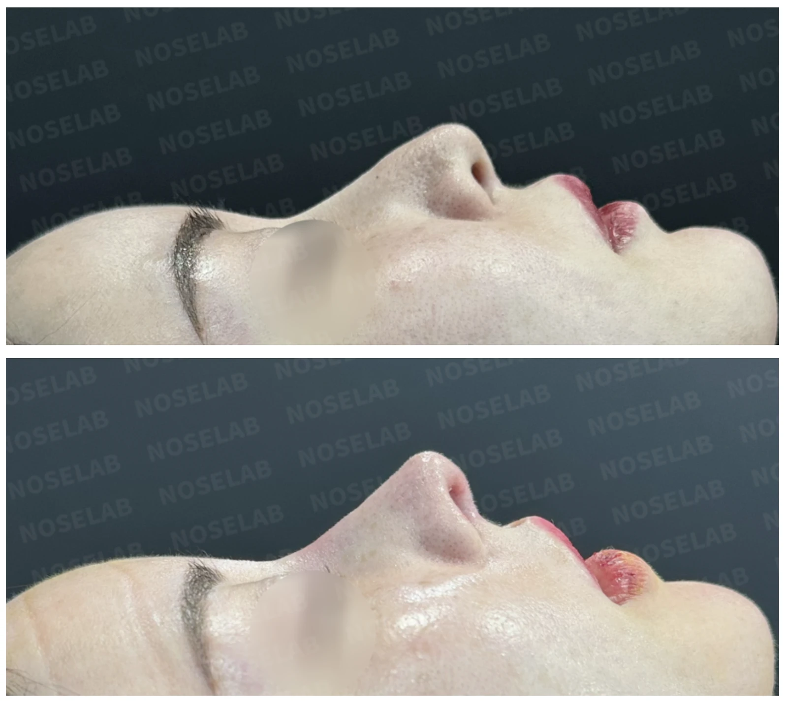
Looking at the basal view, by shifting the septal cartilage to the right during septal reconstruction, the columella moved toward the right, resulting in noticeable improvement in nostril asymmetry.
Here are the photos taken immediately after surgery on Day 0. Please note that immediate postoperative photos reflect swelling and may differ from the final result.
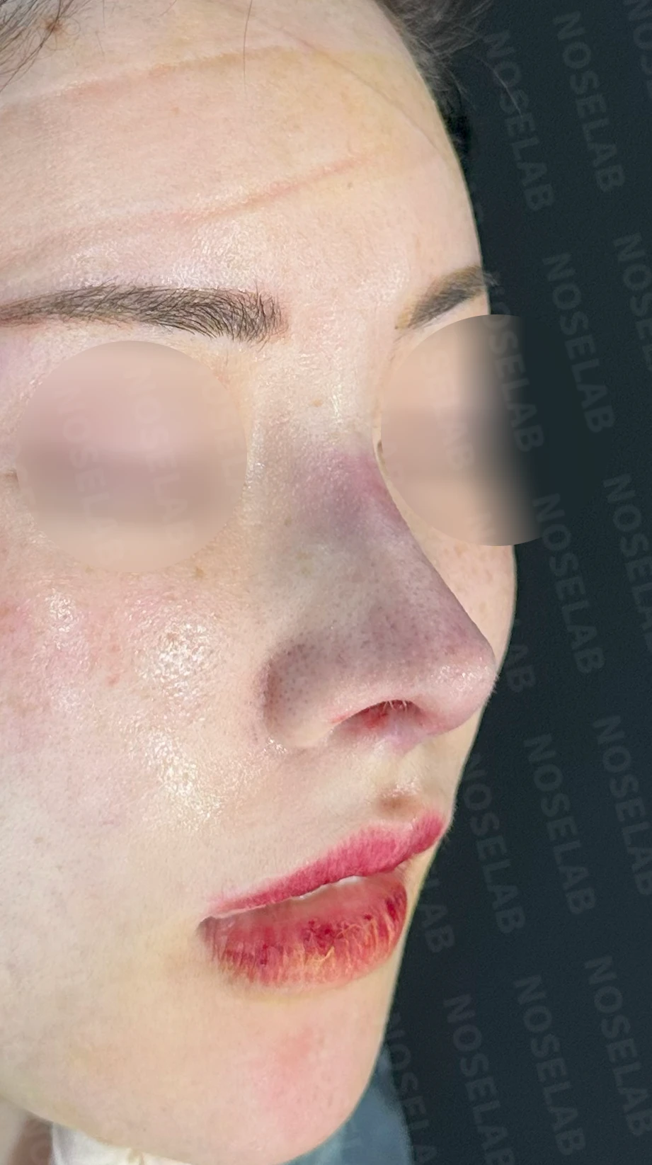
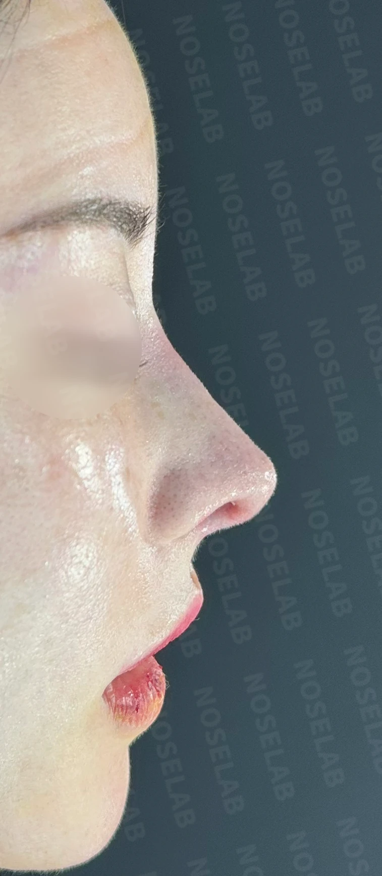
Front, oblique, and profile views immediately after surgery on day 0. Photos taken immediately after surgery show swelling and may differ from the final result.
Compared to before surgery, the nasal proportions have improved, and the added definition creates a more balanced, refined contour. The nostril asymmetry has also noticeably improved, providing better overall harmony.
Basal (nostril) view immediately after surgery (Day 0)Immediate postoperative photos show swelling and may differ from the final result.
Rhinoplasty is not merely about making the nose aesthetically pleasing; its fundamental purpose is to harmonize the overall facial image. I hope sharing this case helps deepen your understanding of these considerations.
Revision rhinoplasty for nostril asymmetry and upturned nose correction
At a trusted clinic
Choosing the right clinic for revision rhinoplasty to correct nostril asymmetry and an upturned nose is essential. Careful thought should be given to selecting a medical facility, ensuring the operating surgeon has substantial experience and the clinic is fully prepared to manage any situation that may arise during surgery or recovery.
In particular, surgery for nasal obstruction and septal reconstruction is an area of expertise for a board-certified otolaryngologist (ENT specialist), making the choice of clinic all the more critical.
I hope this case study has been helpful to those facing similar concerns. NOSELAB will continue to do our best to support healthy, balanced noses for all our patients.
This was Dr. Peter C. Kang. Thank you.
Thank you.
Blog posts related to nostril asymmetry
#closedrhinoplasty #closedrevisionrhinoplasty #autologousribrhinoplasty #autologousribcartilage #upturnednose #nostrilasymmetry #nasalobstruction #nasalobstructionsurgery #septalreconstruction #ENTspecialist

Limitations and risks
When autologous rib cartilage is used, scarring and pain may remain at the donor site on the chest, and pneumothorax (pleural injury) can occur rarely. Over time, the grafted cartilage may warp or undergo partial resorption.
When revising after implant removal, contracture may progress again after removal, the nasal height and length may decrease, and staged surgery may be necessary depending on the skin condition.
When the septum is corrected at the same time, nasal obstruction may persist or recur, and in rare cases, septal perforation or changes in smell may occur.
Common potential risks include infection, bleeding, asymmetry, scarring, skin necrosis, and insufficient improvement; results vary depending on individual skin condition and previous surgical history.