Hello.
I am Dr. Peter C. Kang, medical director.
Today, I would like to talk about septal reconstruction, a procedure frequently performed for contracted noses.
Severe retraction of the columella is very common in noses deformed by contracture. A common underlying issue observed in most of these cases is severe damage to the septal cartilage.
When the septal cartilage is damaged, columellar retraction occurs as the columella curls inward, often accompanied by nasal obstruction.
Columellar retraction, contracted nose, and septal reconstruction surgery?
Today's patient also developed a contracted nose deformity due to severe inflammation. At the time of the initial visit, all implants had been removed following the onset of inflammation. To allow for an earlier revision surgery, active stem cell therapy was administered, and nasal reconstruction using autologous rib cartilage was performed a little over 2 months later.
Preoperative design consultation photo analysis
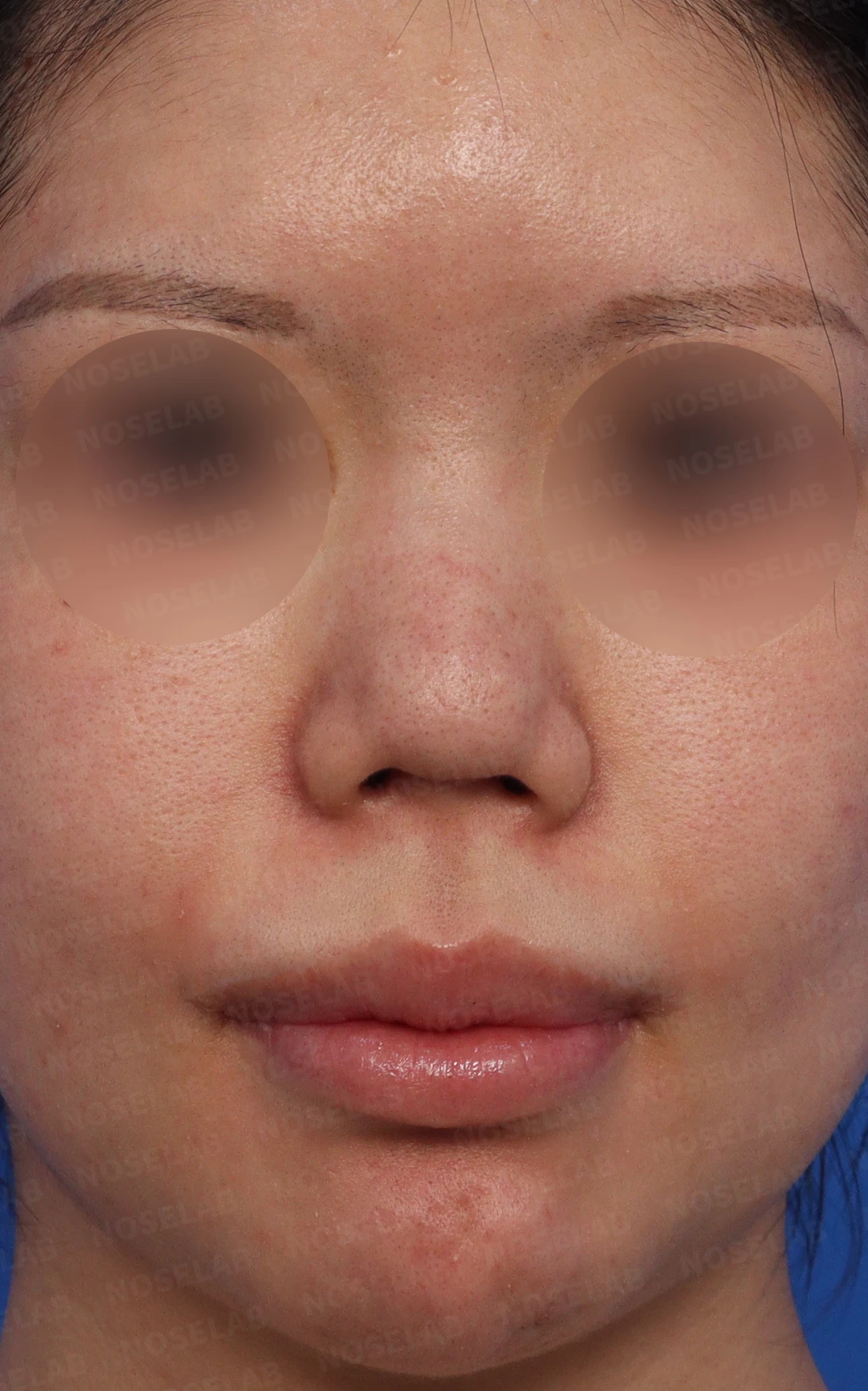
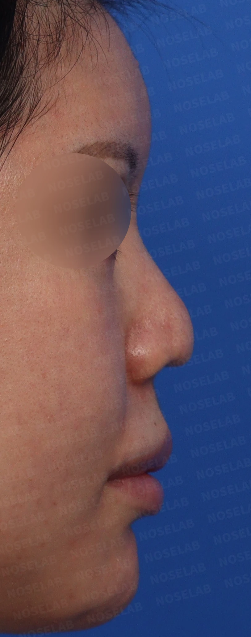
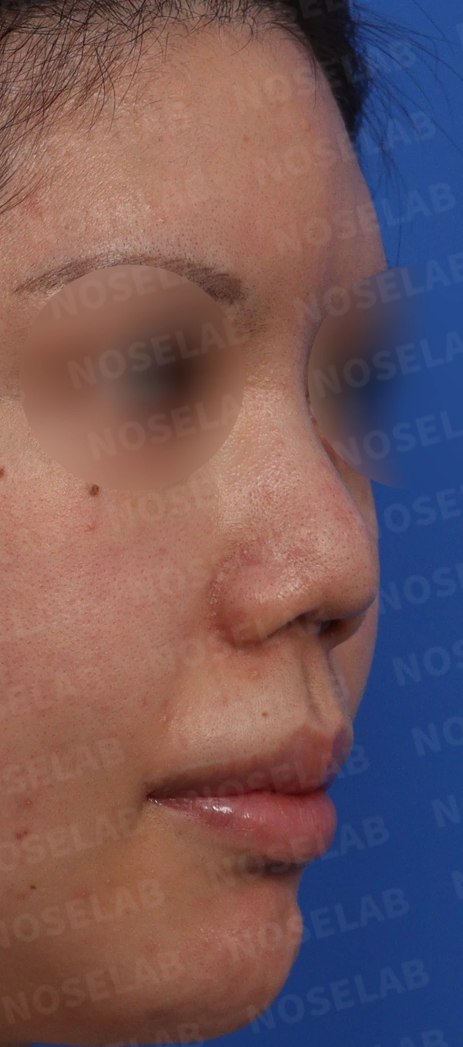
Preoperative design consultation photos: front, right profile, oblique view
Looking at the preoperative photos, the profile view showed a drooping tip, the columella was completely invisible, and findings of a protruding mouth were present. The oblique view revealed columellar retraction and an overall flat appearance. On the front view, the nasal bridge was wide, the nose appeared crooked, and the retracted columella gave the alae a drooping appearance. In the nostril view, an indented columellar scar from a previous open surgery and inward curling of the columella were observed.
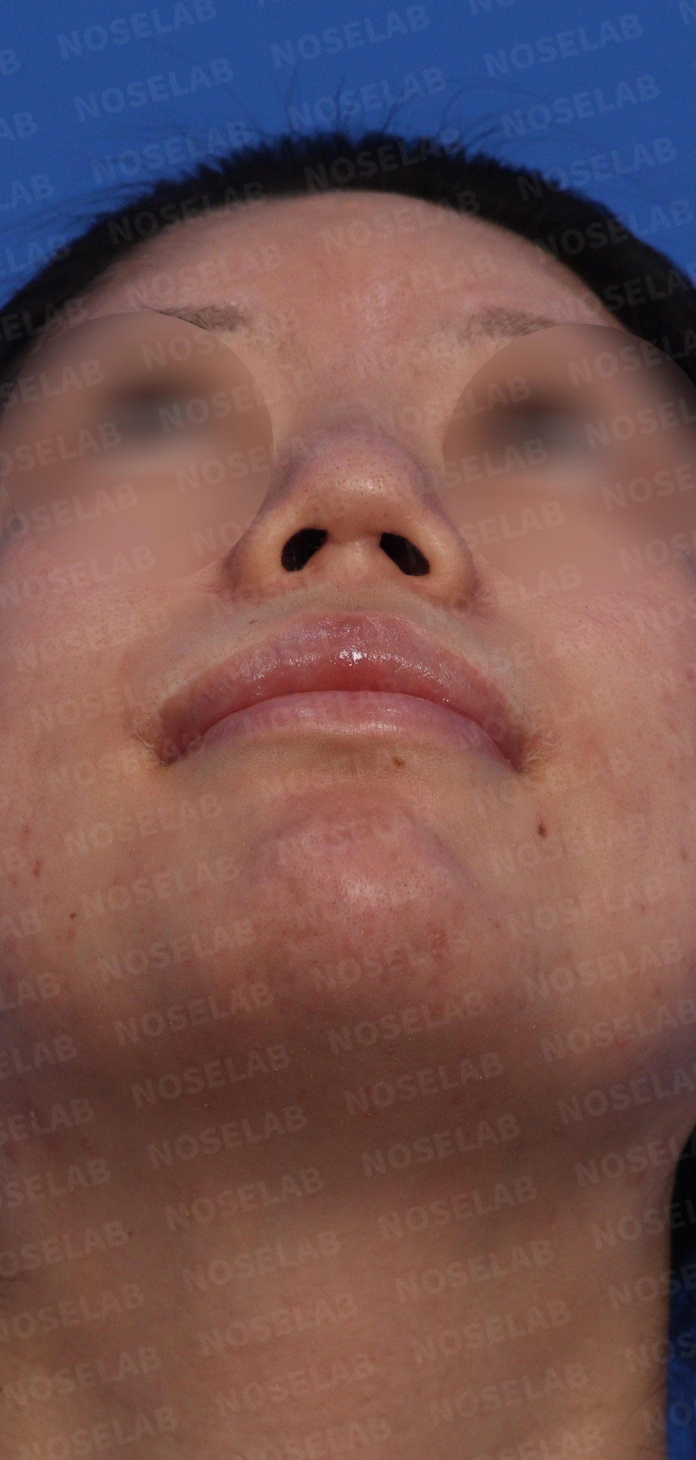
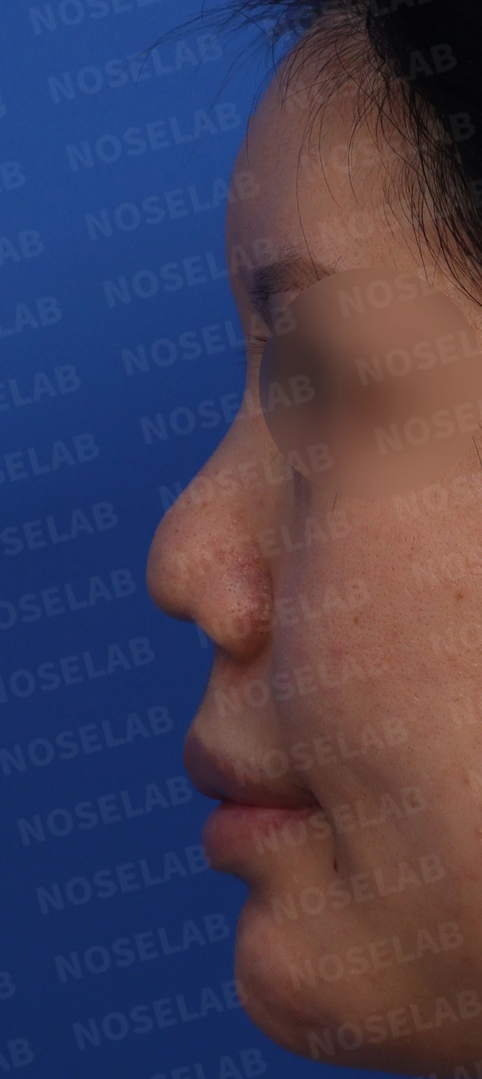
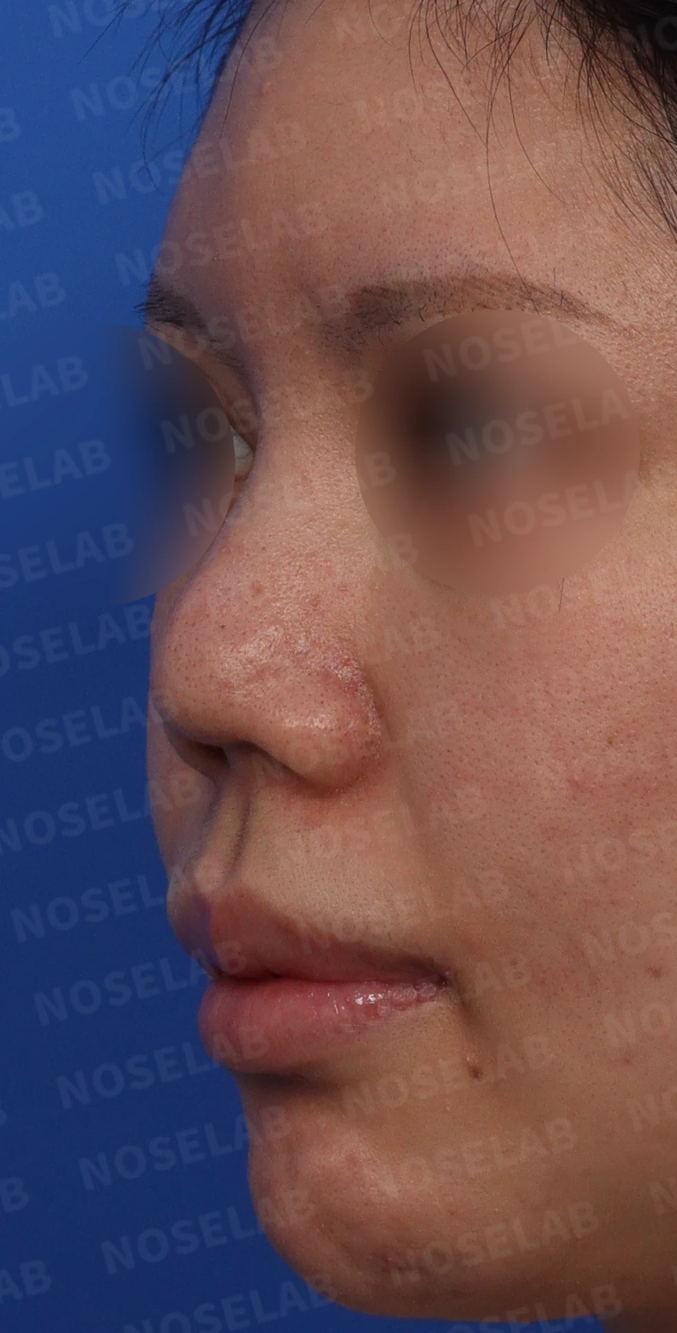
Preoperative design consultation photos: nostrils, left profile, oblique view
Surgical plan
Before proceeding with surgery, any residual inflammation must be resolved completely. Stem cell injection therapy was administered 4 times at 2-week intervals. Because the response to treatment was favorable, surgery was planned after 2 months had passed.
In the case of columellar retraction, damage to the septal cartilage should be suspected, and in such instances, septal reconstruction using autologous rib cartilage is planned. Lateral osteotomies were also planned to correct the wide, crooked nasal bridge, along with placing a silicone implant along the nasal dorsum provided no signs of inflammation were observed inside the nose during surgery.
When the patient visited the clinic, right-sided nasal obstruction was reported, so diagnostic examinations related to nasal obstruction were performed. Based on the following findings, surgery to correct the deviated septum was planned.
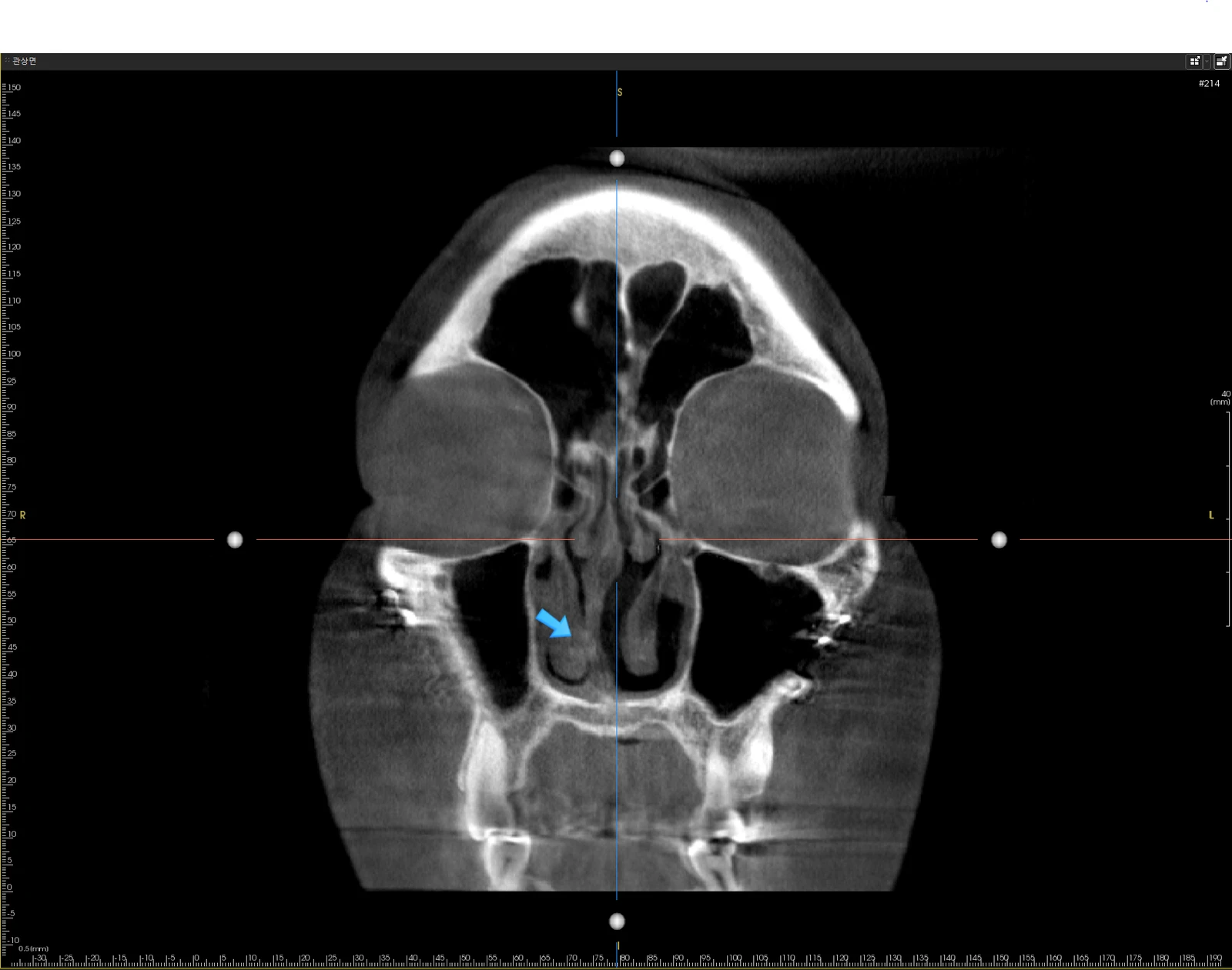
Coronal CT view
On the coronal CT view, deviation of the septal cartilage to the right (arrow) can be seen. It was determined that this narrowing of the right nasal cavity could be the cause of nasal obstruction, so septal deviation correction was planned simultaneously with septal reconstruction.
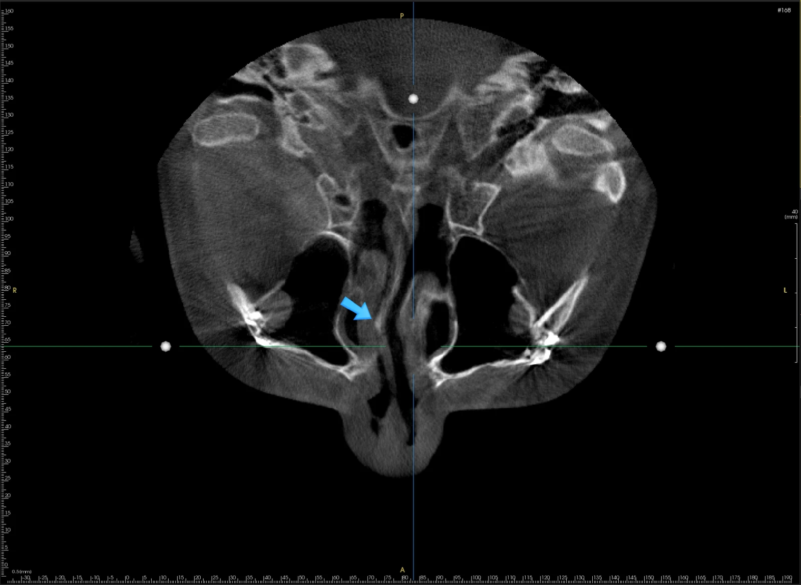
Axial CT view
The axial CT view also confirms that the bony portion of the septum is significantly deviated to the right (arrow) and that the airway is notably narrowed.
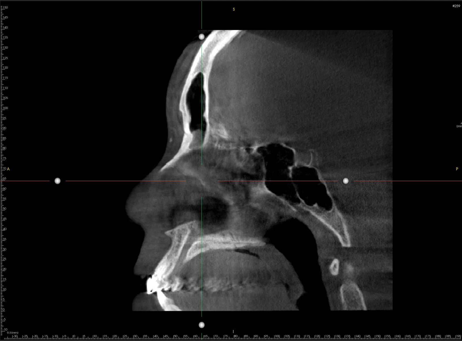
Sagittal CT view
The sagittal CT view revealed a structure where, due to previous two-jaw (orthognathic) surgery, the maxilla (near the incisors) projected higher into the nasal cavity than the palatal bone, raising suspicion that the septal cartilage might be weakened or damaged.
Surgical details and operative findings
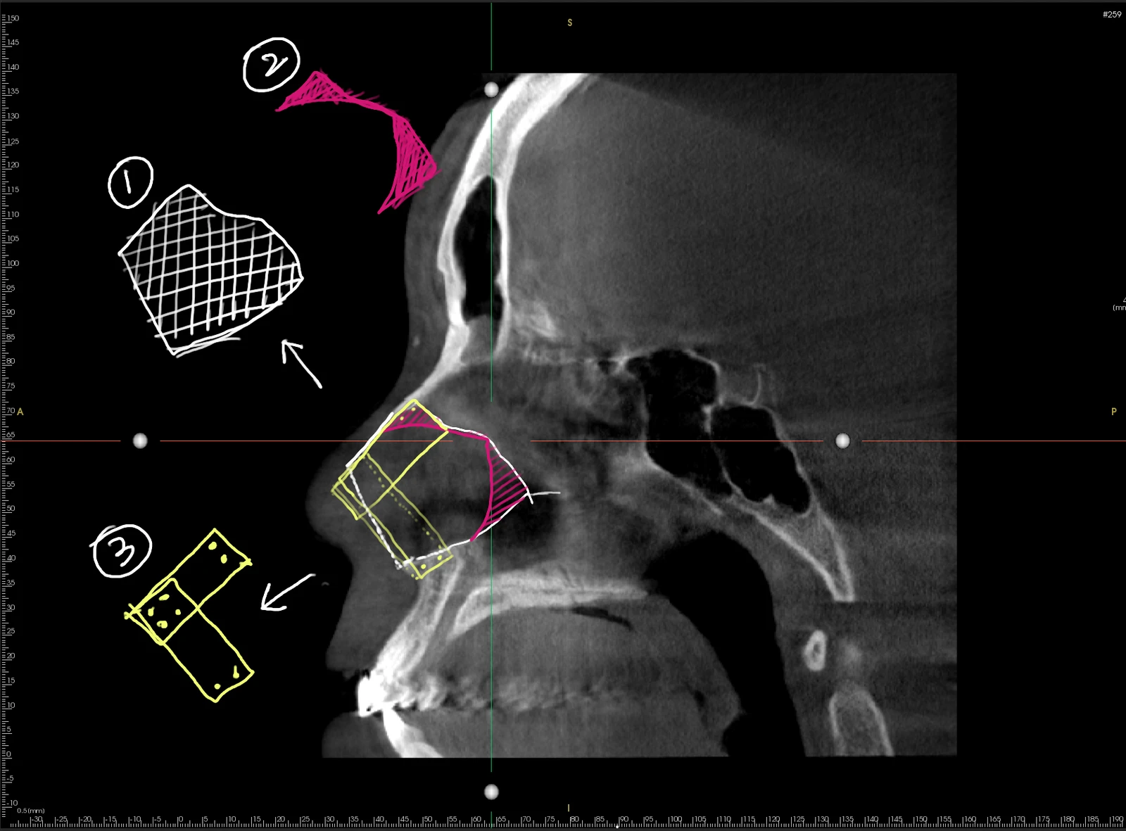
Illustration of septal reconstruction
Upon intraoperative examination, the damage to the septal cartilage was more severe than anticipated, with less than 30% of the total cartilage remaining. In the illustration above, ① represents the estimated size of normal septal cartilage. ② shows the actual remnants of septal cartilage identified during surgery. Only a small portion near the nasal bones and another small piece attached to the deep bony septum remained; most of the cartilage was missing. Consequently, there was no foundation available to anchor structural supports to elevate the drooping nose.
Therefore, drill holes were placed at the anterior nasal spine and the nasal bones, and the septal cartilage was reconstructed using autologous rib cartilage (③). At the same time, the rightward-deviated septal cartilage was straightened, and a septal spur protruding to the right was removed.
To support the drooping tip, a septal extension graft was performed on the reconstructed septum using autologous rib cartilage. Because the lower lateral cartilages had been entirely removed in previous operations, this area was also reconstructed, and nostril elongation was performed concurrently to address the upturned appearance of the nostrils.
In this type of case, the skin over the nasal tip had contracted considerably due to the contracture deformity, resulting in insufficient skin for closure. To resolve this, skin and dermis were harvested from behind the ear and placed as composite grafts on both sides of the columella. In particular, lowering the columella often leads to skin deficiency that makes primary closure difficult, and skin grafting serves as a useful solution in such situations.
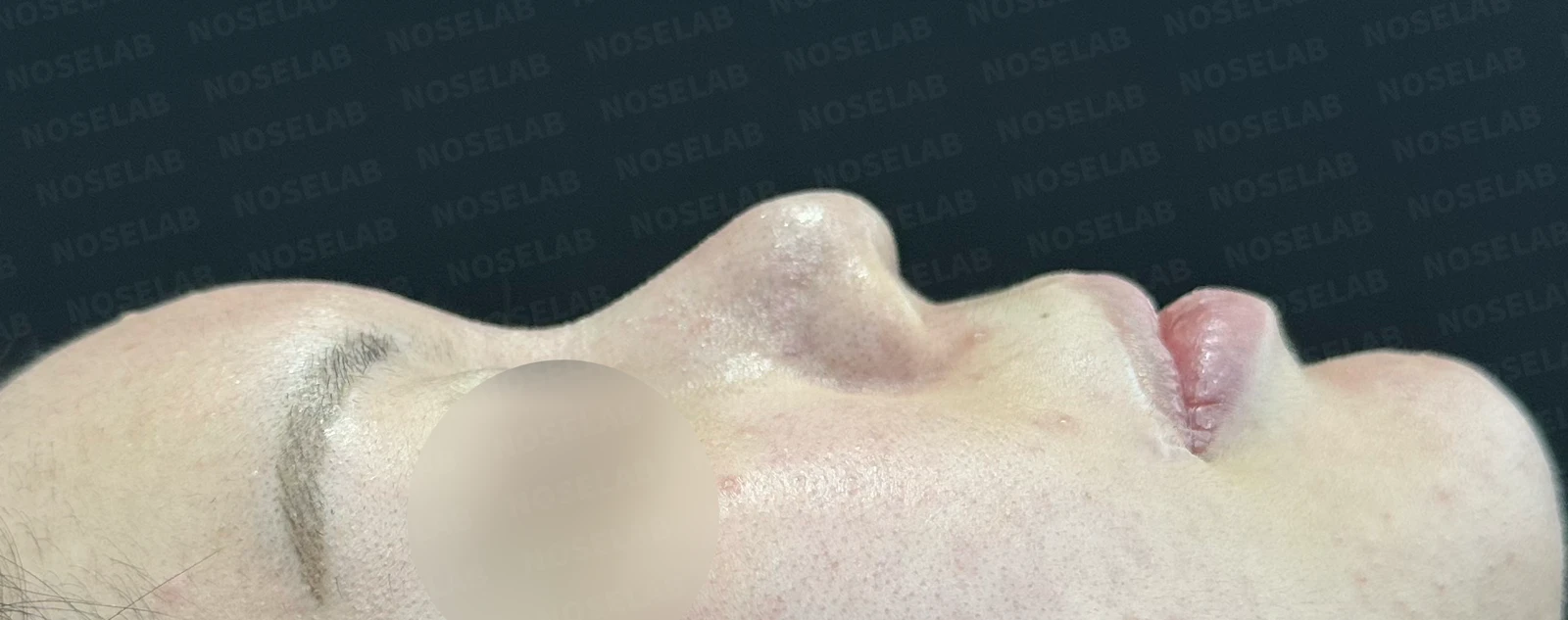
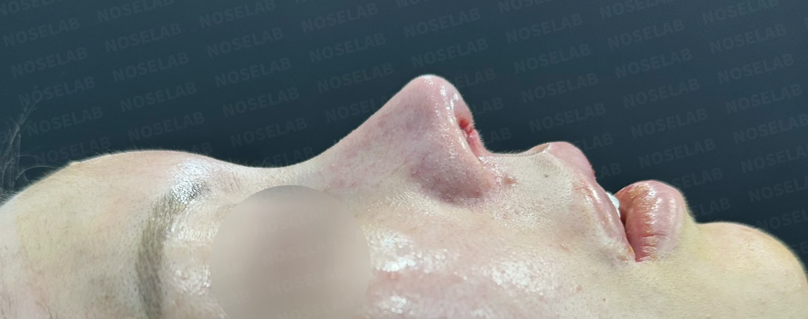
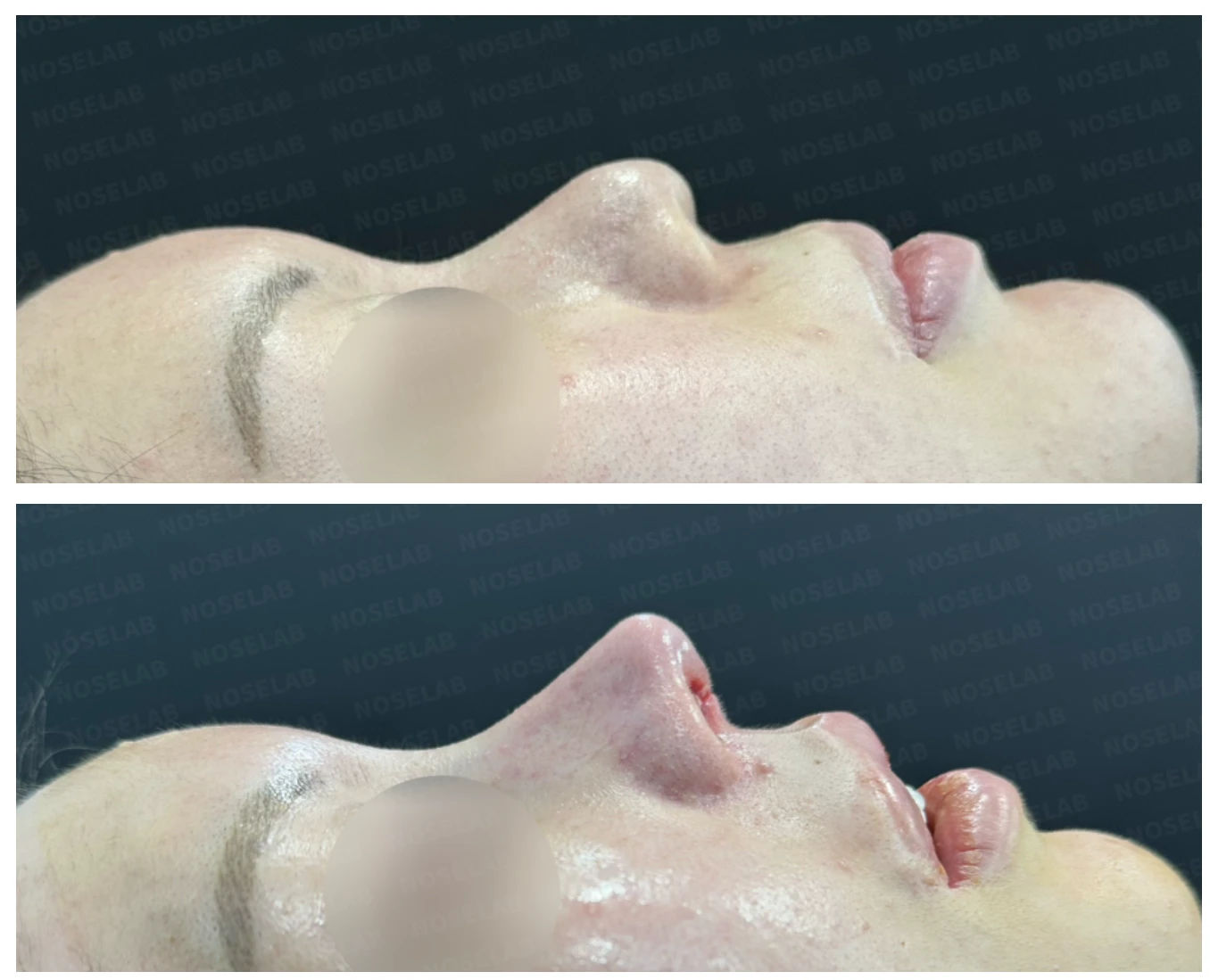
Before surgery / Day 0 immediately after surgery Photos taken immediately after surgery show swelling and may differ from the final result.
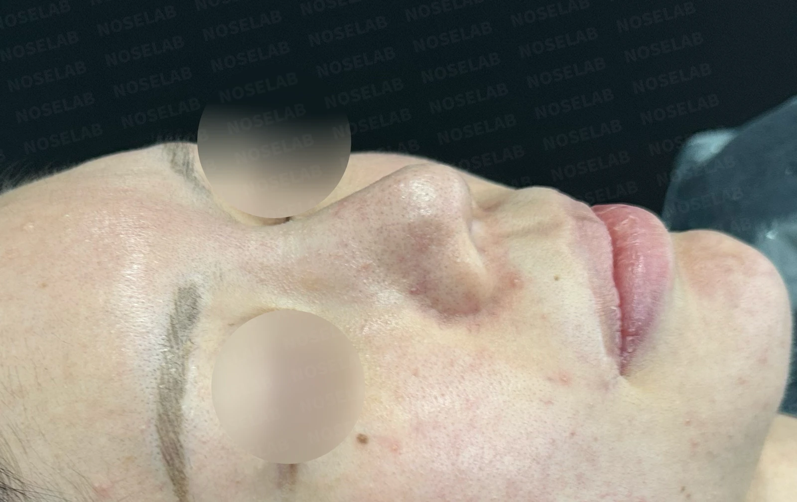
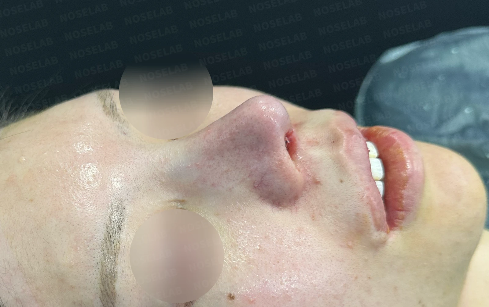
Before surgery / Day 0 immediately after surgery Photos taken immediately after surgery show swelling and may differ from the final result.
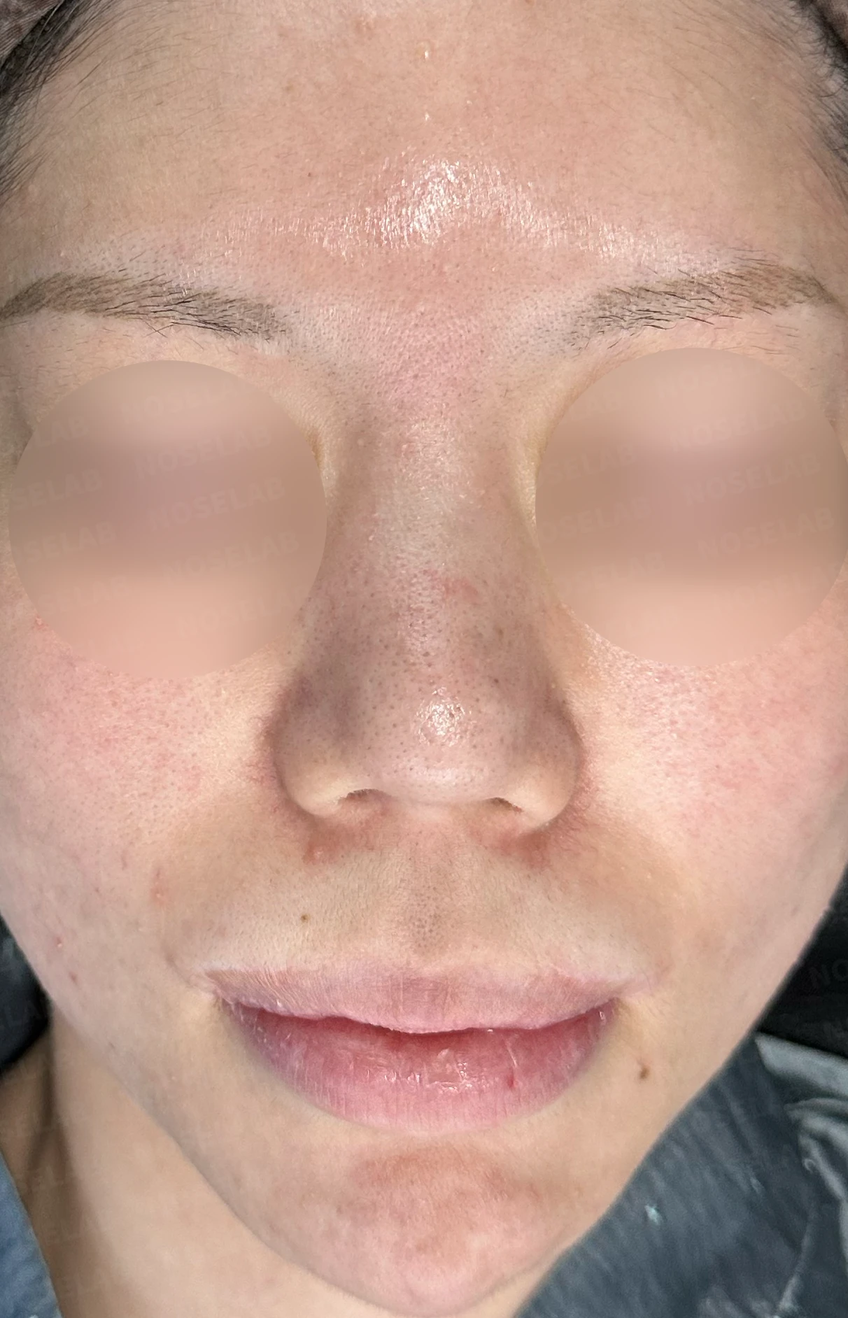
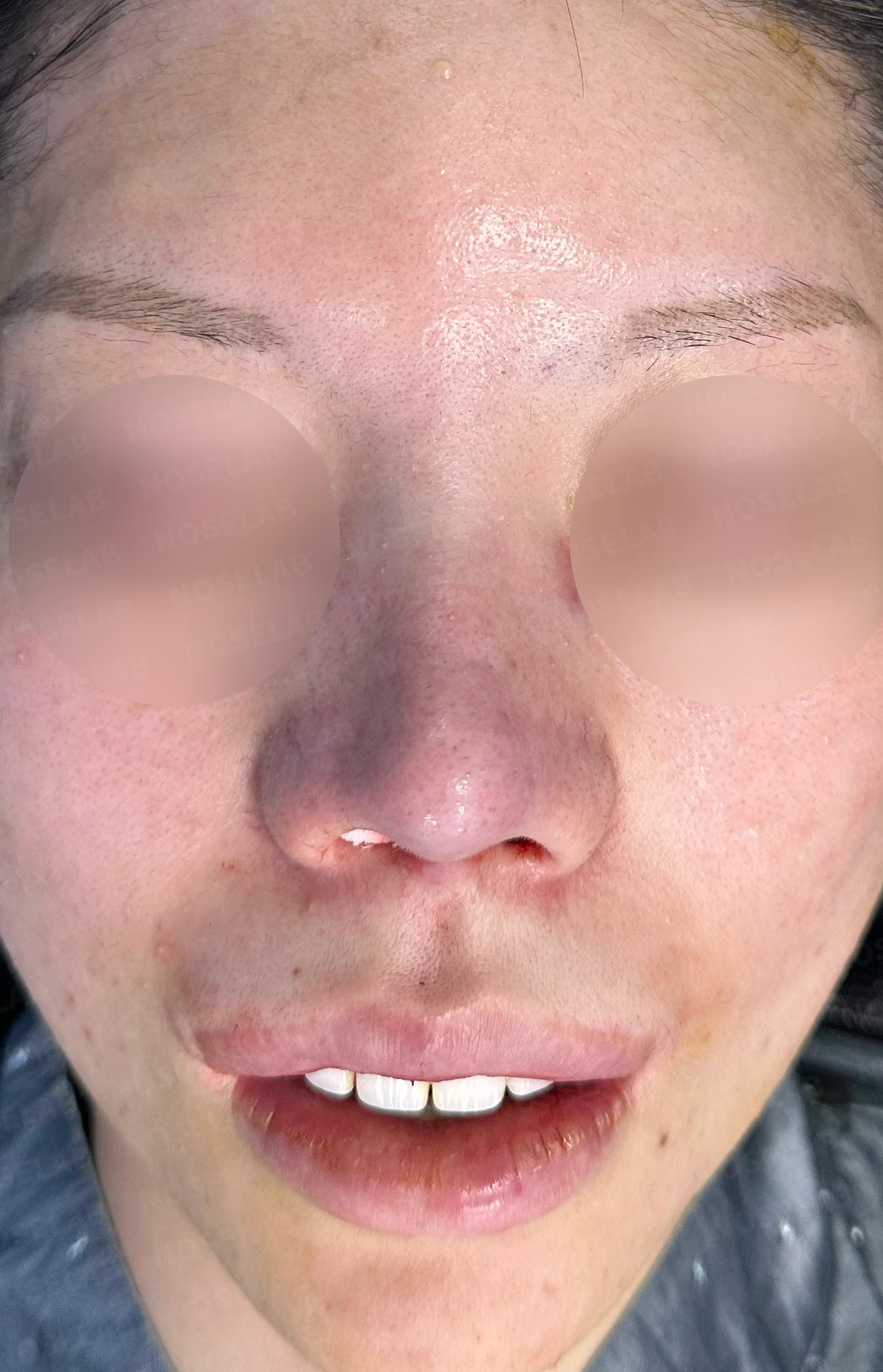
Before surgery / Day 0 immediately after surgery Photos taken immediately after surgery show swelling and may differ from the final result.
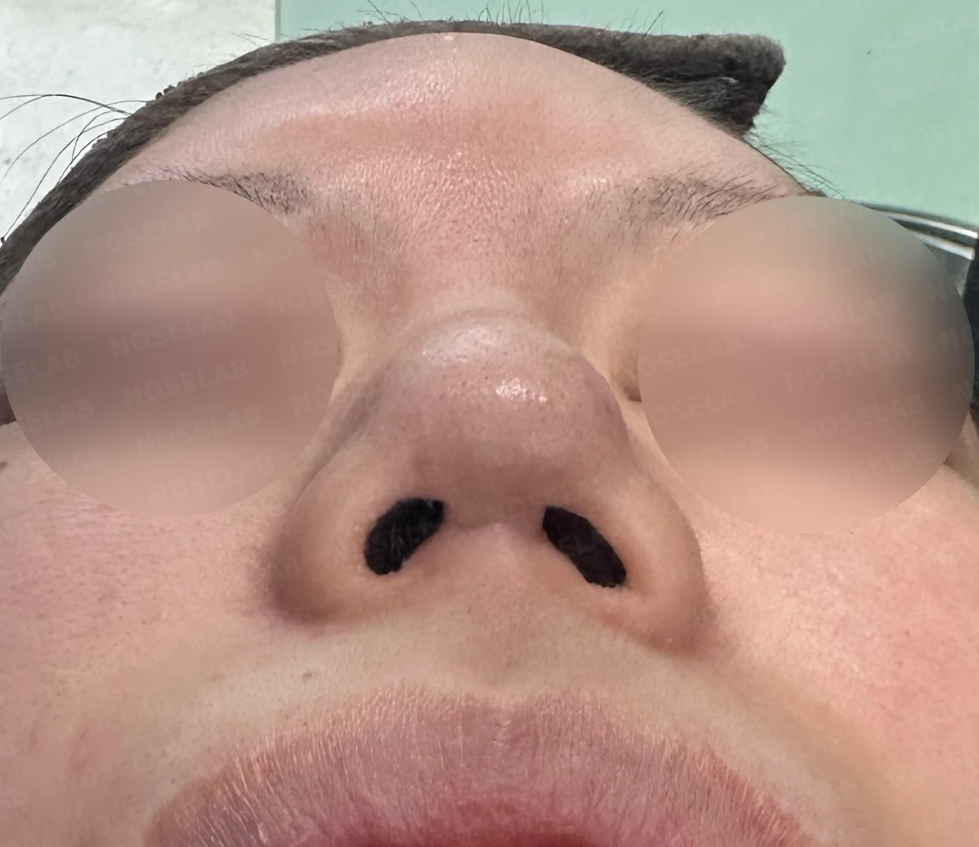
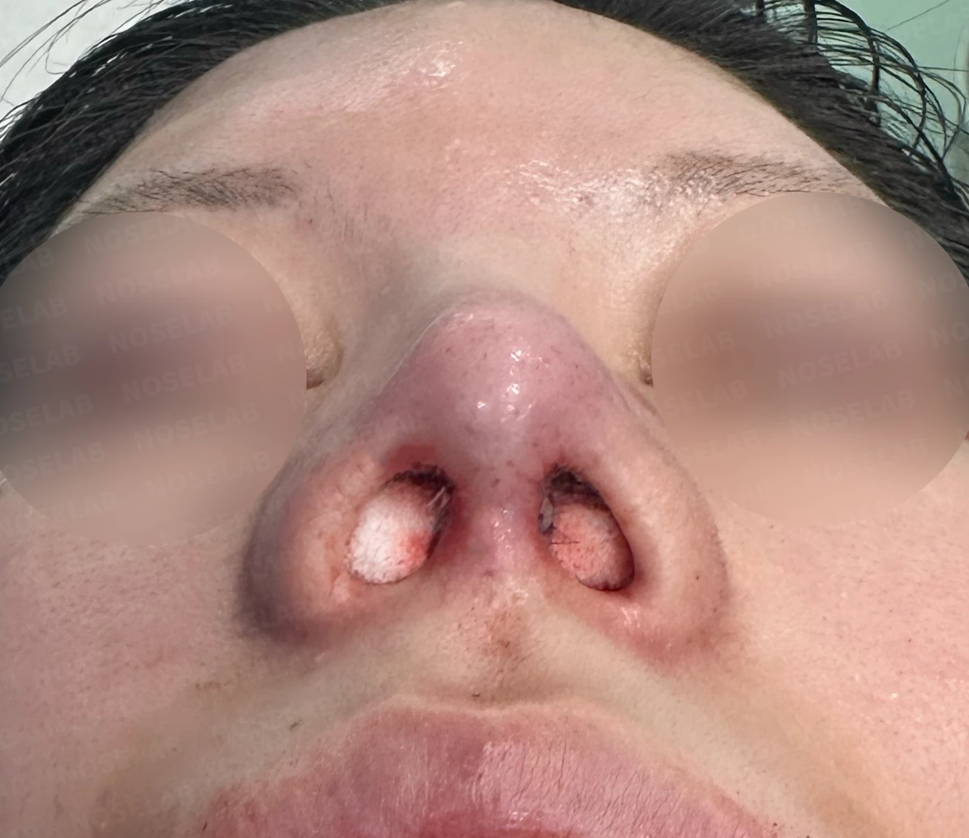
Before surgery / Day 0 immediately after surgery Photos taken immediately after surgery show swelling and may differ from the final result.
In the photographs taken immediately after surgery, the profile view demonstrates that the retracted columella has been lowered naturally, bringing the nasolabial angle to approximately 95 degrees and producing an improvement in the appearance of the protruding mouth. On the oblique view, the radix has been lowered and the drooping nasal tip is upright. On the front view, the crookedness has improved, the nasal bones have been narrowed for a natural contour, and lowering the retracted columella has made the drooping alae appear elevated. Finally, the nostril view shows marked improvement in the columellar retraction caused by the previous open transcolumellar scar, and the narrowed nostrils have regained much of their shape.
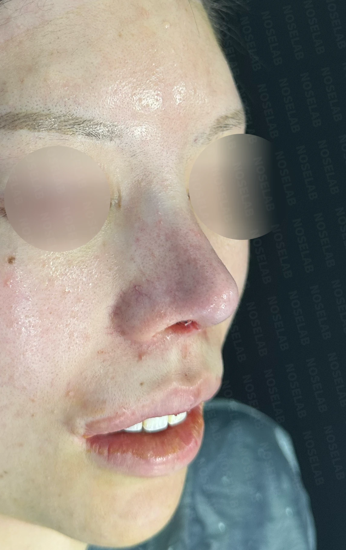
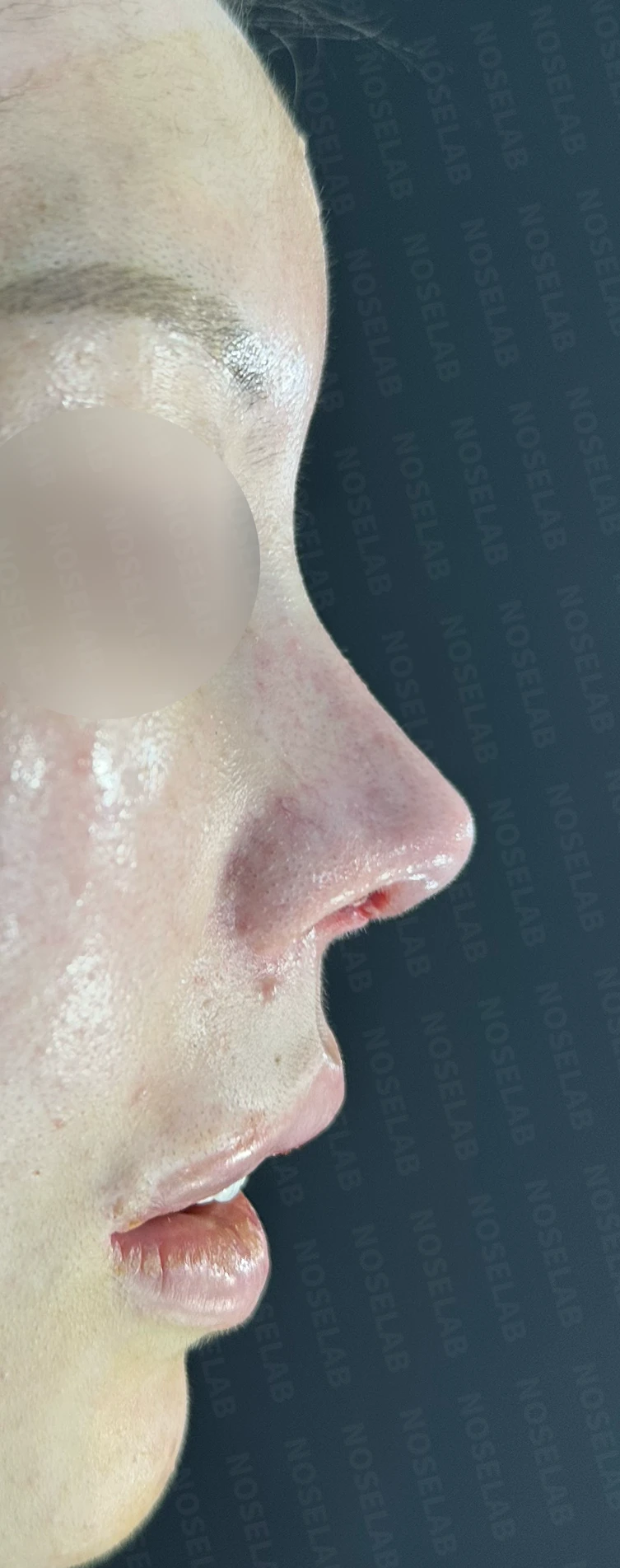
Front, oblique, and profile views immediately after surgery on day 0. Photos taken immediately after surgery show swelling and may differ from the final result.
Base view (nostril) photo
Closing remarks
Septal reconstruction is a demanding and intricate procedure, but it is essential in contracted noses with severe septal damage. Following complications from prior rhinoplasty, columellar retraction is very common, and septal cartilage damage is frequently observed in such scenarios. Furthermore, correcting this condition is far from straightforward. If one attempts correction simply by packing cartilage or dermis into the columella, it may initially appear corrected, but over time the columella gradually recedes again. This occurs because the root cause of columellar retraction is often not the columella itself, but underlying septal damage. Unless this fundamental structural defect is addressed, the columella will ultimately retract again. For this reason, septal reconstruction is often necessary. I believe this type of operation is best undertaken by a board-certified otolaryngologist (ENT specialist).
Today, I shared a case involving the correction of a contracted nose with columellar retraction resulting from severe post-rhinoplasty inflammation, repaired through septal reconstruction using autologous rib cartilage.
I hope this article provides helpful insight for those dealing with contracture and columellar retraction after rhinoplasty.
This was Dr. Peter C. Kang. Thank you.
Thank you.
#contractednose #retractedcolumella #closedrhinoplasty #closedrevisionrhinoplasty #closedcontractednose #autologousribcartilage #ribcartilagerevisionrhinoplasty #closedautologousribcartilage #rhinoplasty #septalreconstruction #columellarretraction

Limitations and risks
When autologous rib cartilage is used, scarring and pain may remain at the donor site on the chest, and pneumothorax (pleural injury) can occur rarely. Over time, the grafted cartilage may warp or undergo partial resorption.
When the septum is corrected at the same time, nasal obstruction may persist or recur, and in rare cases, septal perforation or changes in smell may occur.
Common potential risks include infection, bleeding, asymmetry, scarring, skin necrosis, and insufficient improvement; results vary depending on individual skin condition and previous surgical history.