Hello, I am Dr. Peter C. Kang.
Today, I would like to share a case of nasal reconstruction for an inflamed nose caused by silicone implant exposure.
This patient had undergone rhinoplasty 12 years earlier using a silicone implant and ear cartilage, and visited our clinic after the nose became red and developed signs of inflammation starting 5 months ago.
1. Preoperative design consultation
Let us analyze the patient's preoperative condition in detail:
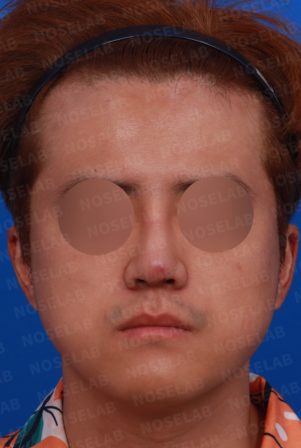
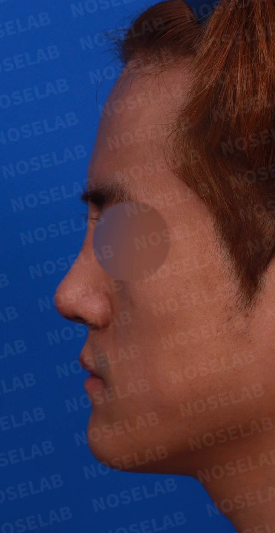
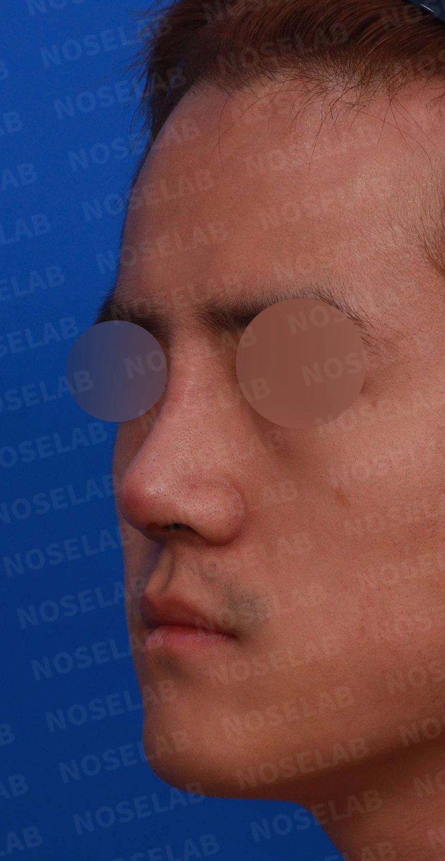
Preoperative design consultation: front, left profile, and oblique views.
Front view:
- The nasal tip was red, and the skin had thinned
- The nasal bridge was swollen
- Nostril asymmetry observed
- The nasal tip appears wide and spread out
Profile view:
- The nose appeared bulky overall
- The radix was positioned high
Oblique view:
- The radix was positioned high
- The entire nose was swollen
- Localized red inflammatory response on the nasal tip
Nostrils:
- Redness observed on the left side of the nasal tip
- Nostril asymmetry is evident
- Granulation tissue observed inside the left nostril
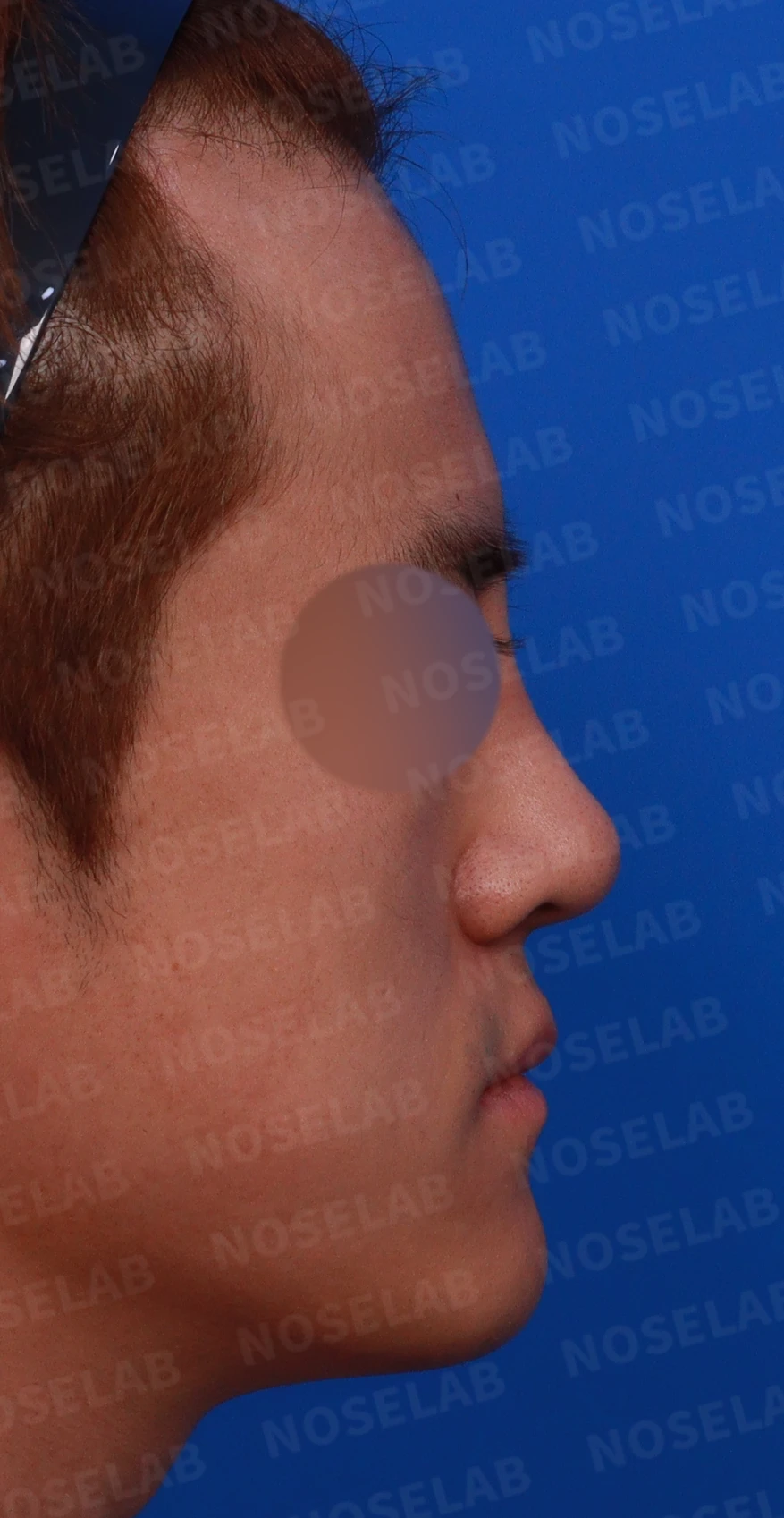
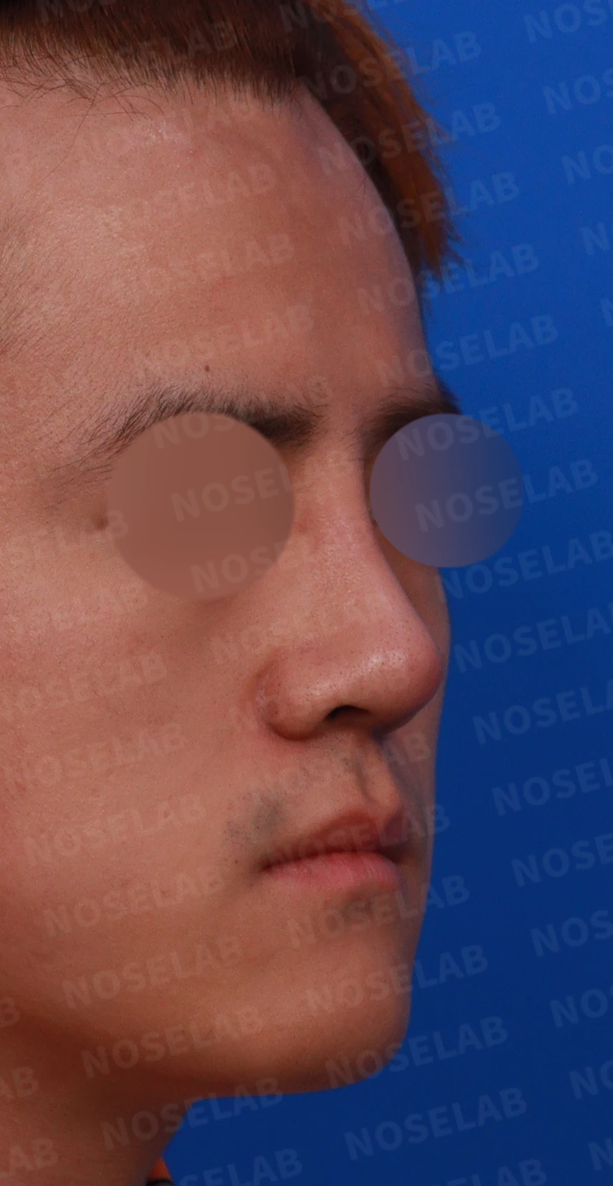
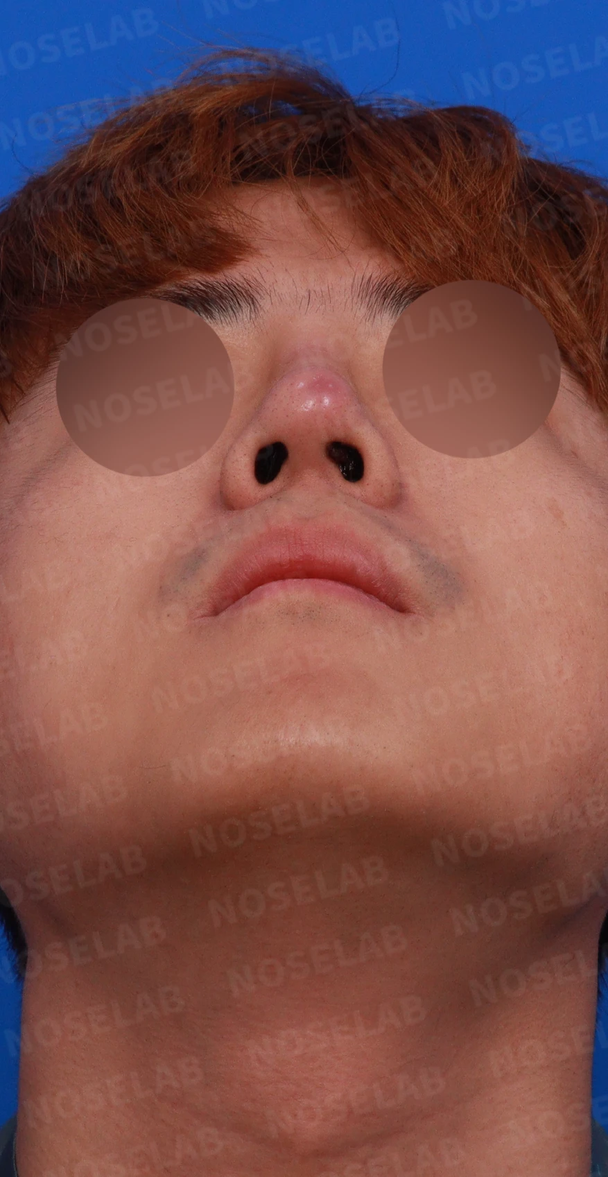
Right profile, right oblique, and basal (nostril) views from the preoperative design consultation.
The patient's main symptoms and requests:
1. The nose began turning red in May
2. Symptoms had been managed with ongoing medication
3. Desired removal of the silicone implant
4. Occasional bleeding from inside the nose
2. Surgical plan
Taking into account the patient's condition and requests, the following surgical plan was established:
a) Removal of the existing silicone implant and capsule
b) Complete removal of inflamed tissue
c) Septal extension using autologous rib cartilage
d) Reconstruction using autologous rib cartilage if damaged cartilage is present
e) Irrigation with saline containing antibiotics
3. Surgical procedure
The following procedures were performed using a closed approach:
a) Identification of inflammation and the area of silicone exposure:
- Granulation tissue resulting from inflammation was protruding along the membranous septum in the left nostril.
- Localized bleeding was observed at that site.
- The silicone implant was confirmed to be exposed directly beneath the granulation tissue.
b) Removal of inflamed tissue and silicone implant:
- All inflammatory granulation tissue inside the nose was thoroughly removed.
- The capsule enclosing the silicone implant was removed alongside it.
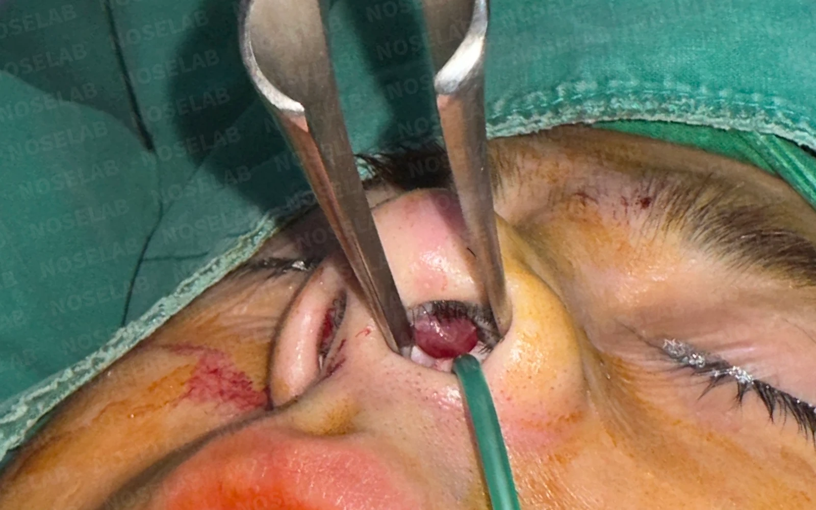
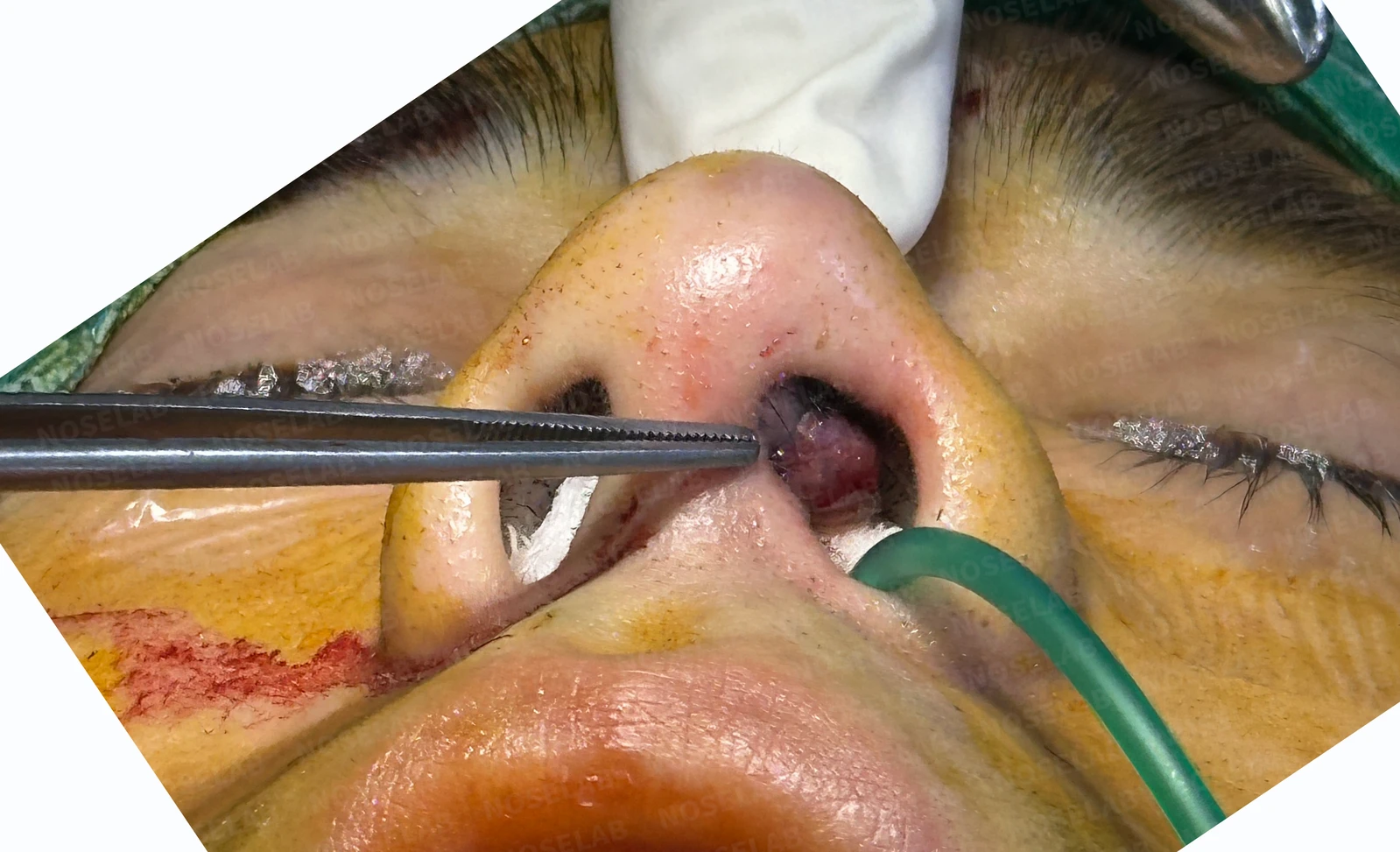
Photograph showing the granulation tissue observed in the left nostril.
c) Irrigation:
- The surgical site was thoroughly irrigated with saline containing antibiotics.
d) Septal extension:
- Septal extension using autologous rib cartilage was performed to prevent contracture deformities that could develop after surgery.
e) Lower lateral cartilage reconstruction:
- The lower lateral cartilage, which had been damaged by acute inflammation, was partially reconstructed using autologous rib cartilage.
All procedures were performed using a closed approach, completing the surgery without external scars.
4. Comparison of before-and-after photos
Comparing photos taken immediately after surgery on day 0 shows the following improvements: Photos taken immediately after surgery show swelling and may differ from the final result.
Front view:
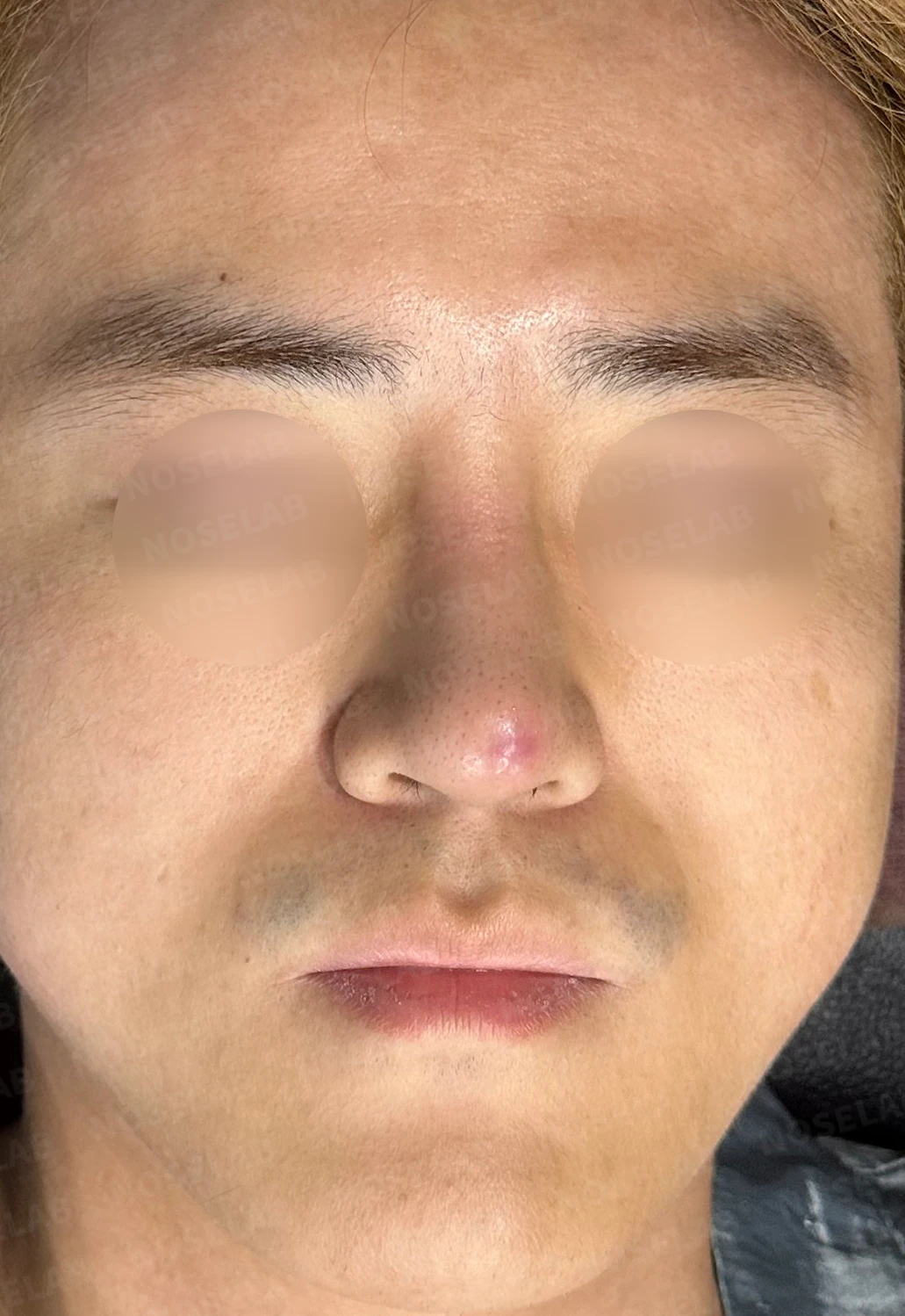
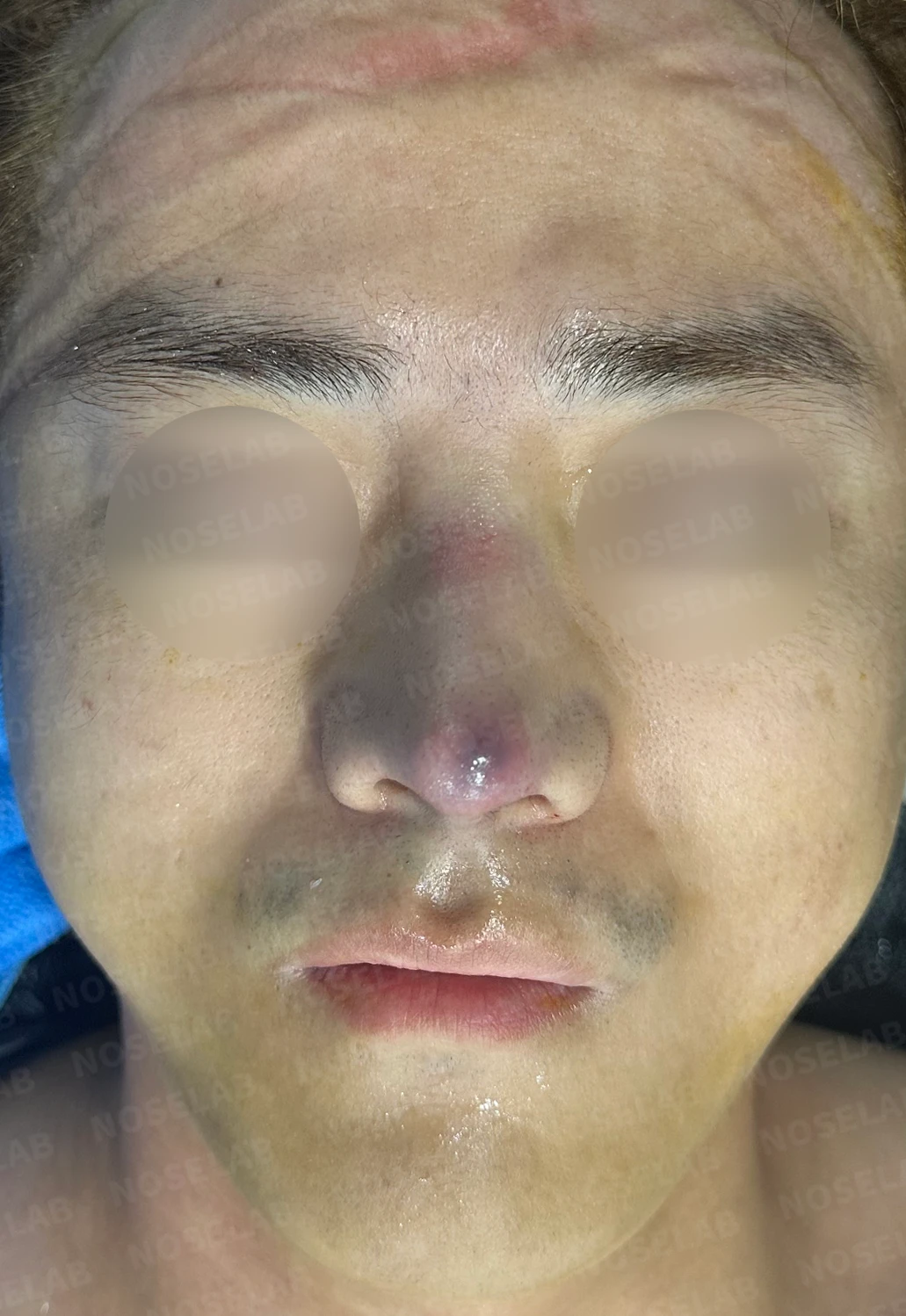
Before surgery / Day 0 immediately after surgery Photos taken immediately after surgery show swelling and may differ from the final result.
- The overall blunt nose became more natural
- The wide-looking nasal tip became more natural
- Nostril asymmetry has been corrected.
Profile view:
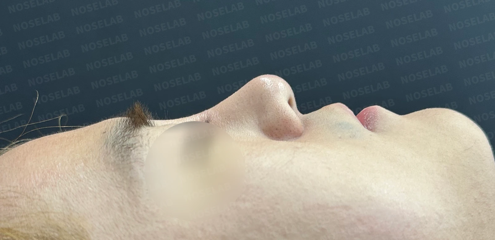
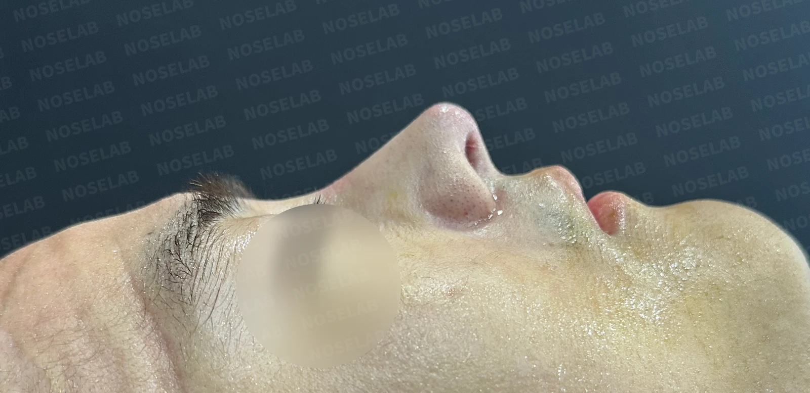
Before surgery / Day 0 immediately after surgery Photos taken immediately after surgery show swelling and may differ from the final result.
- The radix was lowered, creating a natural look
- The blunt-looking nasal tip changed to a slimmer contour
- The columella was lowered naturally, correcting the nasolabial angle
Oblique view:
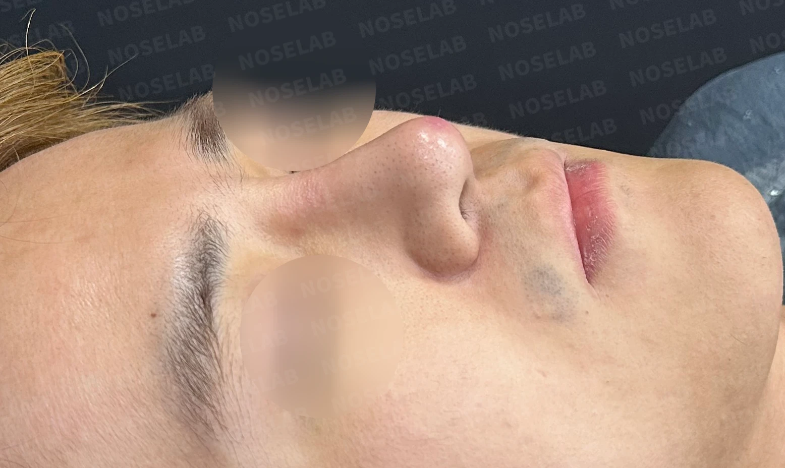
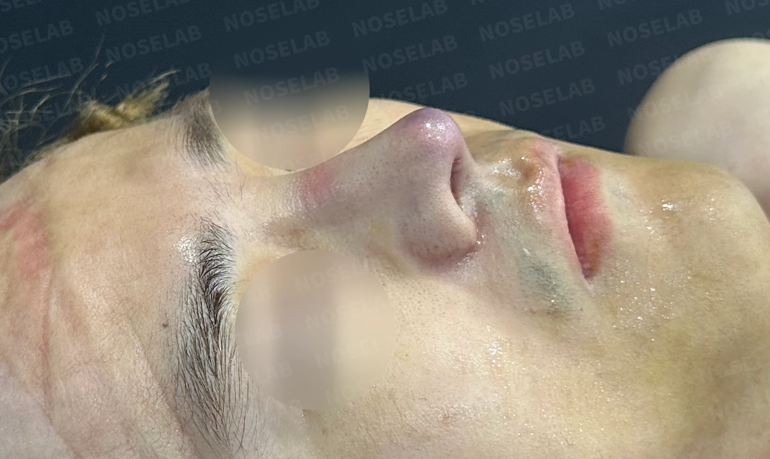
Before surgery / Day 0 immediately after surgery Photos taken immediately after surgery show swelling and may differ from the final result.
- The diffusely swollen, bulky nose transformed into a natural, straight line
Nostrils:
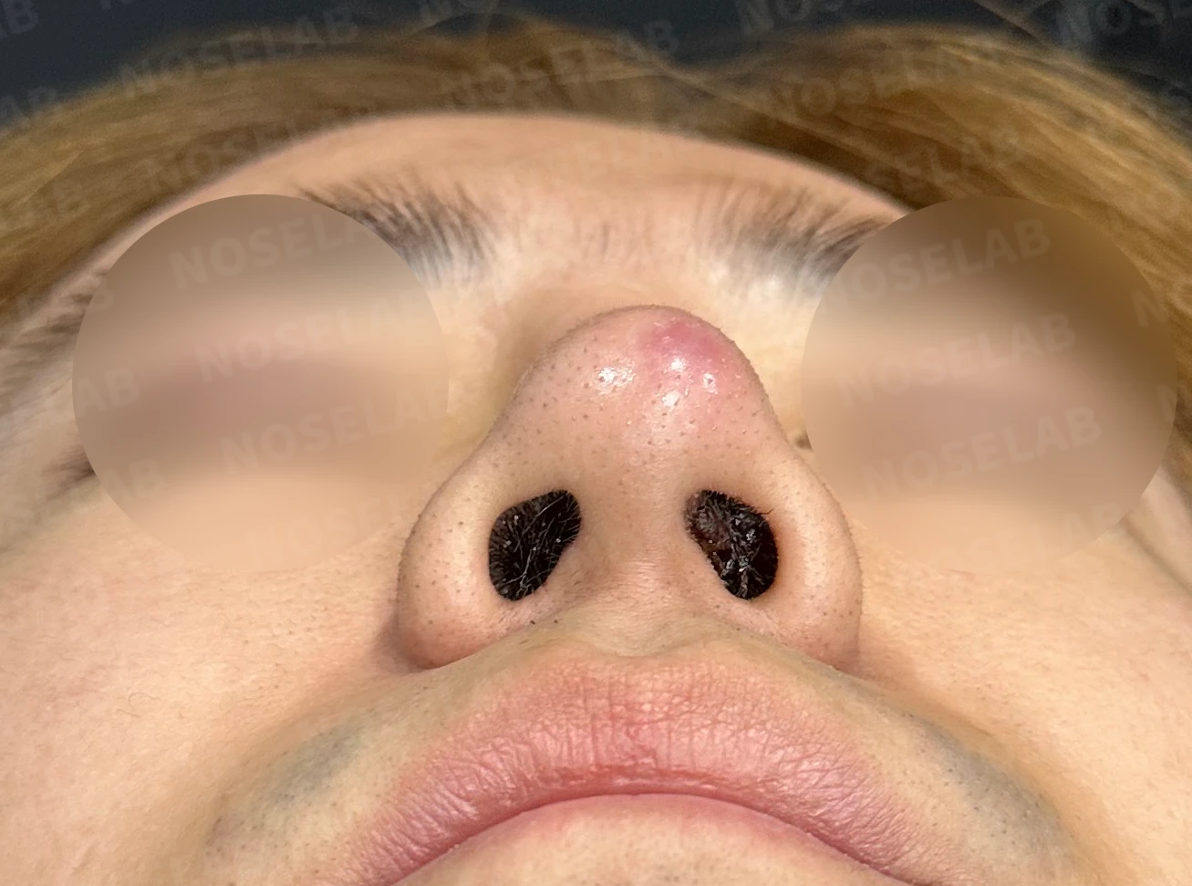
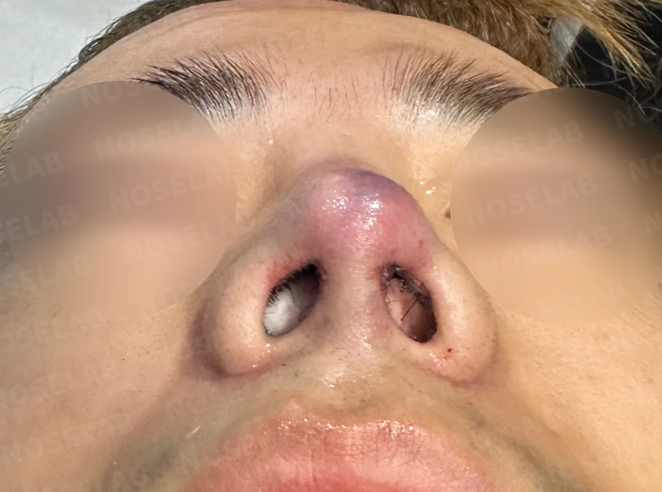
Before surgery / Day 0 immediately after surgery Photos taken immediately after surgery show swelling and may differ from the final result.
- Nostril asymmetry has been corrected.
- The proportion between the nasal tip and nostrils changed naturally
- The granulation tissue was removed, restoring a clean nasal cavity
5. Photos immediately after surgery on day 0 Photos taken immediately after surgery show swelling and may differ from the final result.
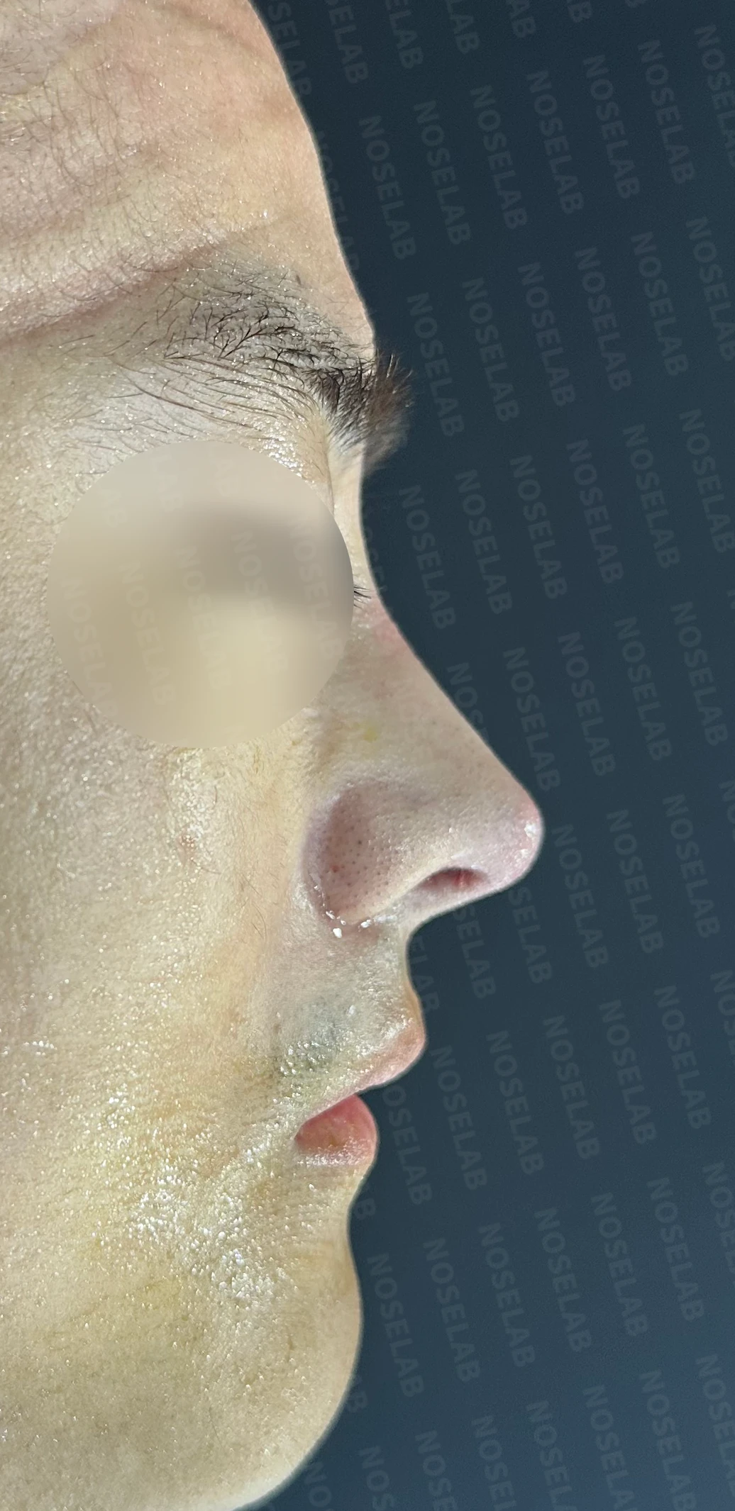
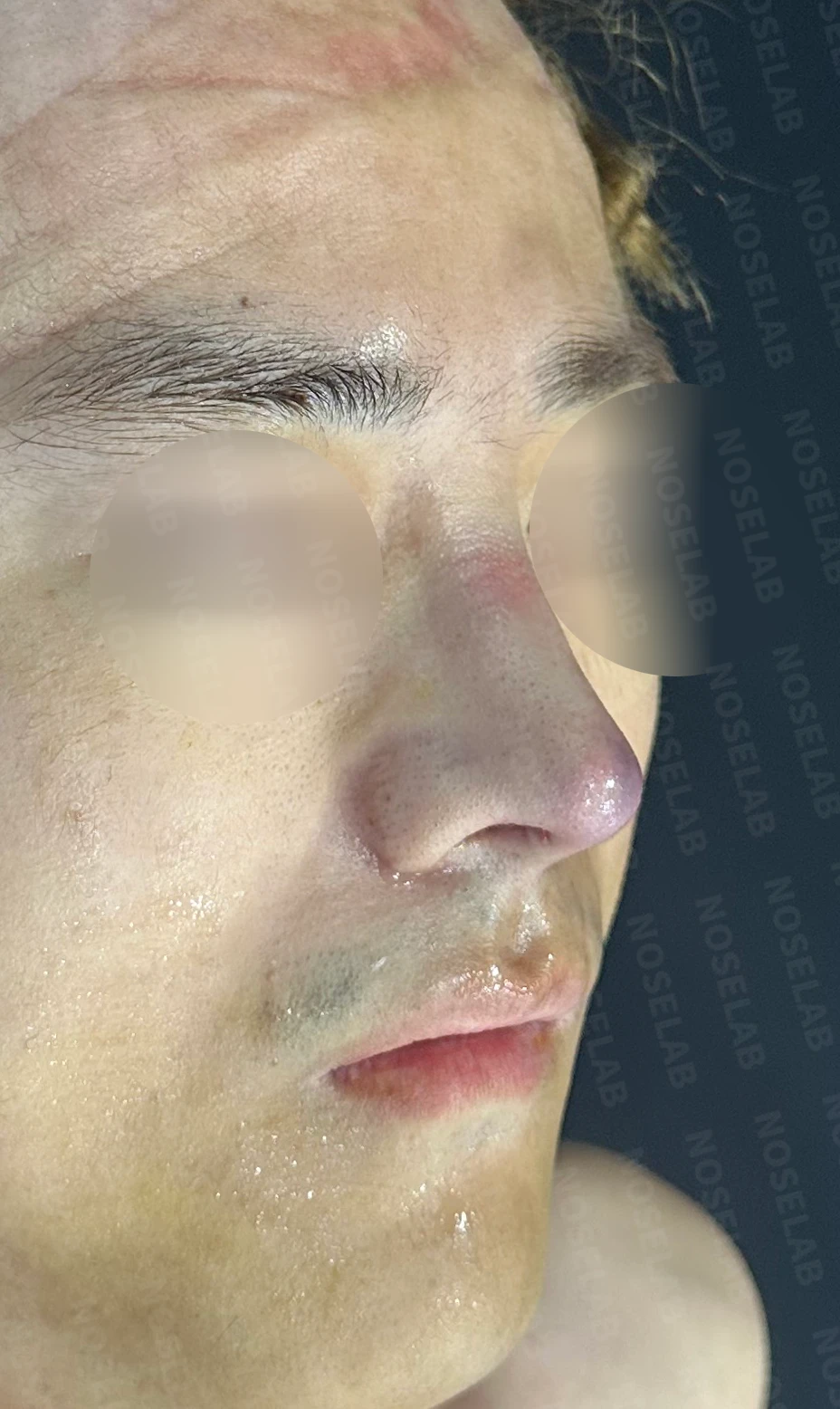
Front, profile, and oblique views taken immediately after surgery (day 0).Immediate postoperative photos show swelling and may differ from the final result.
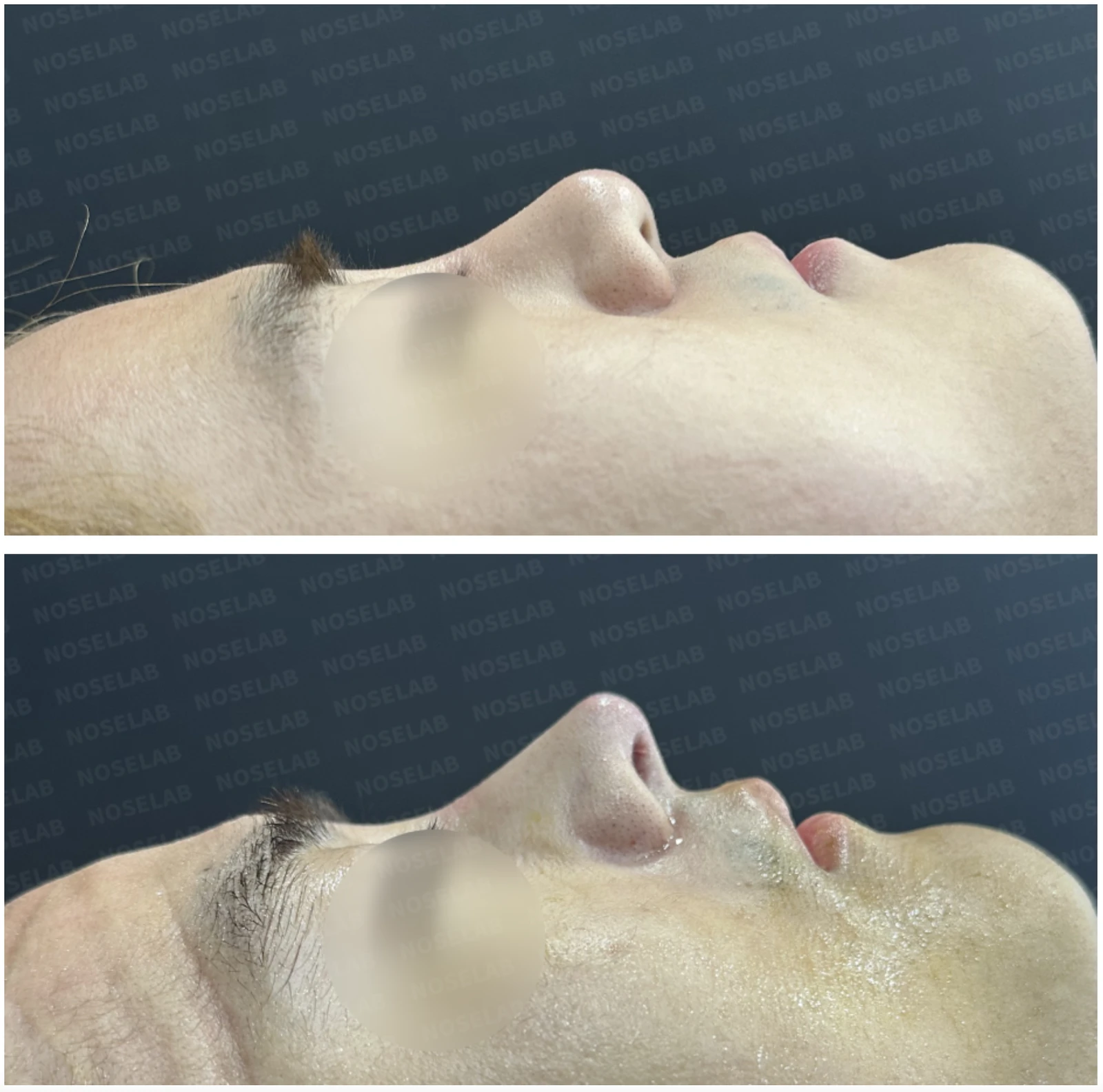
Basal (nostril) view immediately after surgery (Day 0).Immediate postoperative photos show swelling and may differ from the final result.
6. Overall evaluation
This is a case in which the inflammation and exposure issues resulting from a long-standing silicone implant were resolved. The use of L-shaped silicone implants can trigger a persistent foreign body reaction, which may cause the implant to push forward toward the nasal tip and eventually penetrate the skin.
In such situations, pimple-like red, swollen, and painful symptoms appear at the nasal tip; if left unaddressed, discharge mixed with blood and pus may develop, and contracture can progress. This patient presented with these classic symptoms, requiring prompt intervention.
The core of the surgery was to completely remove the silicone implant causing the inflammation, thoroughly irrigate the inflamed area, and reconstruct the nasal framework using autologous rib cartilage. In particular, septal extension and lower lateral cartilage reconstruction secured structural stability and prevented potential contracture deformities from developing in the future.
Even though it was day 0 immediately after surgery, the swelling caused by the inflammation was noticeably reduced, and the overall shape of the nose was improved into a natural and balanced appearance. We plan to support a full recovery through ongoing antibiotic therapy and regular follow-up visits. Photos taken immediately after surgery show some swelling and may differ from the final result.
7. Conclusion
Revision rhinoplasty, especially when accompanied by inflammation, is very complex and requires a meticulous approach. In performing numerous revision procedures, complications where an L-shaped implant is pushed down toward the nasal tip due to chronic foreign body reaction and eventually punctures through the skin are encountered with some regularity.
When these symptoms appear, it is important to consult a rhinoplasty specialist as early as possible. If early intervention is delayed, the severity of the inflammation worsens and contracture progresses, which increases surgical complexity and prolongs the recovery period.
The primary initial steps in treatment are antibiotic therapy and implant removal. Following that, it is essential to reconstruct the nasal contours using autologous tissue to the greatest extent possible. Using autologous tissue, particularly rib cartilage, allows for a stable and natural outcome without foreign body rejection.
For those considering rhinoplasty, especially if revision surgery is required, please be sure to consult a physician with extensive experience and expertise. Based on anatomical knowledge as a board-certified otolaryngologist and extensive surgical experience, Dr. Peter C. Kang provides surgical plans tailored for each individual patient with safety in mind.
I will be with you every step of the way to achieve a beautiful and healthy nose.
This was Dr. Peter C. Kang. Thank you.
Thank you.
#rhinoplasty #inflamednoserevision #siliconeimplantexposure #autologousribcartilagegraft #closedrhinoplasty #revisionrhinoplasty #entrhinoplasty #rhinoplastysafety #contractednoserevision #nasalinflammationtreatment

Limitations and risks
When autologous rib cartilage is used, scarring and pain may remain at the donor site on the chest, and pneumothorax (pleural injury) can occur rarely. Over time, the grafted cartilage may warp or undergo partial resorption.
When revising after implant removal, contracture may progress again after removal, the nasal height and length may decrease, and staged surgery may be necessary depending on the skin condition.
When the septum is corrected at the same time, nasal obstruction may persist or recur, and in rare cases, septal perforation or changes in smell may occur.
Common potential risks include infection, bleeding, asymmetry, scarring, skin necrosis, and insufficient improvement; results vary depending on individual skin condition and previous surgical history.