Hello, I am Dr. Peter C. Kang.
Today, I would like to share a surgical case of a patient who sought treatment for right-sided nasal obstruction, a drooping nasal tip, and lowering the columella.
In cases like this, the patient had already undergone two prior rhinoplasty procedures, making this their 3rd surgery.
1. Preoperative design consultation
Let us take a detailed look at the patient's preoperative condition and requests:
Patient's surgical history:
July 2022: Rhinoplasty using ear cartilage and a silicone implant
April 2023: Rhinoplasty using autologous rib cartilage and a silicone implant
Patient's main requests:
Lowering the columella
Refining and elevating the drooping nasal tip
Creating a natural nasolabial angle
Improvement of right-sided nasal obstruction
Analysis of preoperative condition:
Front view:
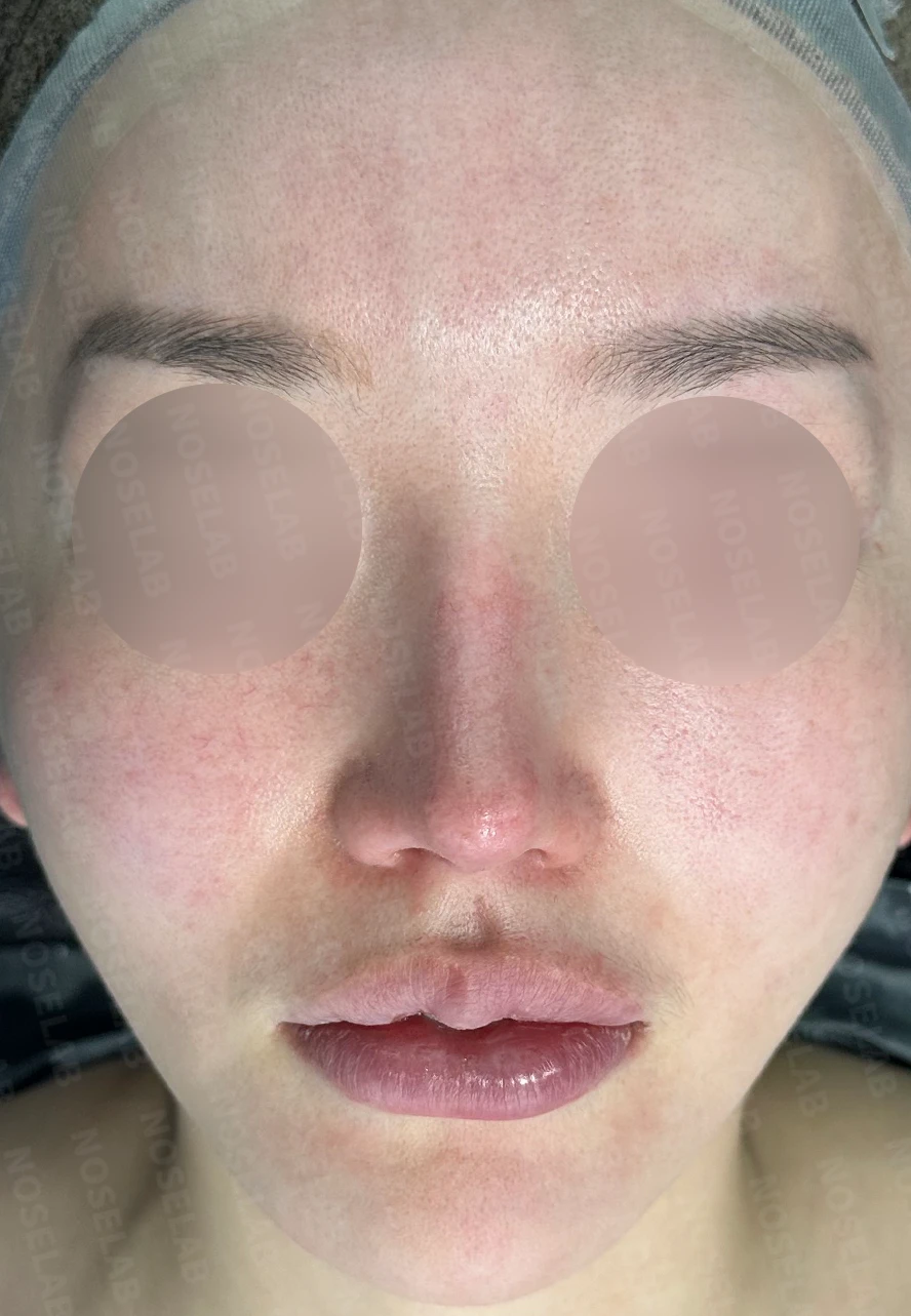
Front view
The nose appears crooked
The nasal bridge implant shows through
Profile view:
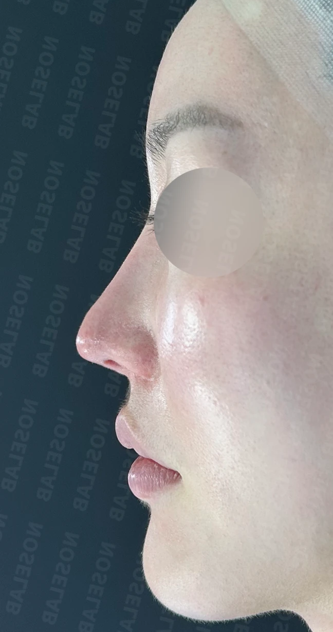
Profile view
The nasal tip is drooping
The columella is retracted
The nasolabial angle looks unnatural
Nostrils:
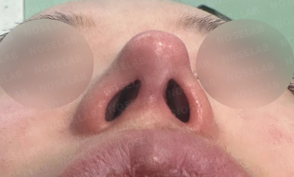
Nostril asymmetry is present
The columella is tilted
Additional findings:
Complaints of right-sided nasal obstruction
CT scan and nasal endoscopy findings:
Deviated septum
Hypertrophied inferior turbinates
Mucosal adhesion in the right nasal cavity
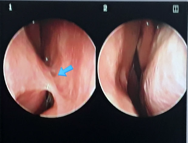
Right intranasal synechia (indicated by arrow) is visible.
2. Surgical plan
Taking into account the patient's condition and requests, the following surgical plan was established:
a) Septal extension using autologous rib cartilage
b) Nasal bridge implant replacement (widening the width)
c) Lowering the columella
d) Nasolabial angle correction
e) Septoplasty
f) Mucosal adhesiolysis
g) Radiofrequency submucosal resection of the inferior turbinates
3. Surgical procedure
a) Autologous rib cartilage harvest and grafting:
The patient's rib cartilage was harvested to secure an adequate amount of cartilage required for nasal reconstruction.
b) Septal extension graft:
Autologous rib cartilage was used to extend and reinforce the septum.
This elevated the drooping nasal tip and reinforced structural support.
c) Nasal bridge implant exchange:
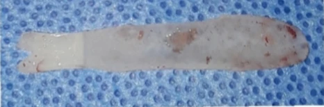
Photograph of the removed silicone implant.
The existing implant was removed and replaced with a wider implant.
This addressed the visible implant show-through and created a natural contour.
d) Lowering the columella:
The retracted columella was lowered appropriately to improve overall nasal balance.
e) Nasolabial angle correction:
Lowering the retracted columella created a natural nasolabial angle.
f) Septoplasty:
The deviated septum was straightened to relieve nasal obstruction.
g) Mucosal adhesiolysis:
Adhesions in the right nasal mucosa were released to relieve nasal obstruction.
h) Inferior turbinate reduction:
Hypertrophied inferior turbinates were reduced using radiofrequency to relieve nasal obstruction.
All procedures were performed using a closed approach, completing the surgery without external scars.
4. Comparison of before-and-after photos
Comparing photos taken immediately after surgery on day 0 shows the following improvements: Photos taken immediately after surgery show swelling and may differ from the final result.
Front view:
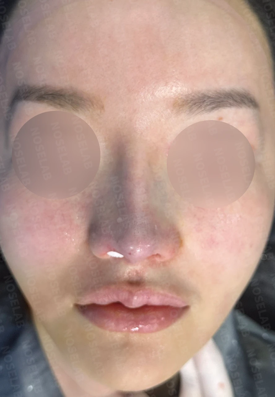
Before surgery / Day 0 immediately after surgery Photos taken immediately after surgery show swelling and may differ from the final result.
The crooked nose was straightened.
The visible implant show-through along the nasal bridge has significantly improved.
The nose presents an overall well-balanced appearance.
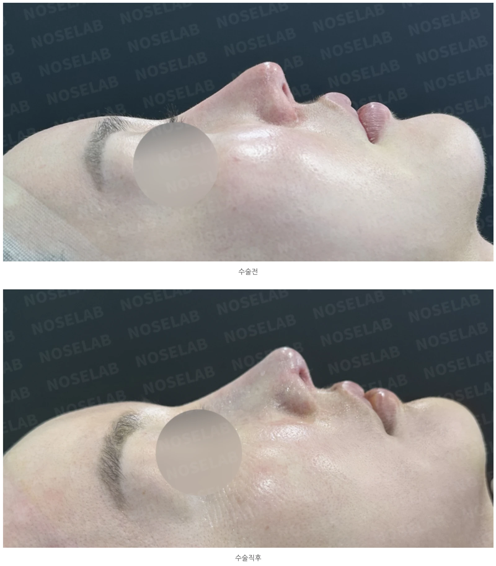
Profile view:
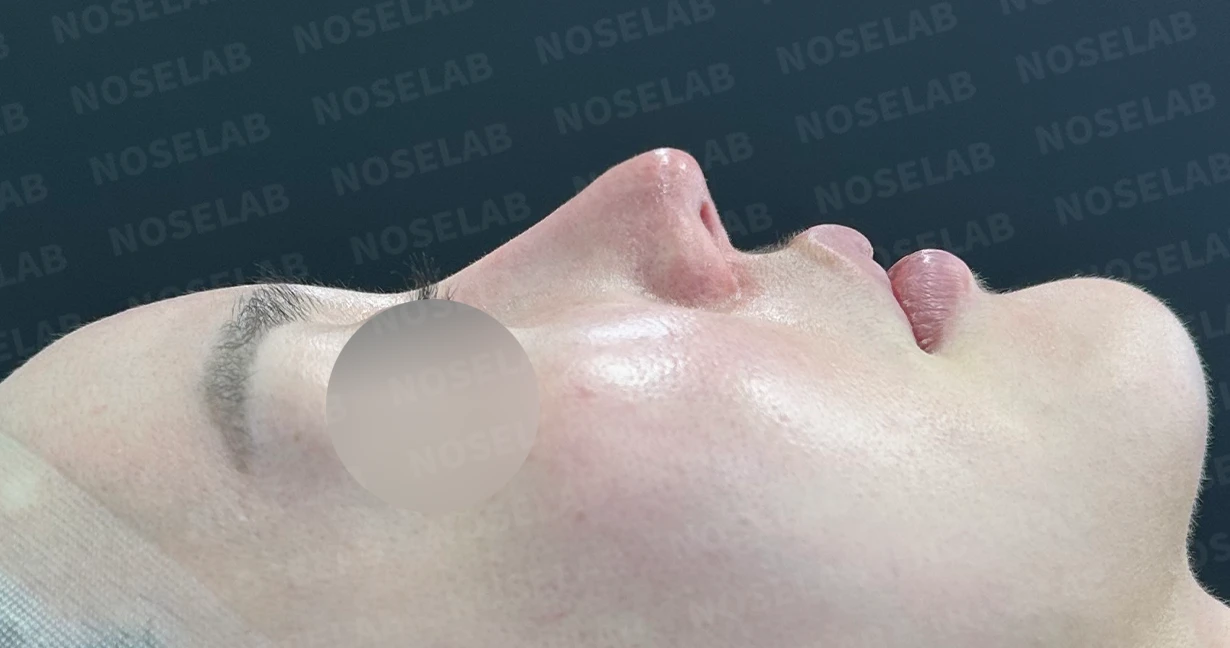
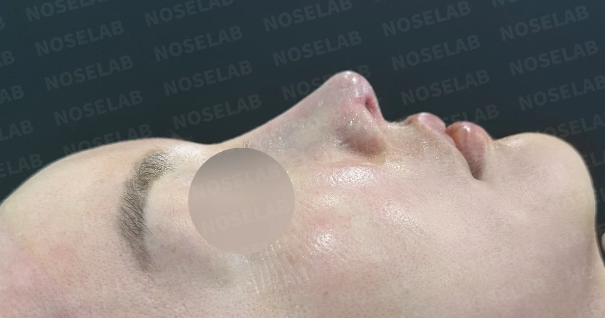
Before surgery / Day 0 immediately after surgery Photos taken immediately after surgery show swelling and may differ from the final result.
The previously drooping nasal tip has been naturally elevated, improving overall facial balance.
The retracted columella was brought down appropriately, forming a natural line.
The unnatural nasolabial angle was corrected, improving harmony between the nose and lips.
Oblique view:
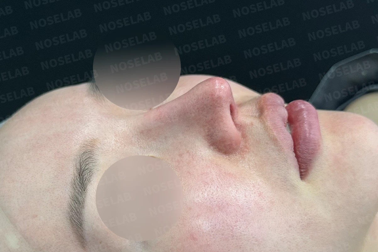
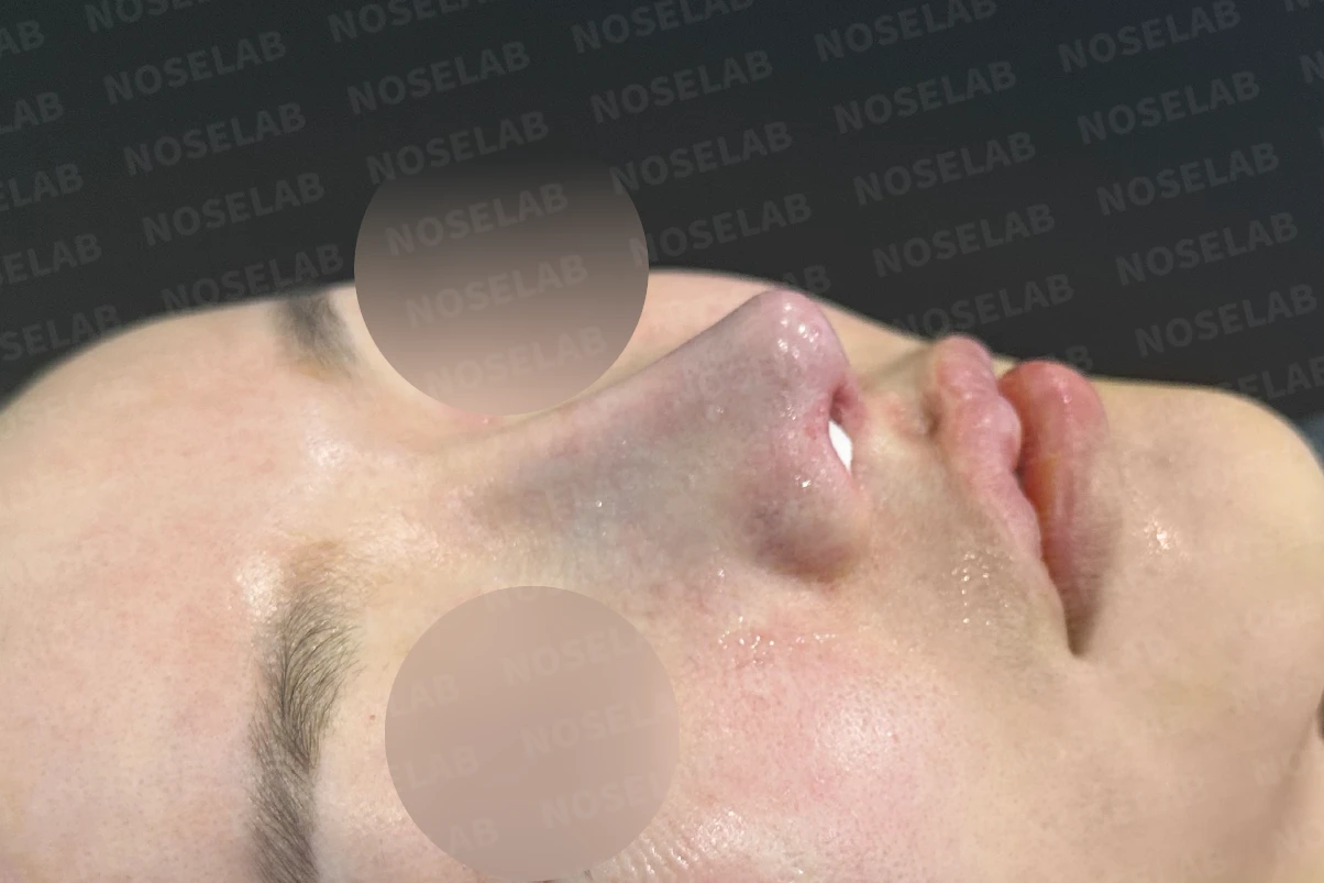
Before surgery / Day 0 immediately after surgery Photos taken immediately after surgery show swelling and may differ from the final result.
The nasal deviation was corrected, creating a smooth line.
The contour of the implant has been naturally improved.
Nostrils:
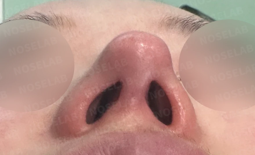
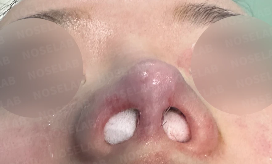
Before surgery / Day 0 immediately after surgery Photos taken immediately after surgery show swelling and may differ from the final result.
Nostril asymmetry has improved, resulting in a balanced appearance.
The tilted columella has been straightened.
5. Photos immediately after surgery (Day 0) Immediate postoperative photos show swelling and may differ from the final result.
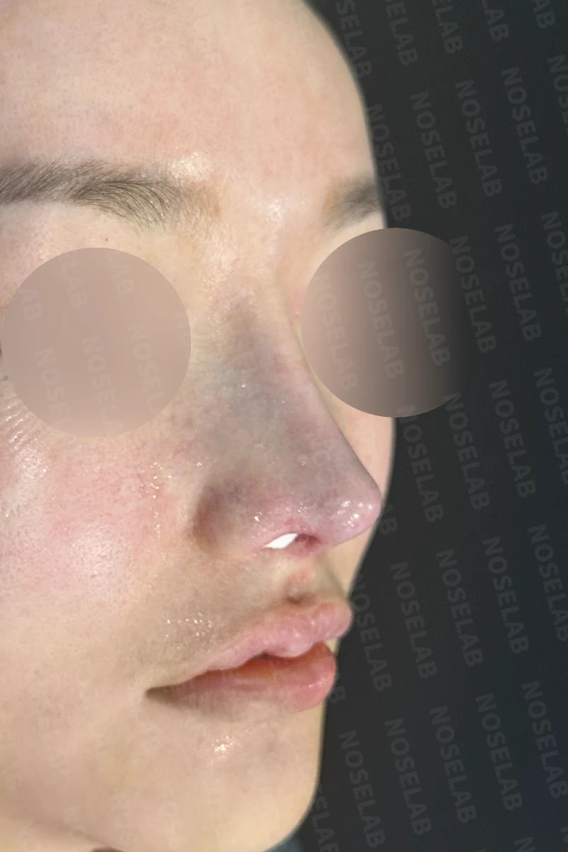
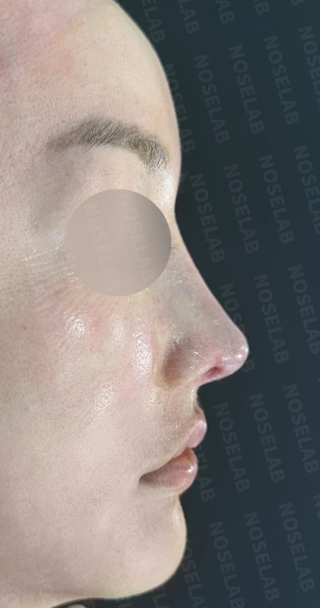
Front, oblique, and profile views on day 0 immediately after surgery Photos taken immediately after surgery show swelling and may differ from the final result.
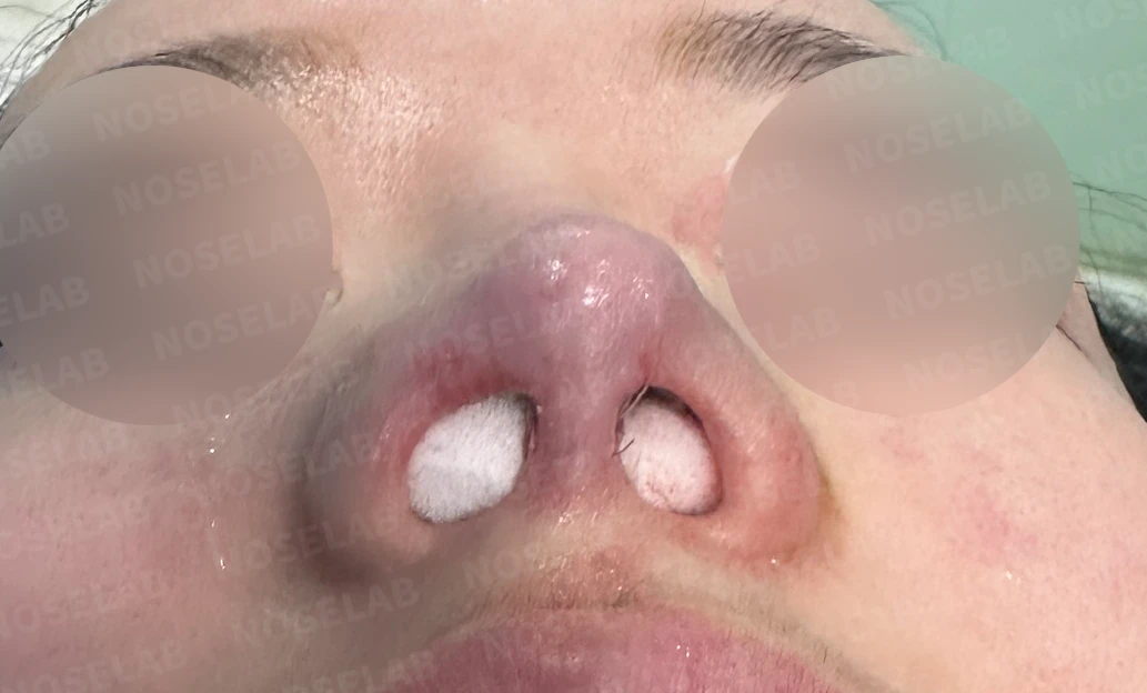
Basal (nostril) view immediately after surgery (Day 0)Immediate postoperative photos show swelling and may differ from the final result.
6. Overall evaluation
This case is a good example of achieving aesthetic and functional improvements simultaneously. Despite two previous rhinoplasty surgeries, the patient remained dissatisfied and continued to experience functional discomfort from nasal obstruction.
Septal extension using autologous rib cartilage played a major role in correcting the drooping nasal tip and reinforcing the overall nasal framework. Additionally, replacing the bridge implant resolved the implant show-through and achieved a much more natural contour.
Lowering the columella and correcting the nasolabial angle improved overall balance and harmony between the nasal tip and perioral region. Beyond altering nasal shape, this played a significant role in softening the patient's overall facial impression.
Particularly noteworthy in this surgery was the comprehensive approach to resolving nasal obstruction. Septoplasty, mucosal adhesiolysis, and inferior turbinate reduction together addressed functional airway issues. This is an area where expertise as a board-certified otolaryngologist proved essential.
Using a closed approach shortened the postoperative recovery period and allowed surgery to be completed without external scars. This contributed significantly to patient satisfaction.
7. Conclusion
I hope this case offers encouragement to those troubled by complex nasal concerns, such as nasal obstruction, a drooping tip, and an unnatural columella. In particular, I hope it helps patients who continue to struggle with unsatisfactory outcomes after previous rhinoplasty procedures.
Revision rhinoplasty requires rebuilding the framework while working around structural changes from prior procedures, making it more complex and demanding than primary surgery. Therefore, guidance from a surgeon with extensive experience and dedicated expertise is essential.
Furthermore, rhinoplasty should never be approached solely from an aesthetic standpoint. Because the nose is an essential organ directly tied to breathing, functional aspects must be evaluated alongside cosmetic goals. When nasal obstruction, a deviated septum, or rhinitis are present, addressing them together during rhinoplasty is advisable.
Drawing on anatomical knowledge and extensive surgical experience as a board-certified otolaryngologist, I provide surgical plans tailored to each individual patient. Through a comprehensive approach that considers both functional and cosmetic improvement, I strive to create a beautiful and healthy nose.
I will be with you every step of the way to achieve a beautiful and healthy nose.
This was Dr. Peter C. Kang. Thank you.
Thank you.
#rhinoplasty #nasalobstructionrelief #droopingtipcorrection #loweringthecolumella #nasolabialanglecorrection #revisionrhinoplasty #autologousribcartilagegraft #closedrhinoplasty #septalextension #entrhinoplasty #safetyfocusedrhinoplasty #nasalbridgecorrection #tipplasty #deviatedseptum #turbinatereduction

Limitations and risks
When autologous rib cartilage is used, scarring and pain may remain at the donor site on the chest, and pneumothorax (pleural injury) can occur rarely. Over time, the grafted cartilage may warp or undergo partial resorption.
When revising after implant removal, contracture may progress again after removal, the nasal height and length may decrease, and staged surgery may be necessary depending on the skin condition.
When the septum is corrected at the same time, nasal obstruction may persist or recur, and in rare cases, septal perforation or changes in smell may occur.
Common potential risks include infection, bleeding, asymmetry, scarring, skin necrosis, and insufficient improvement; results vary depending on individual skin condition and previous surgical history.