Hello.
I am Dr. Peter C. Kang, medical director.
Today, I would like to share a rhinoplasty case involving foreign body removal.
In this case, the patient had previously undergone alar reduction surgery and had calcium filler injected into the nasal bridge and nasal tip.
Preoperative design consultation
Let us analyze the patient's preoperative condition:
Front view:
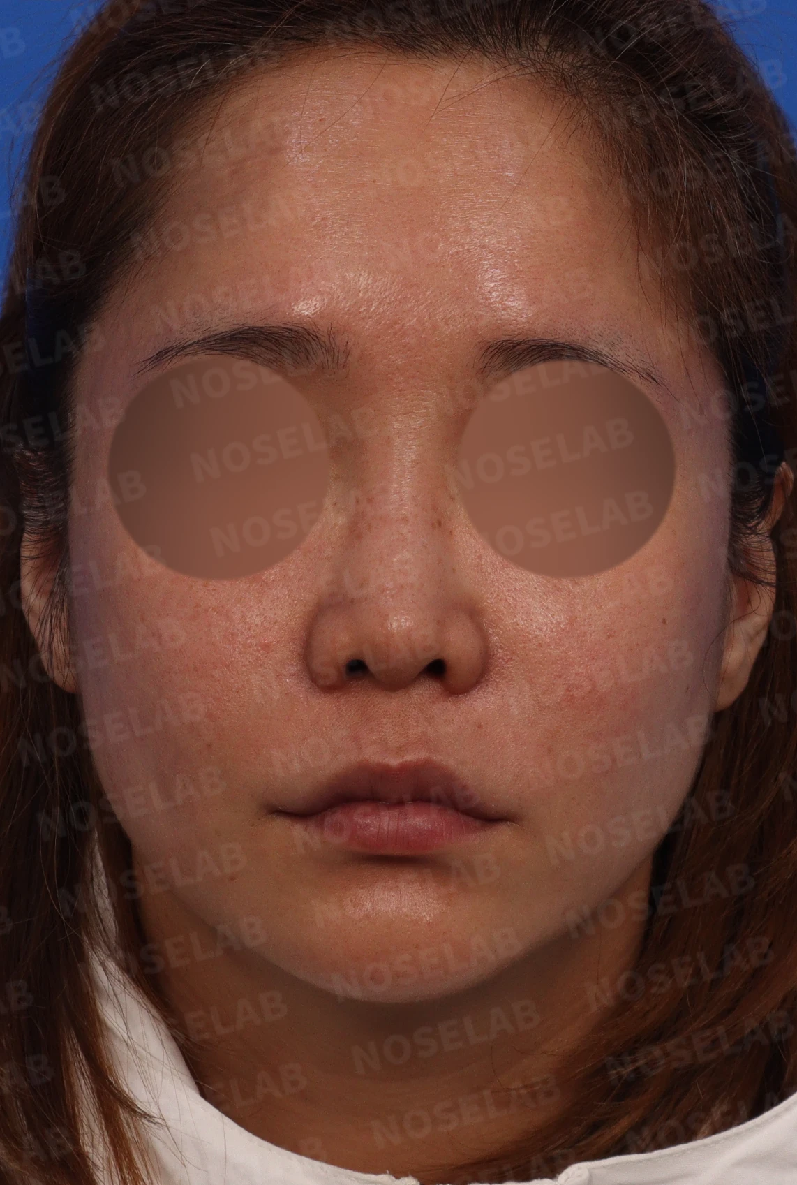
The nose is overall small and short
The glabella is widened and looks unnatural due to the filler
The nasal bridge appears deviated to the right
Noticeable nostril show
Noticeable asymmetry from the prior alar reduction
Profile view:
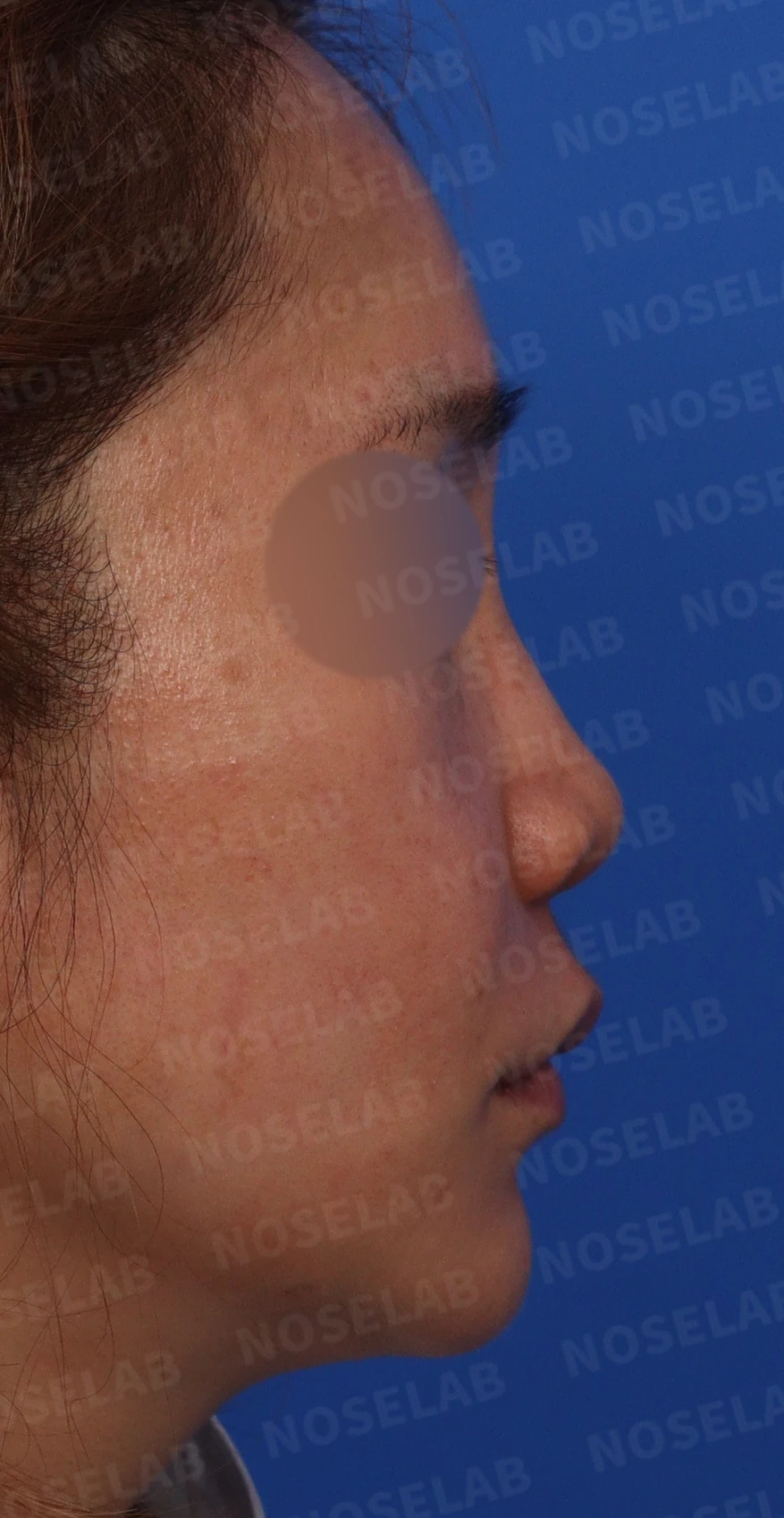
Observed protruding mouth
The nasal tip and bridge height are low, giving an overall flat appearance
Oblique view:
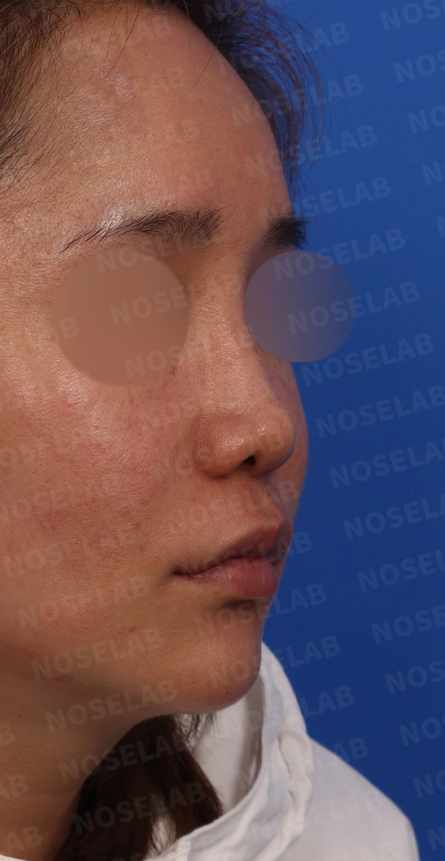
The deviation of the nasal bridge is even more pronounced
The low nasal tip is distinctly visible
Basal (nostril) view:
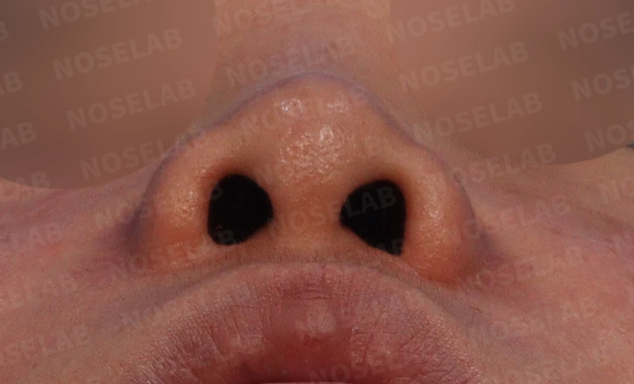
Noticeable nostril asymmetry
The nasal tip looks depressed
Surgical plan
Taking into account the patient's condition and requests, the following surgical plan was established:
a) Complete removal of foreign material (calcium filler) from the nasal bridge and nasal tip
b) Nasal tip reconstruction and septal extension using autologous rib cartilage
c) Repositioning the nasal tip so that the nostrils are less visible from the front
d) Improvement of the protruding mouth by correcting the nasolabial angle
e) Correction of nostril asymmetry through lower lateral cartilage repositioning and suturing
f) Nasal bridge augmentation using a silicone implant
The patient wanted a natural-looking nose, but the reference photos brought to the consultation featured very high, dramatic noses. Therefore, I planned a design that maintained a natural look while providing a well-defined, dramatic profile.
In particular, because the nostrils had become smaller due to previous alar reduction surgery, I explained that raising the height excessively could cause the nostrils to narrow into an unnatural, elongated "11" shape. I informed the patient in advance that the height would be adjusted to maintain a natural look.
Surgical procedure
The intraoperative findings and key surgical steps were as follows:
a) Foreign material removal:
A larger amount of calcium filler than expected was found.
The filler had spread into the upper glabella directly beneath the forehead, and it was entirely removed.
b) Nasal tip reconstruction:
Because the septum was thin and weak, it was solidly reinforced using autologous rib cartilage.
The nasal tip was elevated and refined.
c) Nasal bridge reconstruction:
The nasal bridge, which had become very low after removing the foreign material, was elevated using a silicone implant.
The profile was designed to connect smoothly from the forehead to the nasal tip.
d) Lower lateral cartilage reinforcement:
The bilateral lower lateral cartilages, which were very small and thin, were reinforced with autologous rib cartilage.
e) Nasolabial angle correction:
Using autologous rib cartilage, the nasolabial angle was corrected, creating the effect of improving the protruding mouth.
All procedures were performed through a closed approach (inner-incisional rhinoplasty).
Before-and-after photo comparison
Front view:
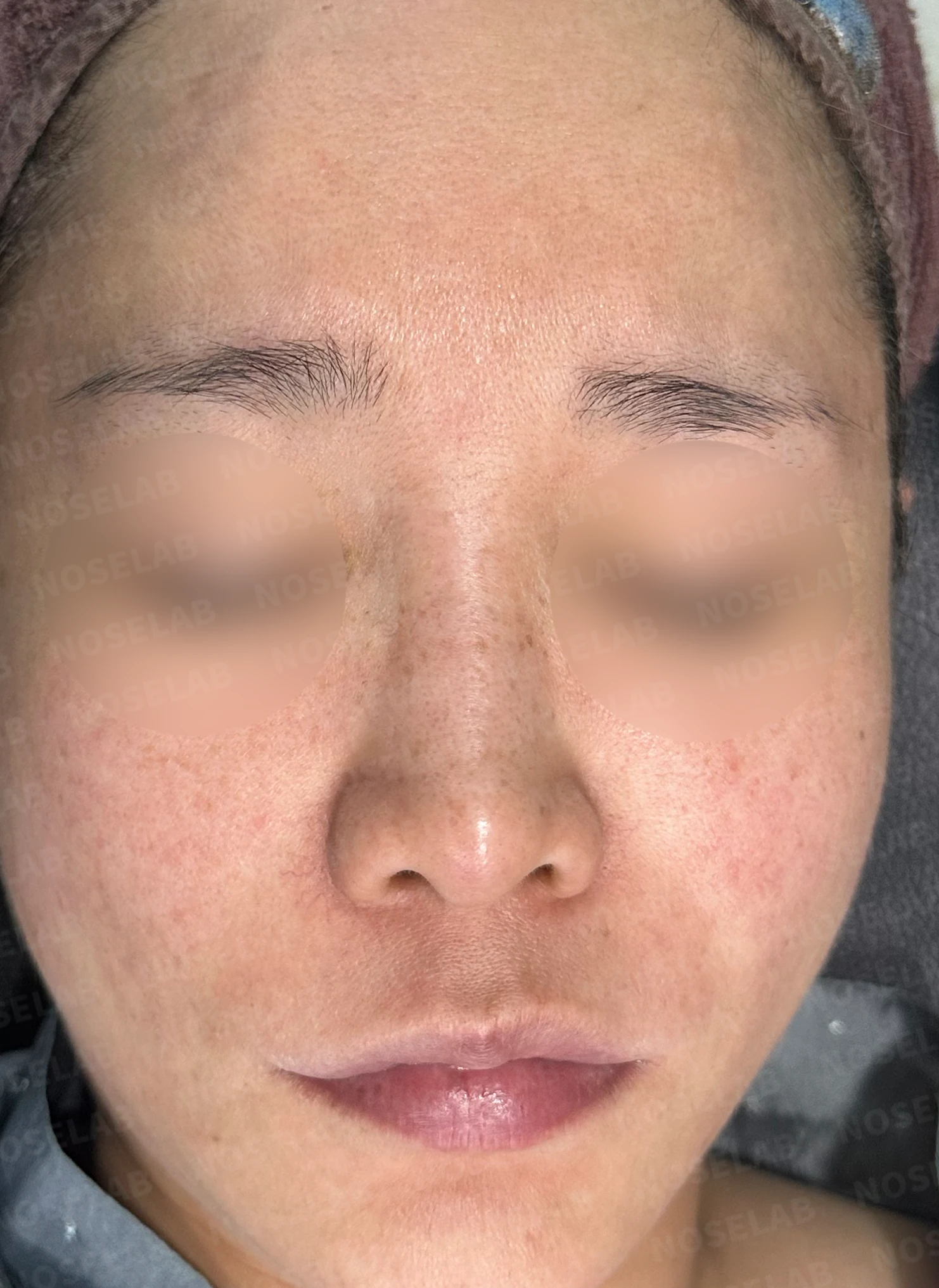
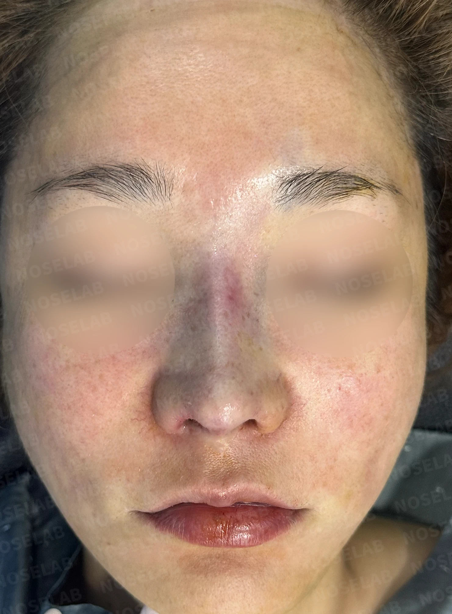
Before surgery / Day 0 immediately after surgery Photos taken immediately after surgery show swelling and may differ from the final result.
The widened glabella was corrected to look natural.
The nasal bridge deviated to the right was straightened.
Nostril asymmetry was improved, though complete symmetry was difficult to achieve due to the influence of the previous alar reduction.
The overall nasal contour has become more distinct.
Profile view:
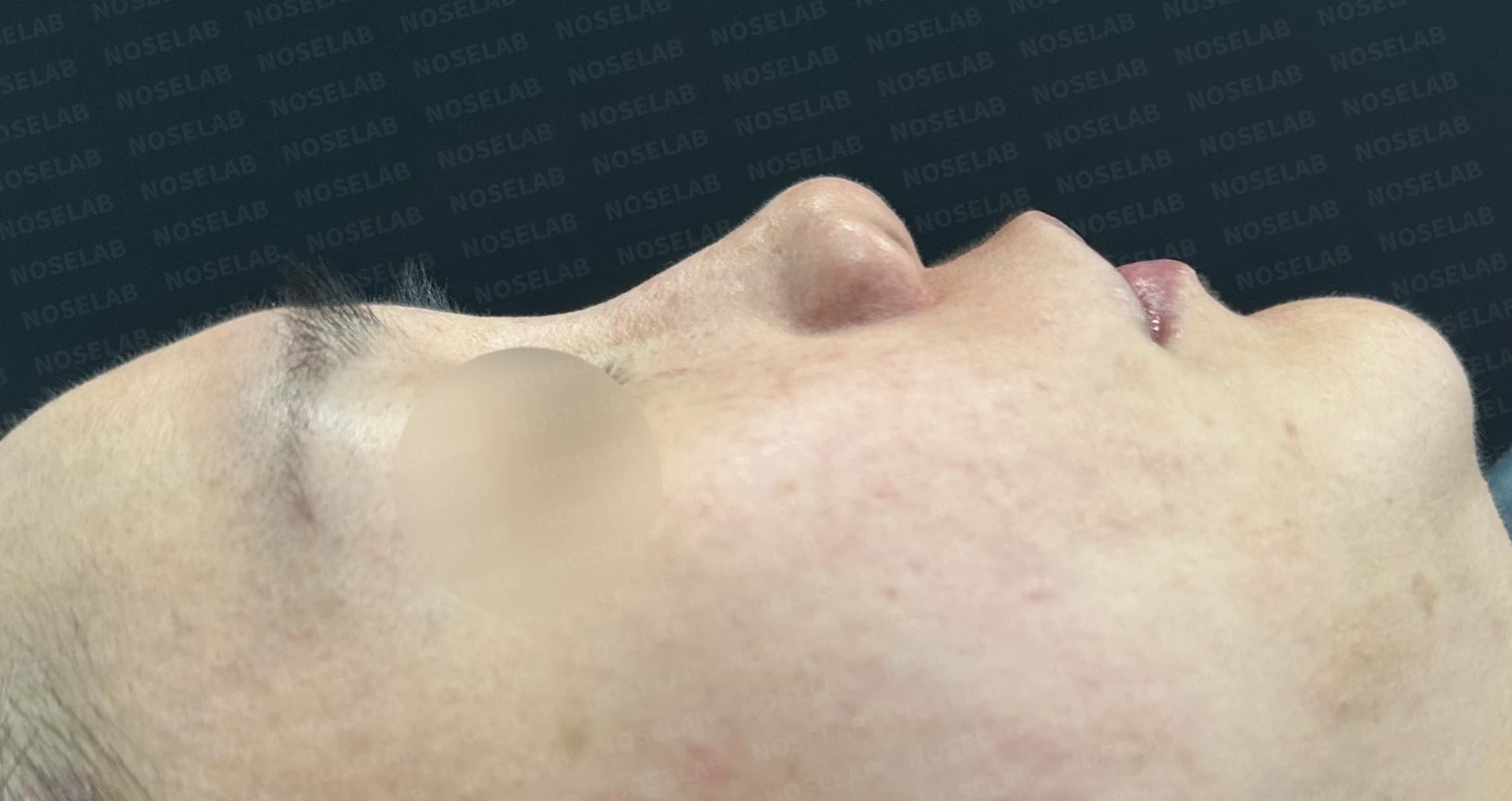
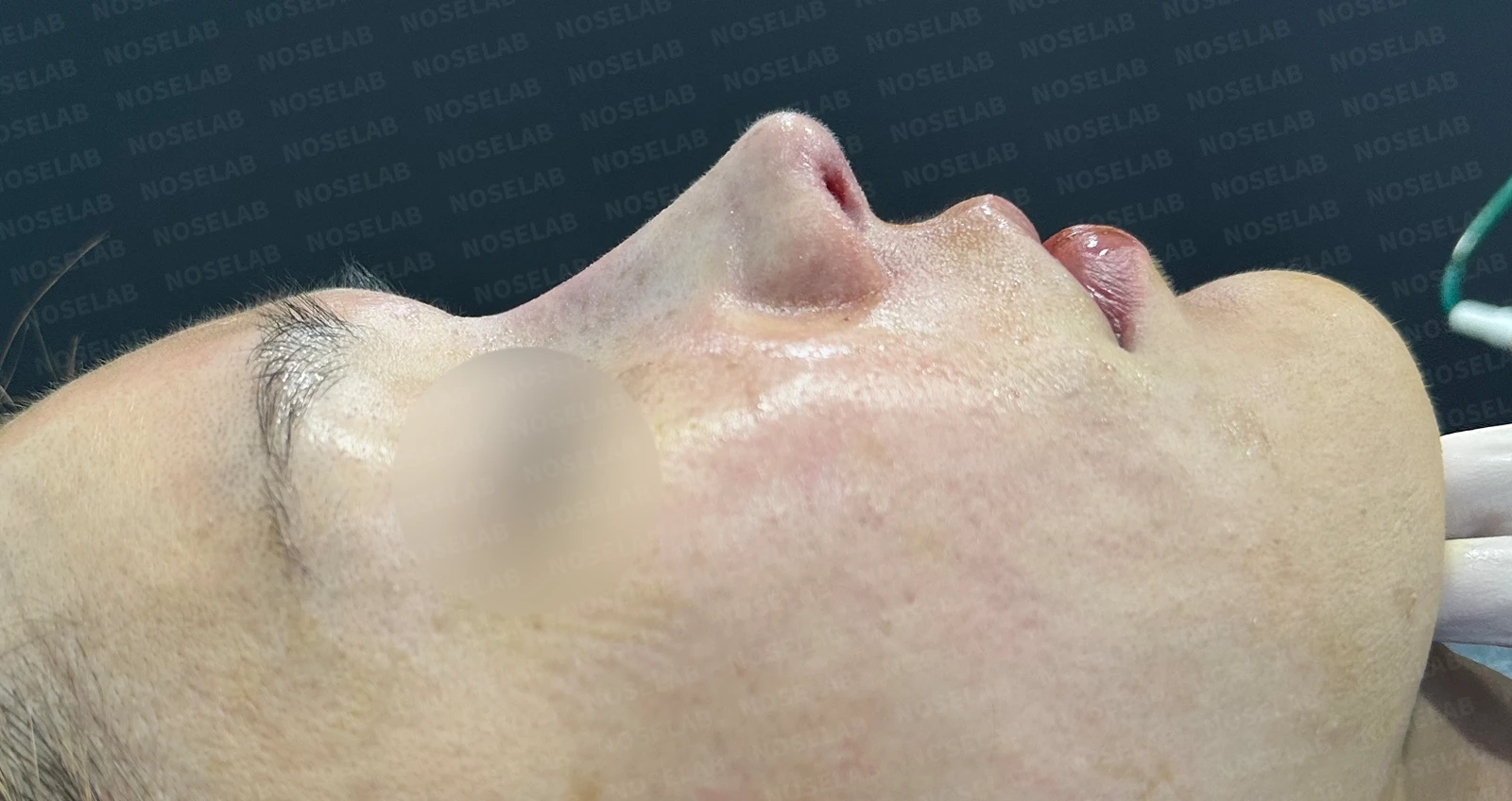
Before surgery / Day 0 immediately after surgery Photos taken immediately after surgery show swelling and may differ from the final result.
The nasal tip was properly elevated, improving the flat appearance.
Harmony between the nasal bridge and tip significantly improved three-dimensional projection.
The appearance of the protruding mouth improved, enhancing overall facial balance.
Oblique view:
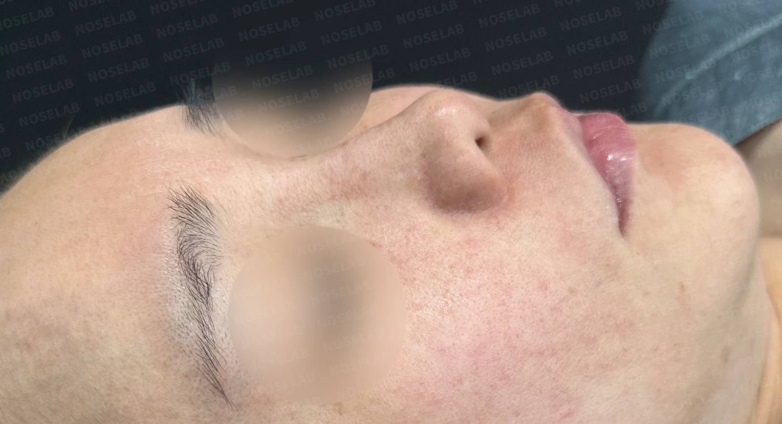
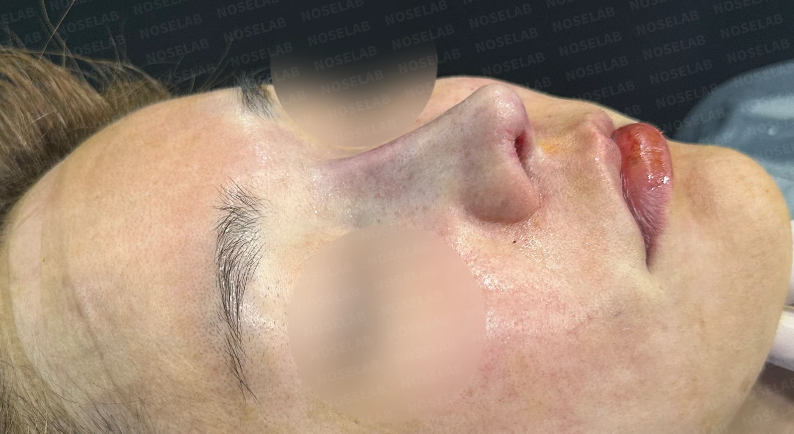
Before surgery / Day 0 immediately after surgery Photos taken immediately after surgery show swelling and may differ from the final result.
The deviation of the nasal bridge was corrected, showing a smooth line.
The height and projection of the nasal tip were naturally improved.
Nostrils:
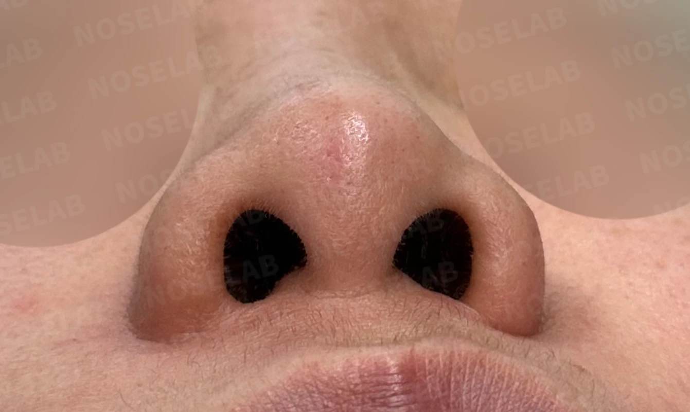
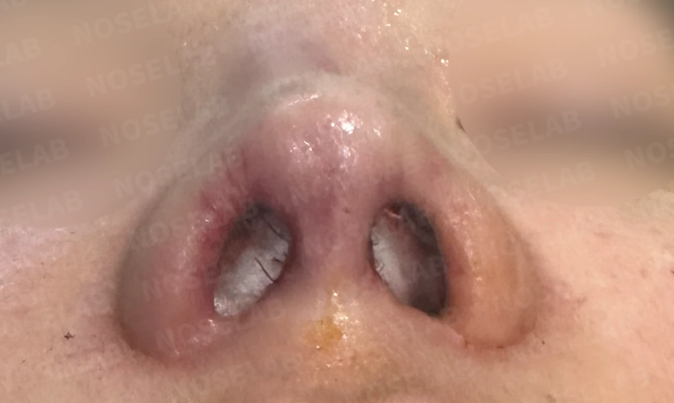
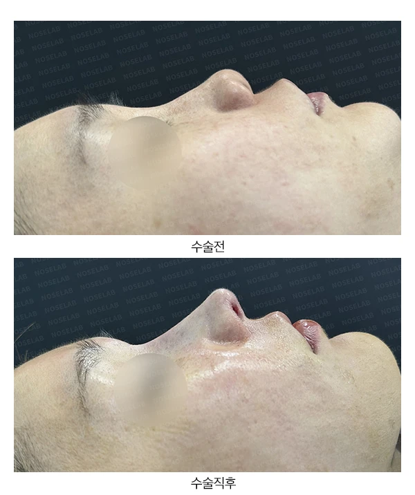
Before surgery / Day 0 immediately after surgery Photos taken immediately after surgery show swelling and may differ from the final result.
The depressed nasal tip was elevated and improved.
Nostril asymmetry showed marked improvement, although a slight difference remains due to the prior surgery.
Photos immediately after surgery
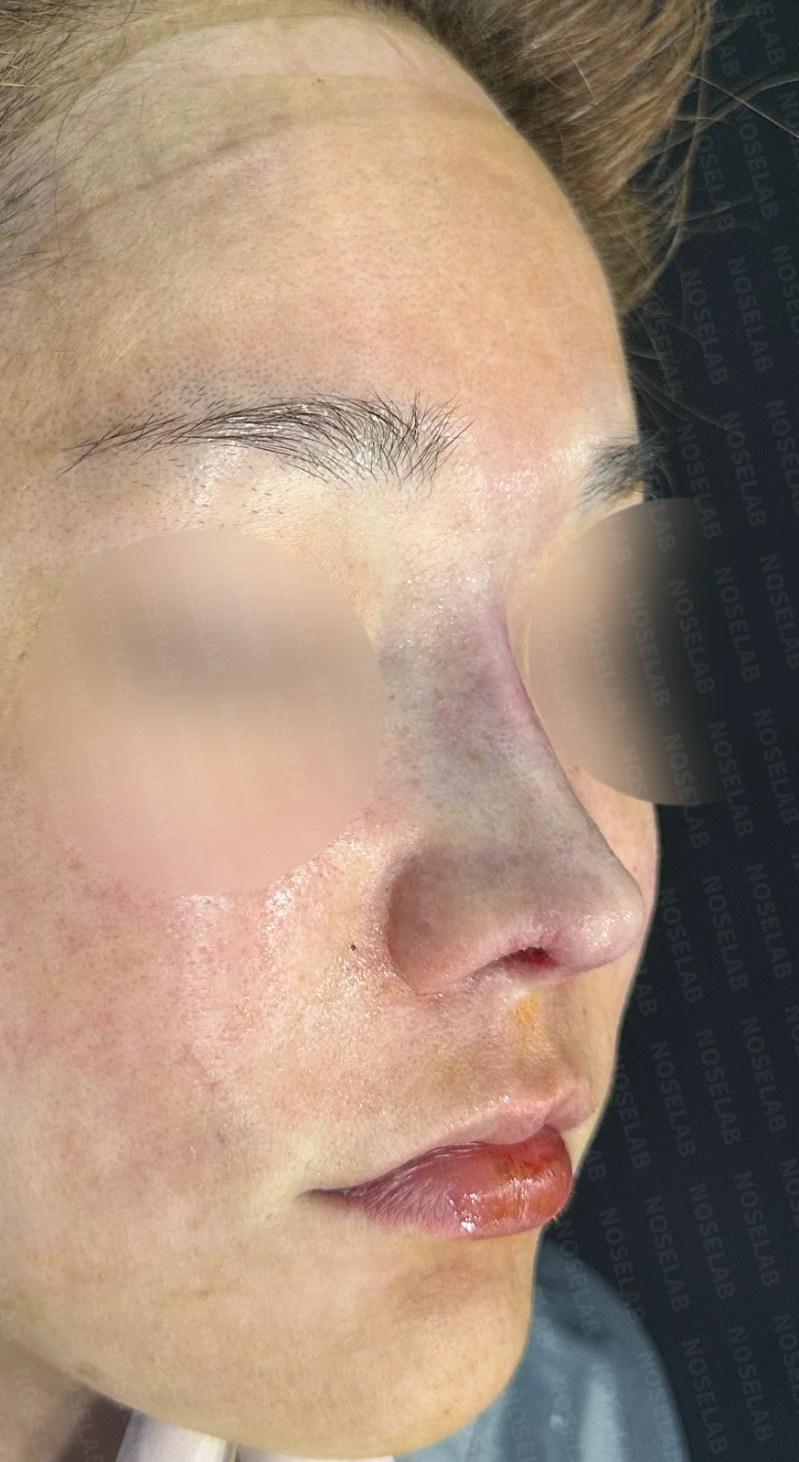
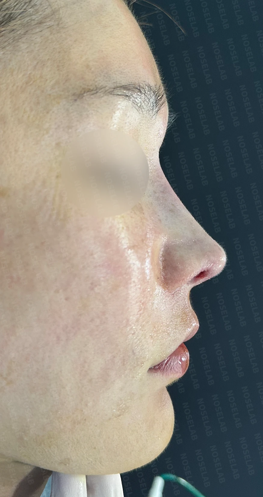
Photos immediately after surgery (Day 0): front, oblique, and profile viewsImmediate postoperative photos show swelling and may differ from the final result.
Basal (nostril) view immediately after surgery (Day 0)Immediate postoperative photos show swelling and may differ from the final result.
Conclusion
This was a complex case requiring foreign body removal combined with nasal reconstruction. As a principle, all foreign materials in the nose should be removed. In particular, non-absorbable semi-permanent fillers and calcium fillers must be completely removed. This is critical for preventing future inflammation.
Because removing foreign materials carries the risk of damaging normal tissues, including blood vessels and nerves, the procedure must be performed by an experienced specialist. In particular, vascular injury can lead to severe complications such as skin necrosis.
Inner-incisional surgery is an approach well-suited to minimizing these risks. Because it preserves the columellar artery, tip necrosis can be prevented even if partial injury occurs to dorsal nasal blood vessels.
In addition, using sturdy, high-quality cartilage is paramount when refining a short, small nose. Therefore, I strongly recommend the use of autologous rib cartilage.
Closing remarks
Rhinoplasty—especially when foreign body removal and reconstruction are required—demands a very meticulous approach. A customized surgical plan tailored to each patient's specific anatomy and high-level surgical expertise are required.
I hope this case helps those considering revision rhinoplasty for complex concerns. For a safe result, thorough consultation and careful selection of surgical technique are essential. NOSELAB always prioritizes patient safety, striving for the intended result.
#foreignbodyremoval #closedforeignbodyremoval #shortnose #smallnose #closedshortnosesurgery #closedsmallnosesurgery #closedrhinoplasty #closedribrhinoplasty #rhinoplasty #revisionrhinoplasty #autologousribcartilagegraft #septalextension #calciumfillerremoval #bridgerevision #tiprhinoplasty

Limitations and risks
When autologous rib cartilage is used, scarring and pain may remain at the donor site on the chest, and pneumothorax (pleural injury) can occur rarely. Over time, the grafted cartilage may warp or undergo partial resorption.
When foreign materials such as injected fillers are removed and reconstructed, contracture may recur, nasal height and length may decrease, and staged surgery may be necessary depending on the skin condition.
When the septum is corrected at the same time, nasal obstruction may persist or recur, and in rare cases, septal perforation or changes in smell may occur.
Common potential risks include infection, bleeding, asymmetry, scarring, skin necrosis, and insufficient improvement; results vary depending on individual skin condition and previous surgical history.