Hello.
I am Dr. Peter C. Kang, medical director.
Today, I would like to share a surgical case where a nose that lacked three-dimensional definition due to a high radix, a high nasal bridge, and a low tip was transformed into a defined, refined profile by lowering the radix and elevating the nasal tip.
Preoperative design consultation
The patient had previously undergone two rhinoplasty procedures along with alar reduction. The following concerns were identified during the preoperative consultation:
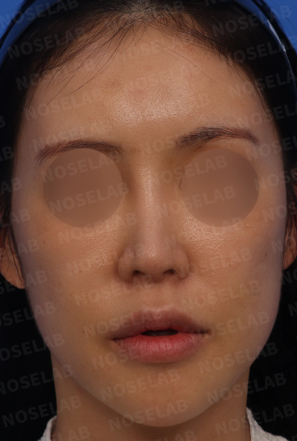
Front view
Front view:
Abnormally high radix
Overall nasal deviation toward the right
Noticeable nostril asymmetry
Depression above the right ala
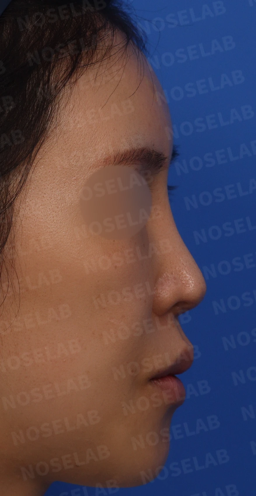
Profile view
Profile view:
Flat overall appearance because the nasal bridge was high while the nasal tip drooped
Lack of three-dimensional definition
Protruding mouth
Retracted columella
Narrow nasolabial angle
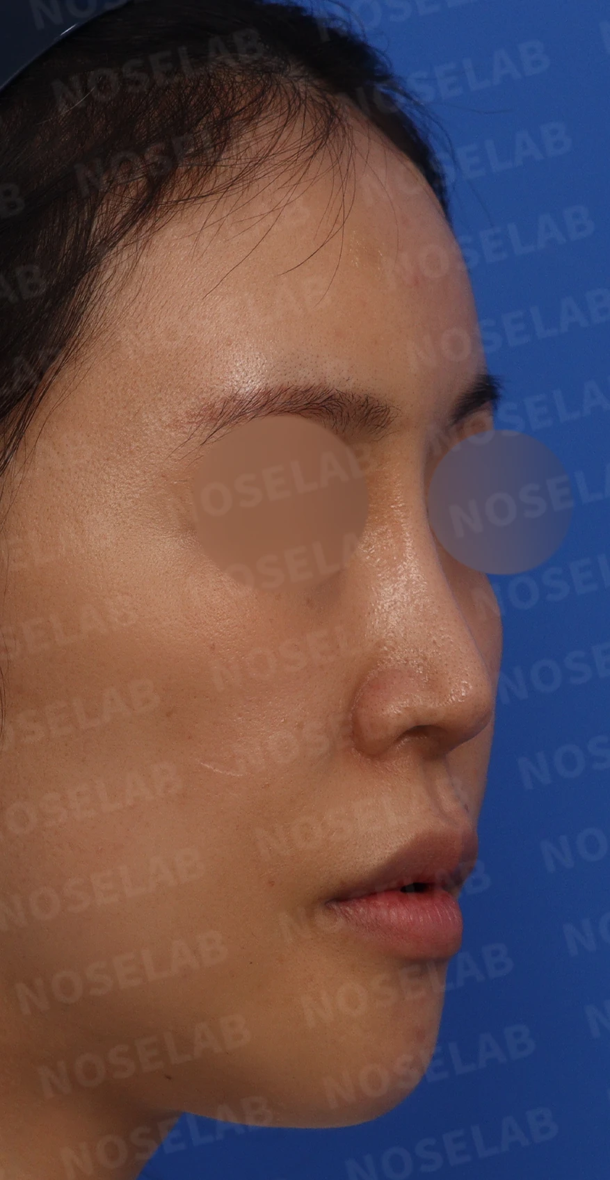
Oblique view:
Very unnatural bridge line due to the high radix
Noticeable drooping of the nasal tip
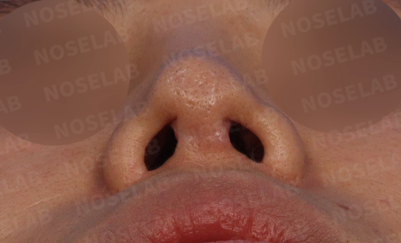
This is the basal (nostril) view.
Basal (nostril) view:
Asymmetry resulting from the previous alar reduction
Identified left nasal cavity stenosis
Surgical plan
Taking into account the patient's condition and requests, the following surgical plan was established:
a) Nasal tip reconstruction and septal extension using autologous rib cartilage
b) Correction of a deviated septum via septoplasty (narrowed left nasal cavity)
c) Lowering the radix and inserting a new implant
d) Creating a natural curve along the nasal bridge
e) Reconstruction of the right lower lateral cartilage
f) Additional cartilage grafting to reinforce the depressed skin above the right ala
The patient desired a high, well-defined nose, but considering the nasal skin envelope, the plan was made to create as defined a look as possible within a natural range. In addition, I thoroughly explained the limitations resulting from the previous alar reduction, such as changes in nostril shape and the risk of tip retraction.
Surgical procedure
During actual surgery, the condition of the tissues was worse than expected. The main surgical steps were as follows:
a) Release of adhesions: Dissection was performed cautiously, as severe adhesions between the cartilage and skin were present from the very start of tip dissection.
b) Septal reinforcement: Judging that the tip drooped because the septal cartilage was extremely thin and weak, the septum was reinforced firmly using autologous rib cartilage.
c) Nasal tip reconstruction: The tip was elevated as much as safely possible and its structural support was reinforced using autologous rib cartilage.
d) Nasal bridge reconstruction:
The overall height was adjusted while lowering the radix, taking into account the nasal bone that had been lowered by previous surgery.
The deviated existing implant was removed, and a new implant was carved to fit the nasal bone precisely before insertion.
e) Alar cartilage reconstruction: Because the right lower lateral cartilage was almost entirely missing, it was completely reconstructed using autologous rib cartilage.
f) Additional correction: The previously used ear cartilage was crushed and grafted into the depressed area on the right side.
g) Submucosal resection of the inferior turbinate using radiofrequency: Hypertrophic rhinitis was corrected to improve symptoms of nasal obstruction.
The entire procedure was performed through a closed approach.
Before-and-after photo comparison
Front view:
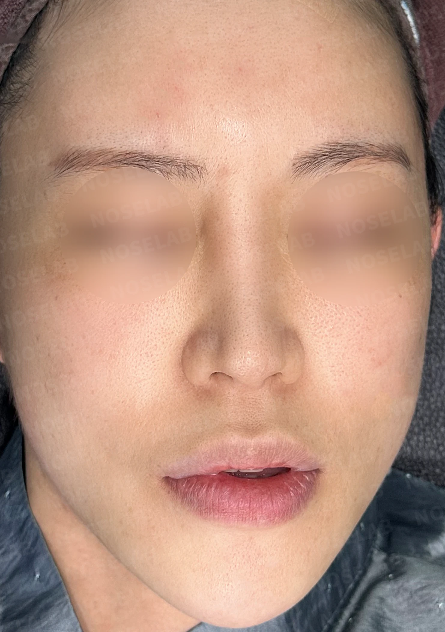
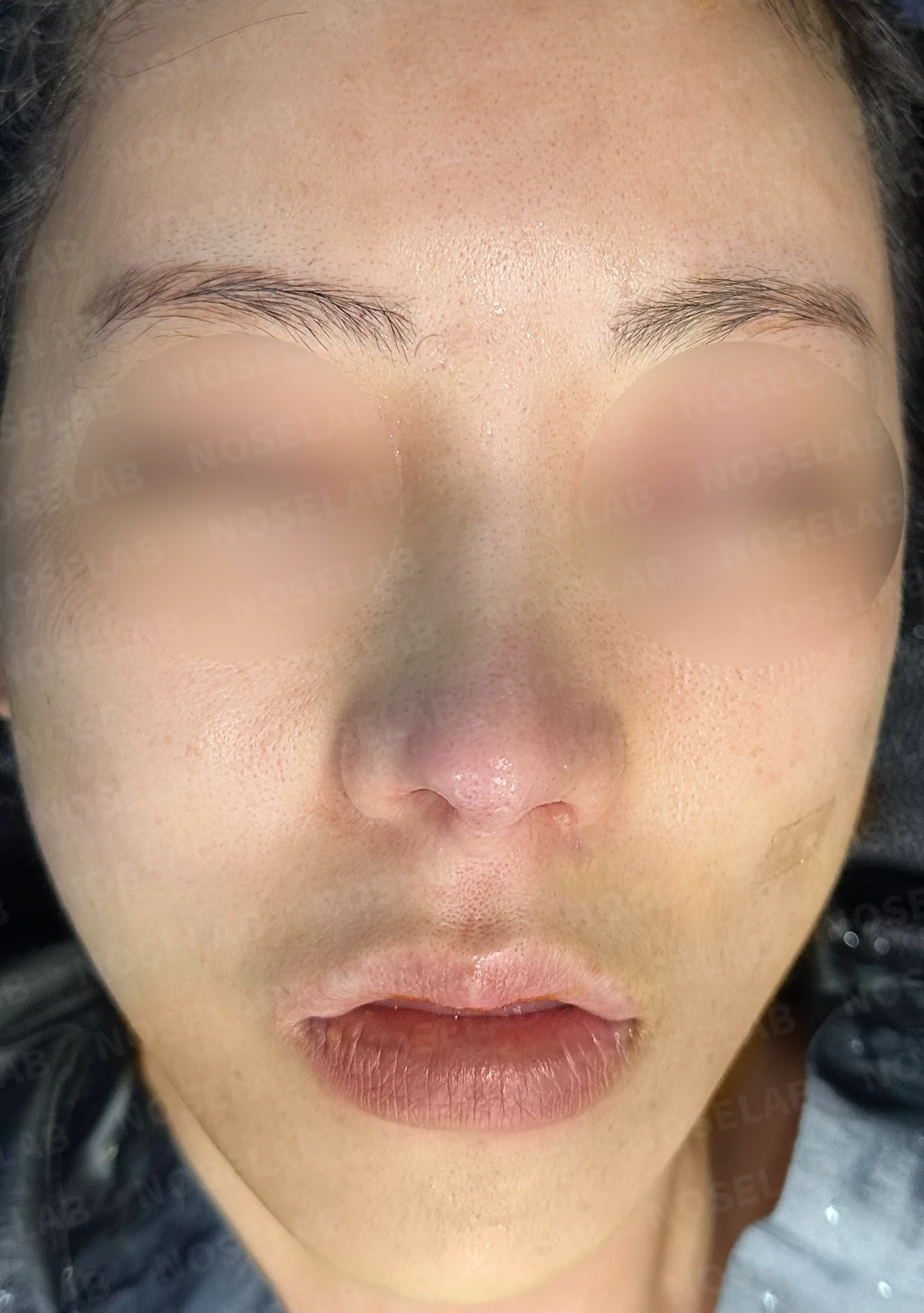
Before surgery / Day 0 immediately after surgery Photos taken immediately after surgery show swelling and may differ from the final result.
The height of the radix was lowered naturally.
The nose that deviated to the right was straightened.
Nostril asymmetry was improved, although complete symmetry was difficult to achieve due to the effects of the previous alar reduction.
The overall nasal contour has become more distinct.
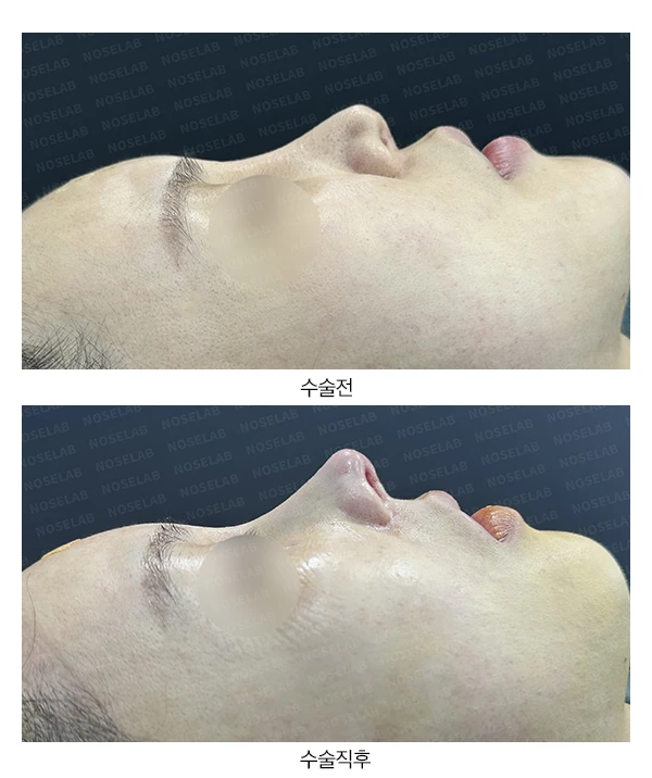
Profile view:
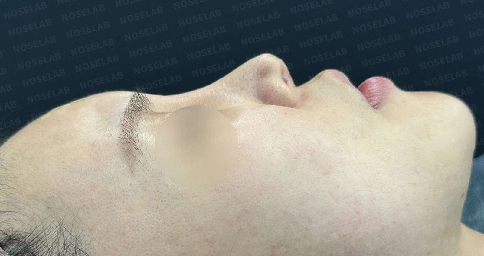
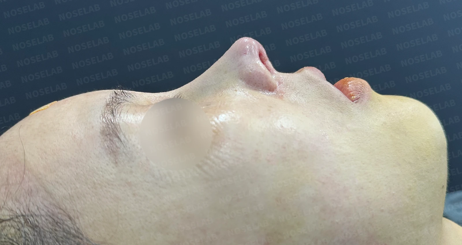
Before surgery / Day 0 immediately after surgery Photos taken immediately after surgery show swelling and may differ from the final result.
The nasal tip was adequately elevated, correcting the drooping appearance.
Harmony between the nasal bridge and tip significantly improved three-dimensional projection.
A natural curve was established along the nasal bridge, creating an elegant line.
Oblique view:
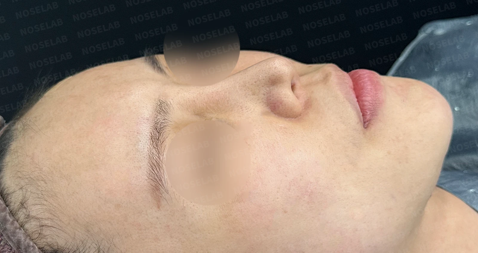
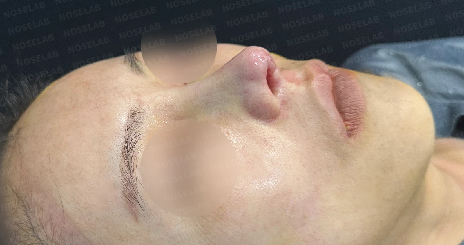
Before surgery / Day 0 immediately after surgery Photos taken immediately after surgery show swelling and may differ from the final result.
The deviation of the nasal bridge was corrected, showing a smooth line.
The drooping tip was corrected, resulting in an overall balanced appearance.
Nostrils:
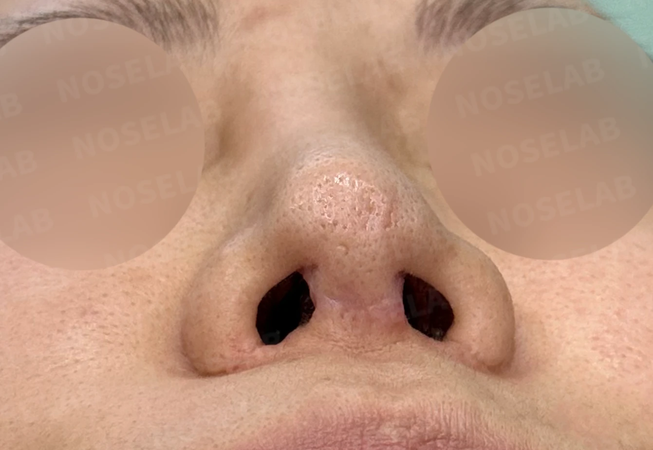
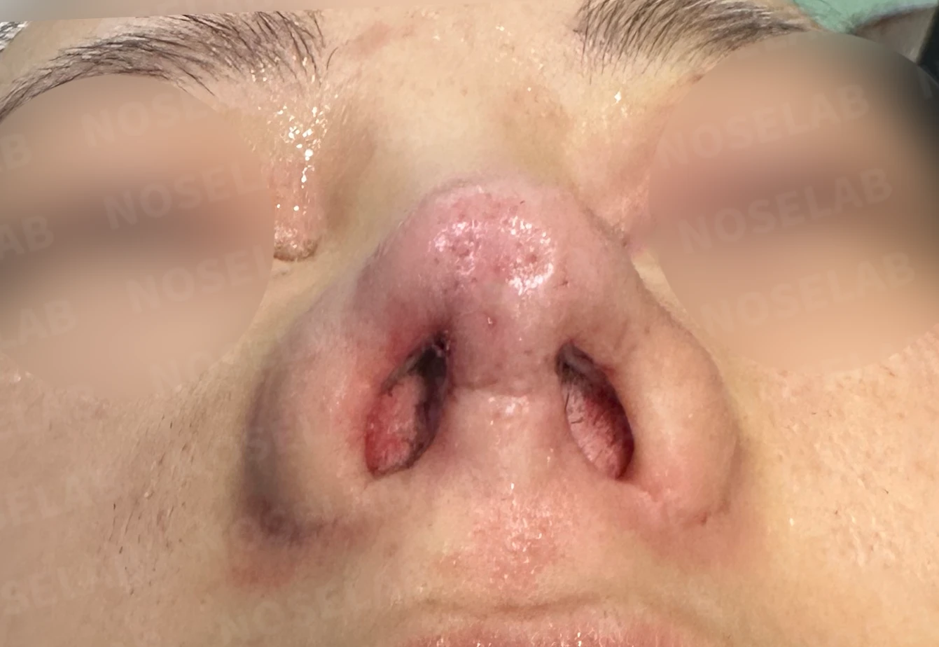
Before surgery / Day 0 immediately after surgery Photos taken immediately after surgery show swelling and may differ from the final result.
The left nasal cavity was widened, improving symptoms of nasal obstruction.
The contour was improved through reconstruction of the right lower lateral cartilage.
Although complete symmetry was limited due to the previous alar reduction, the overall balance was enhanced.
Photos immediately after surgery
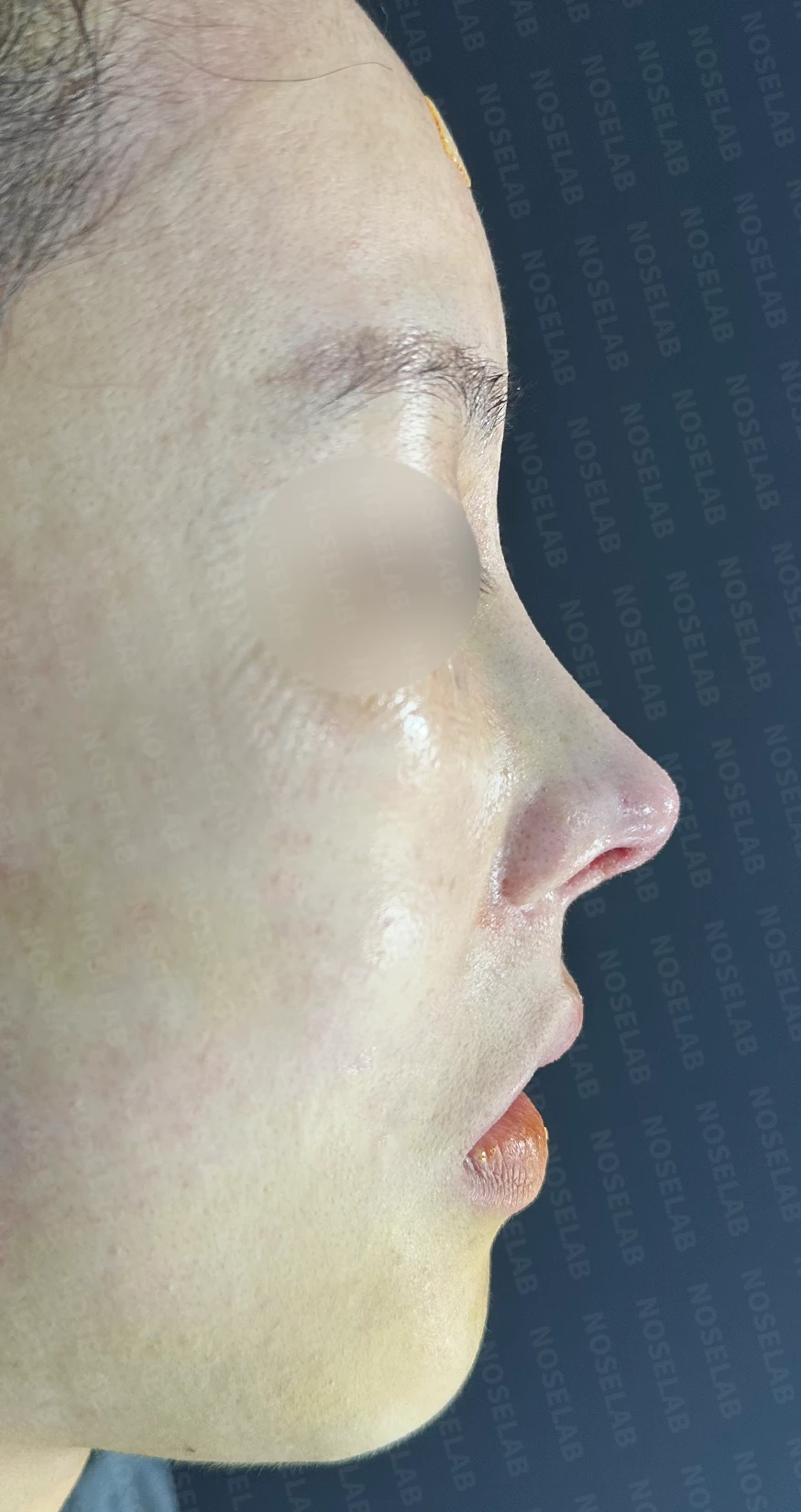
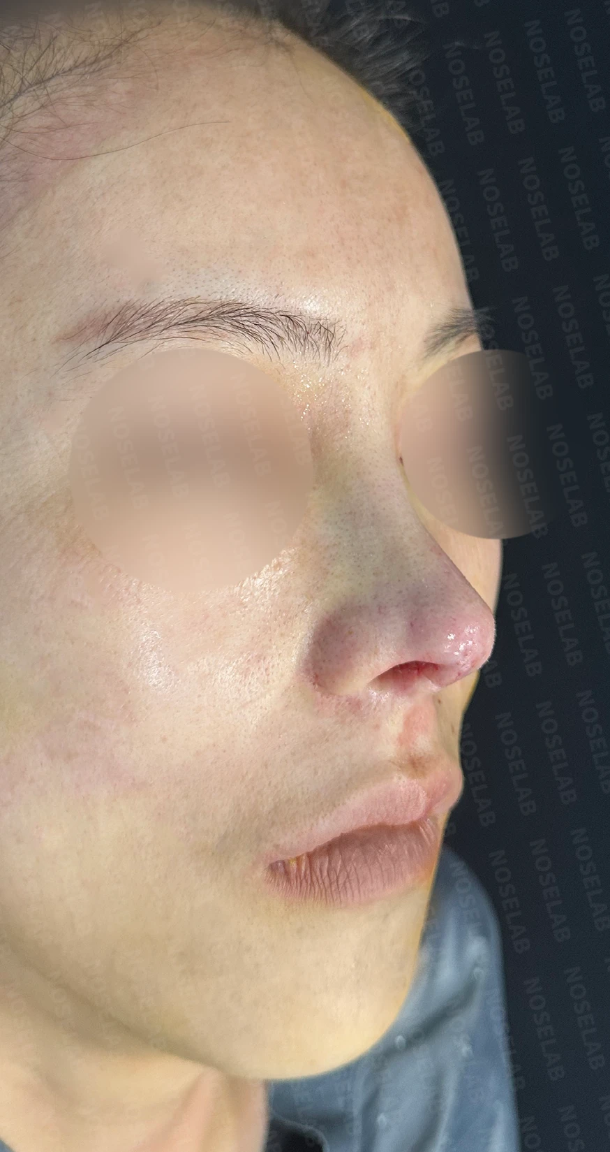
Photos taken on day 0, immediately after surgery. Front, profile, and oblique views. Immediate postoperative photos show swelling and may differ from the final result.
Nostril view photo.
Conclusion
This case illustrates a complex revision rhinoplasty that shows both the benefits and technical demands of the closed approach. Severe adhesions, damaged cartilage structures, and a deviated implant were all successfully addressed. In particular, performing extensive reconstruction using autologous rib cartilage through a closed approach is noteworthy.
Excessive elevation of the nasal bridge carries the risk of the implant shifting off-center and deviating. Therefore, designing the radix area naturally is extremely important. Because the height of the radix greatly influences the overall impression of the face, selecting the appropriate starting point is key.
In this surgery, considering the patient's existing skin condition and the limitations from previous surgery, we created a defined, three-dimensional nasal profile within a natural range. While complete symmetry was limited, significant improvements were achieved both functionally and aesthetically.
Closing remarks
Rhinoplasty, particularly revision surgery, requires a tailored approach suited to each patient's unique anatomy. A closed approach is a viable method to achieve natural outcomes without external columellar scars, but it requires advanced surgical skill and experience.
I hope this case helps those who are considering such complex revision rhinoplasty. A consultation with a specialist is essential to find an appropriate solution suited to each individual situation. I always place patient safety and satisfaction as my priority and strive to achieve the intended results.
#closedrhinoplasty #closedautologousribrhinoplasty #rhinoplasty #revisionrhinoplasty #autologousribcartilagegraft #septalextension #nasalbridgecorrection #tiprhinoplasty #deviatedseptumcorrection #alarcartilagereconstruction #customrhinoplasty

Limitations and risks
When autologous rib cartilage is used, scarring and pain may remain at the donor site on the chest, and pneumothorax (pleural injury) can occur rarely. Over time, the grafted cartilage may warp or undergo partial resorption.
When revising after implant removal, contracture may progress again after removal, the nasal height and length may decrease, and staged surgery may be necessary depending on the skin condition.
When the septum is corrected at the same time, nasal obstruction may persist or recur, and in rare cases, septal perforation or changes in smell may occur.
Common potential risks include infection, bleeding, asymmetry, scarring, skin necrosis, and insufficient improvement; results vary depending on individual skin condition and previous surgical history.