Hello.
I am Dr. Peter C. Kang, medical director.
Today, I will share the case of a patient who had a first surgery using septal cartilage and ear cartilage, and a second surgery using autologous rib cartilage, who visited our clinic to correct a widened, spread-out appearance of the nasal tip and nasal bridge. Following the second surgery, adhesions developed in the internal nasal mucosa, causing severe nasal obstruction on the right side, so the patient also wanted relief from the nasal obstruction. As the nasal tip collapsed toward the left, the tip height decreased, the tip deviated to the left, and nostril asymmetry occurred. In addition, because the patient had undergone alar reduction during a previous surgery, the length of the alae was also asymmetrical.
Let us examine the details of the preoperative design consultation.
Preoperative design consultation photo
These are photos taken before surgery.
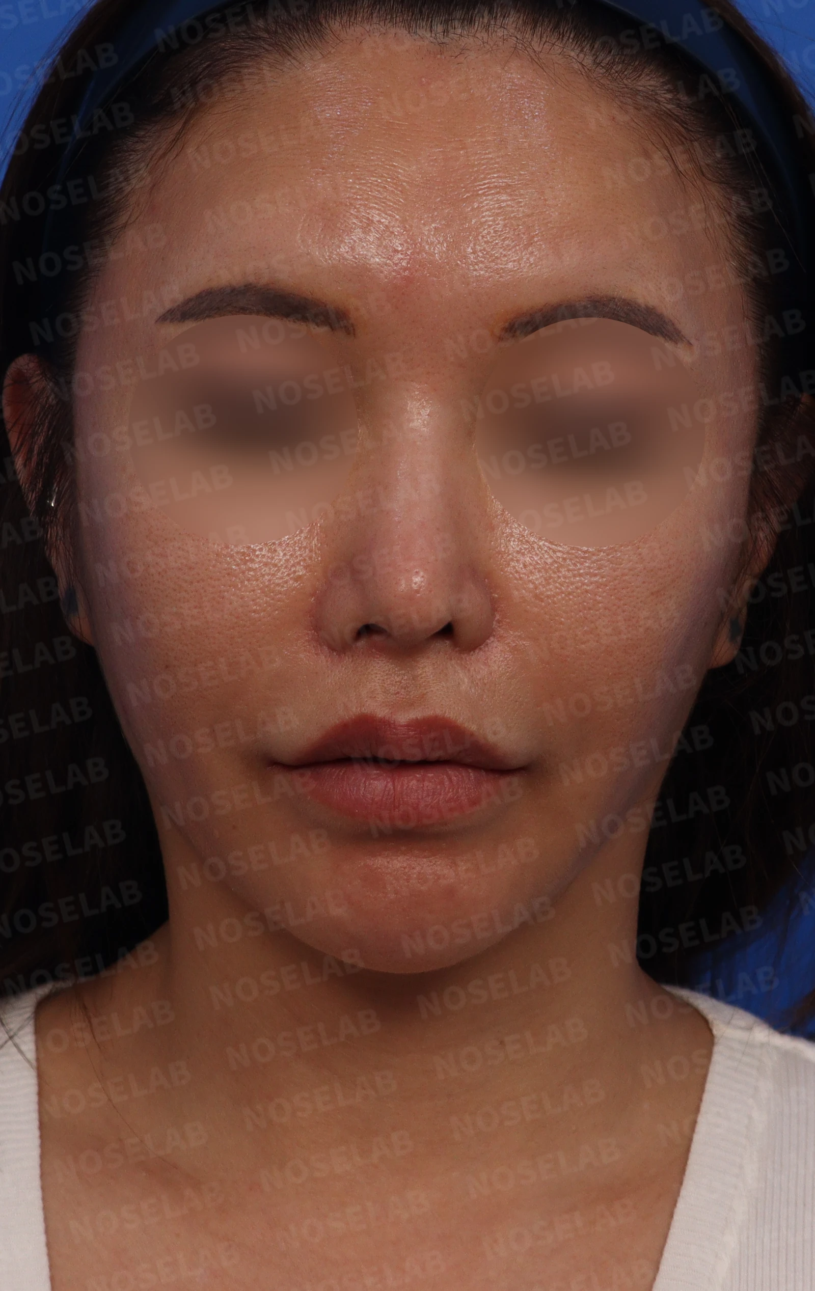
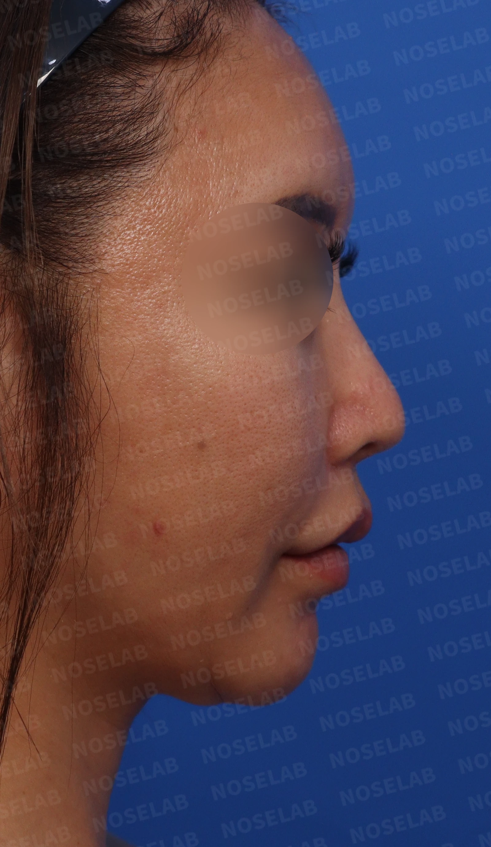
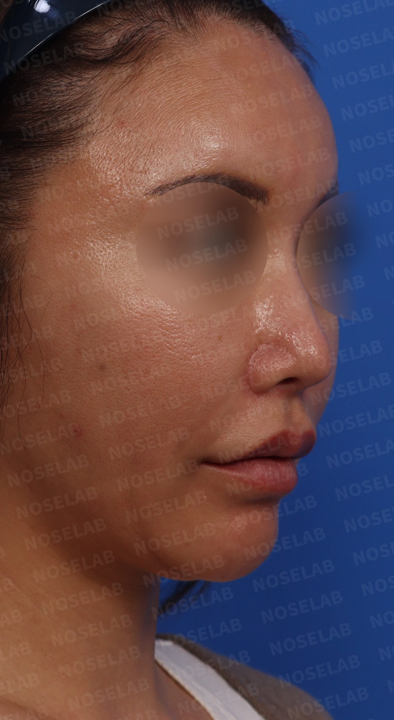
Preoperative design consultation photos: front, 90-degree profile, and 45-degree oblique views.
From the front, the nasal bridge and nasal tip appear spread out, the entire nose looks deviated to the left, and nostril asymmetry is noticeable. The nasal tip is drooping, and findings of a protruding mouth are visible. On the oblique view, a slight dorsal hump is present, and an acute nasolabial angle makes the protruding mouth appear more pronounced.
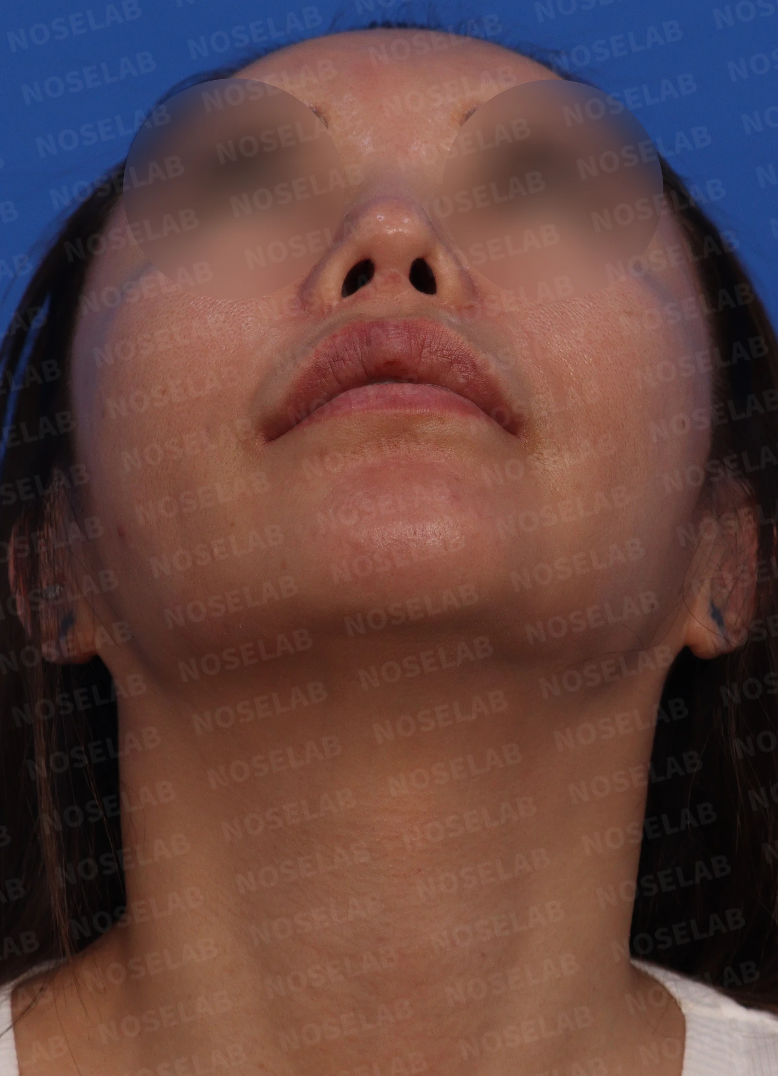
This is the preoperative basal (nostril) view.
The columella is tilted to the left, the nasal tip deviates to the left, and the nostrils differ in both shape and size.
Surgical plan
The nasal obstruction examination revealed severe septal cartilage deviation—to the right anteriorly and to the left deeper inside—along with bilateral inferior turbinate hypertrophy. Stenosis of the right internal nasal valve was observed, as well as right nasal mucosal adhesion. Therefore, we planned septoplasty, submucosal resection of the inferior turbinates using radiofrequency, lysis of intranasal adhesions, and nasal valve reconstruction.
We planned to correct the wide nasal bones using lateral osteotomies and the insertion of a silicone implant.
Believing the leftward-deviated nasal tip was due to damaged tip cartilage, we planned to remove all previously placed autologous rib cartilage, septal cartilage, and ear cartilage. We aimed to reconstruct the remaining septal cartilage as much as possible, and then construct a sturdy support framework using fresh autologous rib cartilage.
We planned to address the nostril asymmetry by performing nostril lowering (alar rim grafting).
We also planned to adjust the nasolabial angle to improve the appearance of the protruding mouth.
Before surgery vs. immediately after surgery
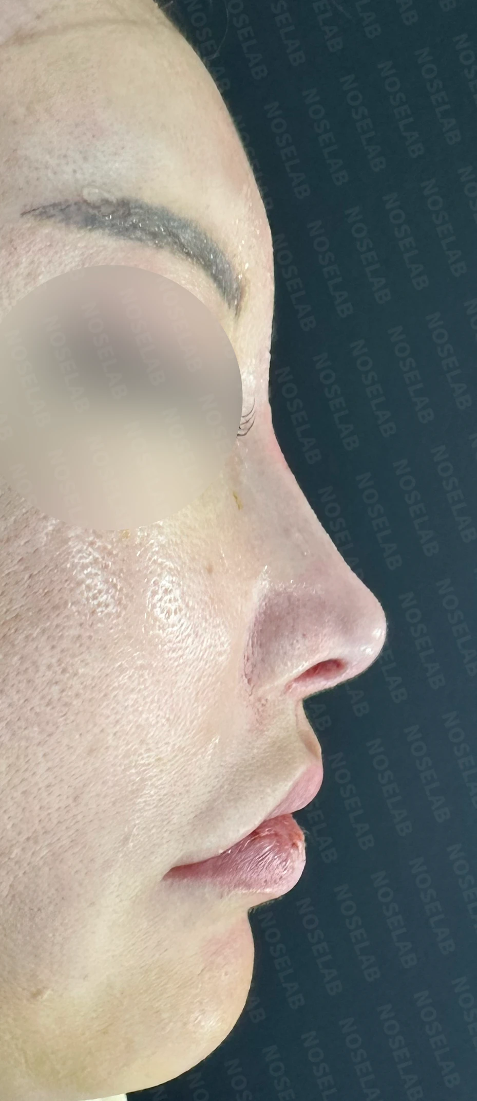
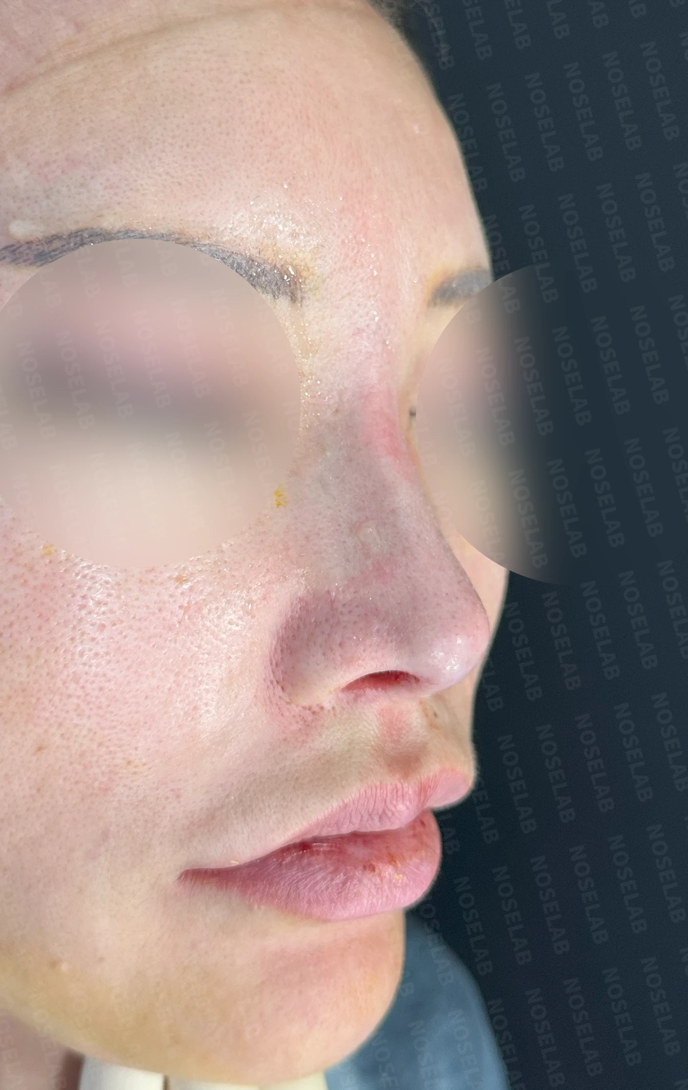
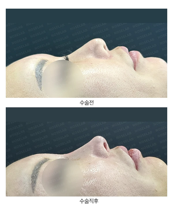
Let us compare the preoperative photos with the photos immediately after surgery (Day 0).Immediate postoperative photos show swelling and may differ from the final result.
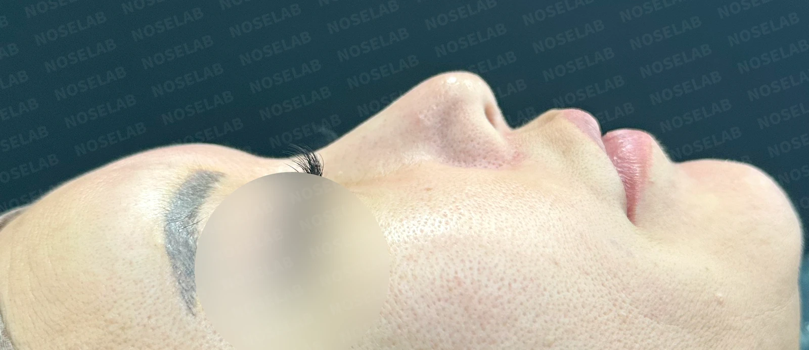
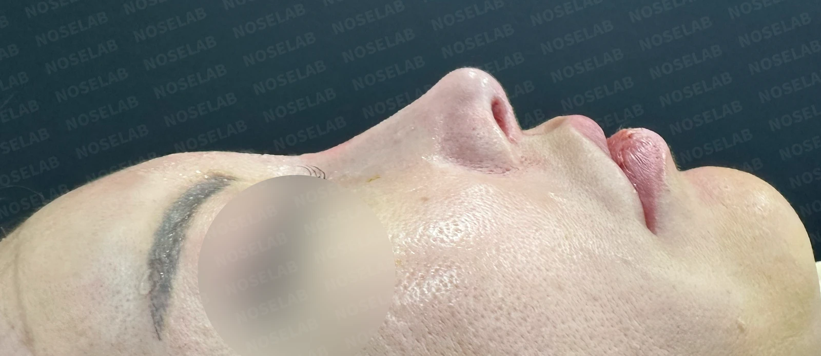
Before surgery / Day 0 immediately after surgery Photos taken immediately after surgery show swelling and may differ from the final result.
Because the patient did not want excessive tip height or a sharp, pinched tip, we raised the nasal tip height only slightly, lowered the radix, and smoothed the dorsum to create a soft, feminine, and refined impression.
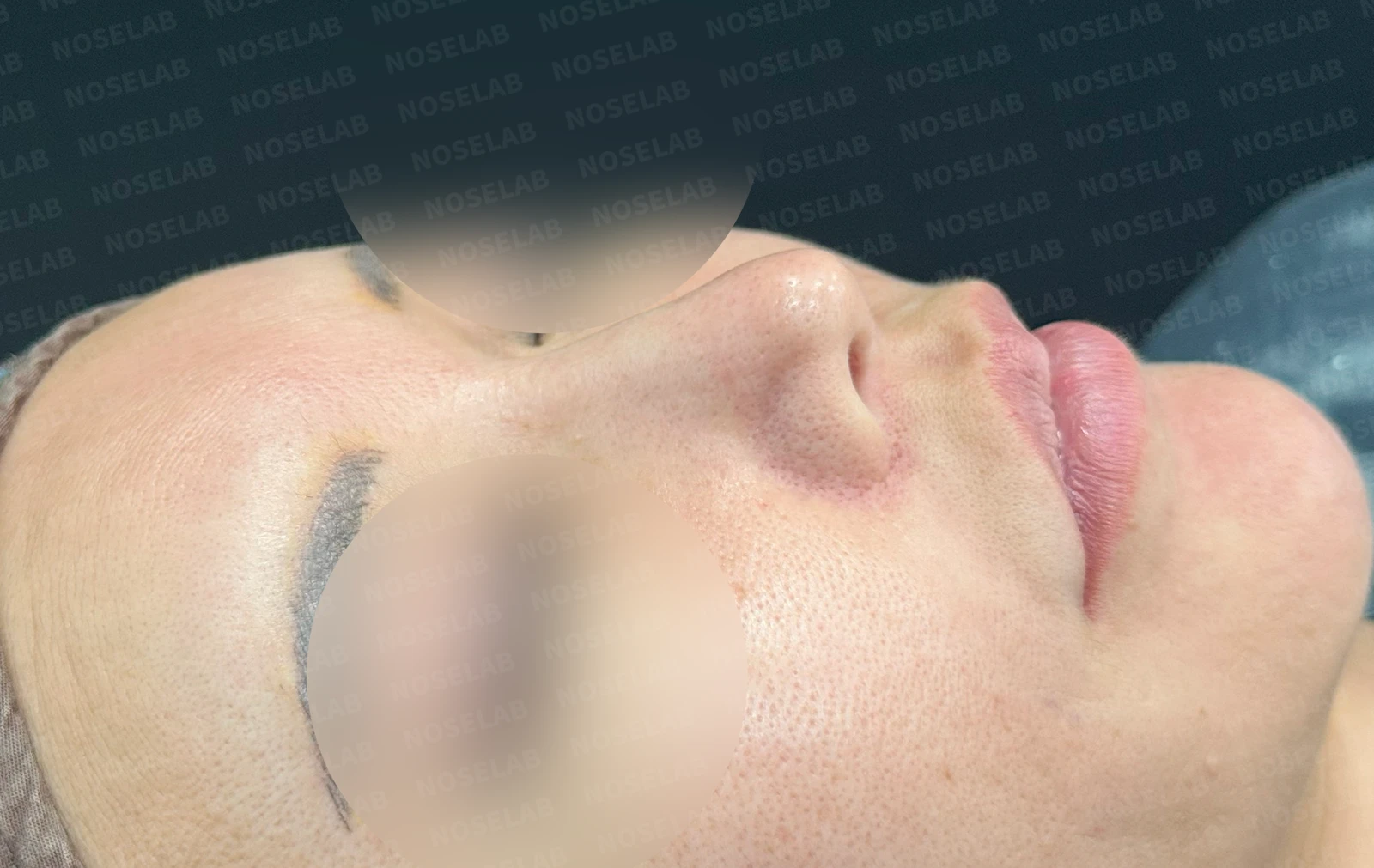
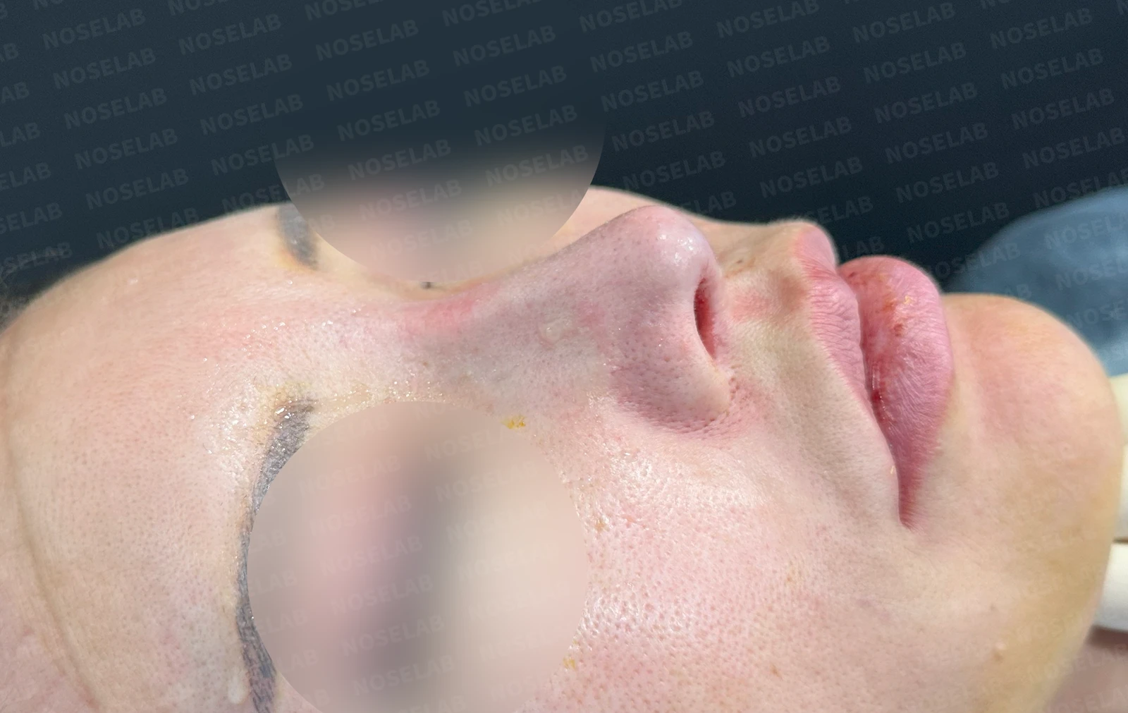
Before surgery / Day 0 immediately after surgery Photos taken immediately after surgery show swelling and may differ from the final result.
The nasal bridge was softly elevated, giving it a feminine contour.
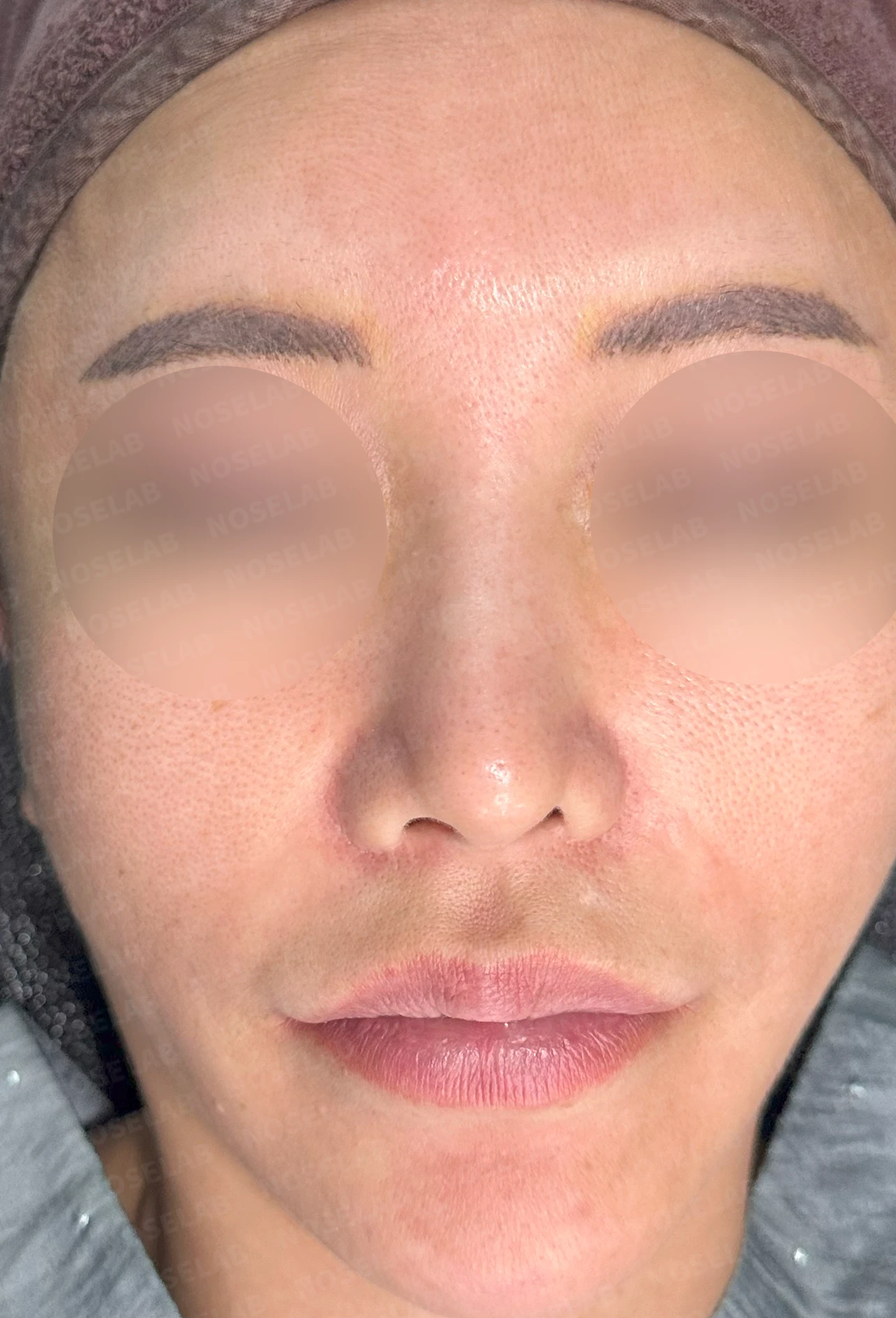
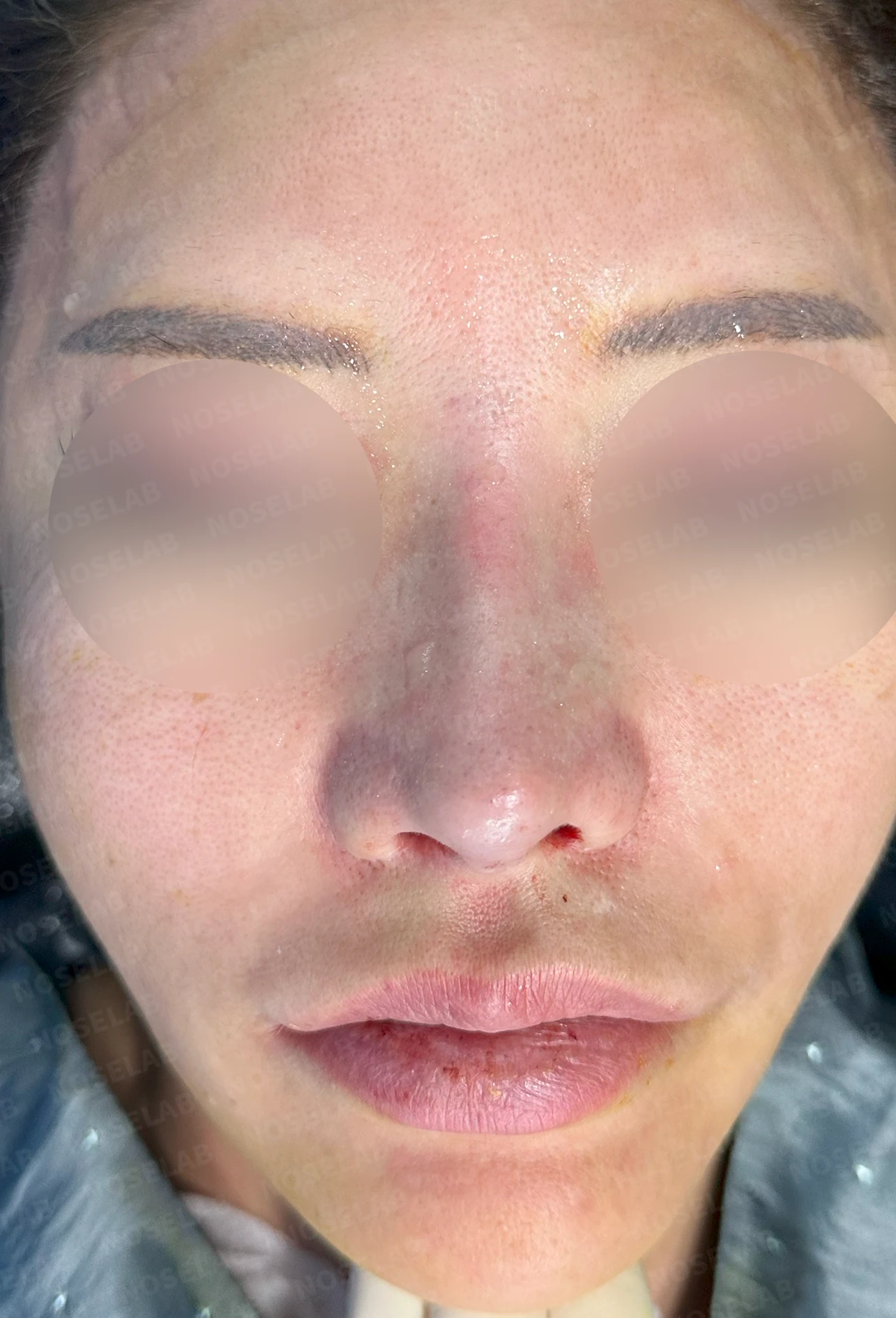
Before surgery / Day 0 immediately after surgery Photos taken immediately after surgery show swelling and may differ from the final result.
From the front, the leftward deviation of the nasal tip, the marked nostril asymmetry, and the pinched appearance have substantially improved, and the wide, crooked look of the nasal bridge has also been corrected considerably.
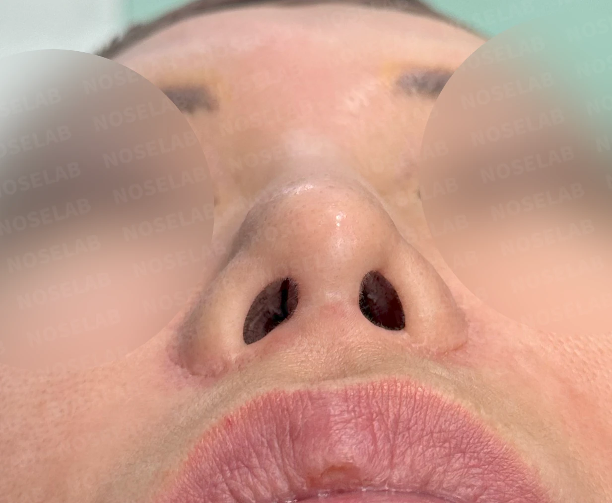
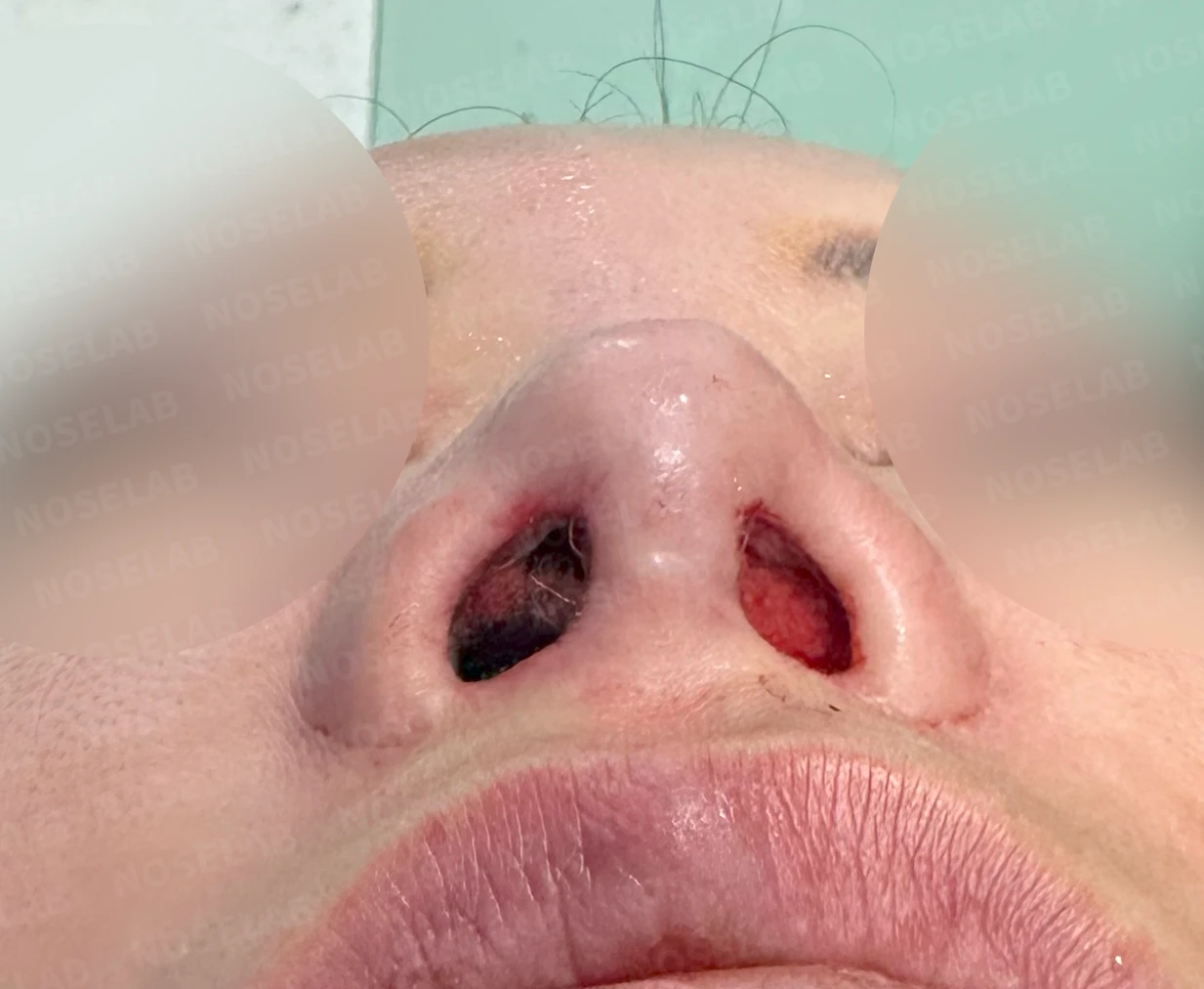
Before surgery / Day 0 immediately after surgery Photos taken immediately after surgery show swelling and may differ from the final result.
A noticeably improved feature is the shape of the nostrils. Although the discrepancy in alar length resulting from the previous alar reduction could not be altered, we aligned the overall contours and straightened the tilted columella, substantially improving nostril asymmetry.
Photos immediately after surgery
Conclusion
The patient had previously undergone surgery to lower an overly high nasal tip, but the tip became excessively low and severely deformed, leading to this revision; consequently, they harbored significant anxiety about having an overly high nose. We therefore gently elevated the nasal tip to a natural height and corrected the spread-out contours. Changing the overall shape without introducing large changes in height is often technically more challenging.
However, in situations where skin surface area has been reduced through procedures such as alar reduction or philtrum reduction, recontouring the nose without drastically elevating height often produces a much more natural result. In this case as well, the height change was within roughly 1 to 2 mm, yet the overall improvement in shape is expected to bring great satisfaction.
We have reviewed the correction of a wide, blunt, and crooked nose. I hope this was helpful to you today.
This was Dr. Peter C. Kang. Thank you.
#droopingtip #bulboustip #nostrilasymmetry #crookednosesurgery #crookednose #closedrhinoplasty #closedautologousribrhinoplasty #advancedclosedrhinoplasty #contractednose

Limitations and risks
When autologous rib cartilage is used, scarring and pain may remain at the donor site on the chest, and pneumothorax (pleural injury) can occur rarely. Over time, the grafted cartilage may warp or undergo partial resorption.
When the septum is corrected at the same time, nasal obstruction may persist or recur, and in rare cases, septal perforation or changes in smell may occur.
Common potential risks include infection, bleeding, asymmetry, scarring, skin necrosis, and insufficient improvement; results vary depending on individual skin condition and previous surgical history.