Hello.
I am Dr. Peter C. Kang, medical director.
Today, I would like to share the case of a patient who endured a truly difficult time due to severe inflammation.
In this type of case, this was the patient's 9th rhinoplasty. Most of the previous 8 surgeries were related to inflammation, and in the most recent surgery, severe inflammation led to partial necrosis of the nasal tip skin, leaving a depressed scar on the nasal tip.
After visiting our clinic, the patient underwent 4 sessions of stem cell therapy followed by a 9th revision rhinoplasty.
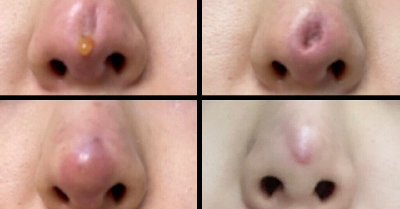
These photographs show partial necrosis of the nasal tip skin resulting from severe inflammation.
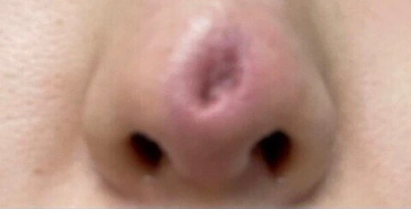
Photograph showing partial necrosis of the nasal tip skin
Preoperative design
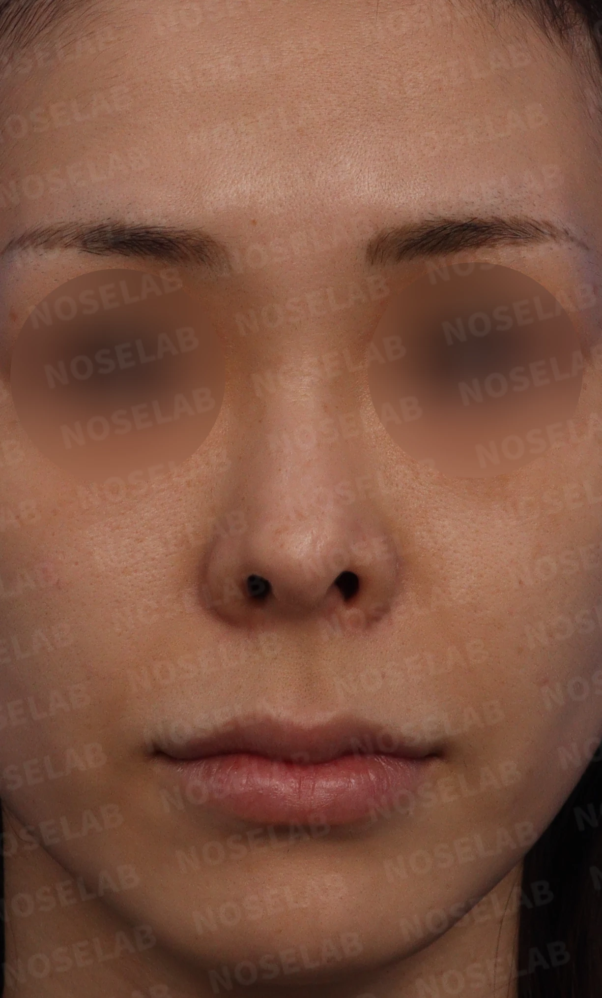
Before surgery: front view
On the front view, significant nostril show is visible, the nasal tip deviates to the right, and severe nostril asymmetry is evident.
The nose appears widened overall. In addition, a scar resulting from previous inflammation can be observed on the left nasal tip.
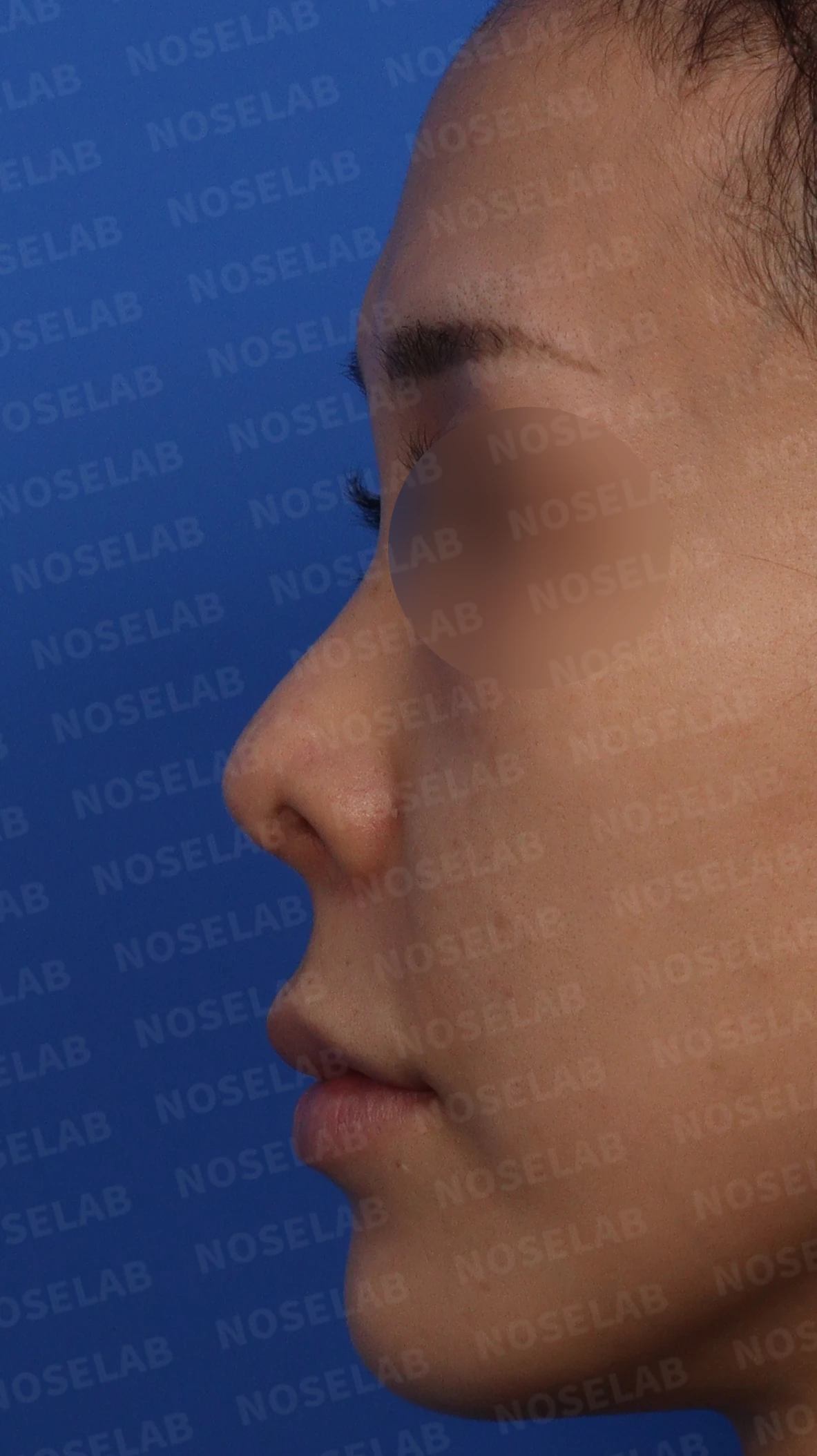
Before surgery: profile view
On the profile view, a slight dorsal hump is present, the nasal tip is drooping yet severely upturned, significant columellar show is noted, and the nasolabial angle is wide.
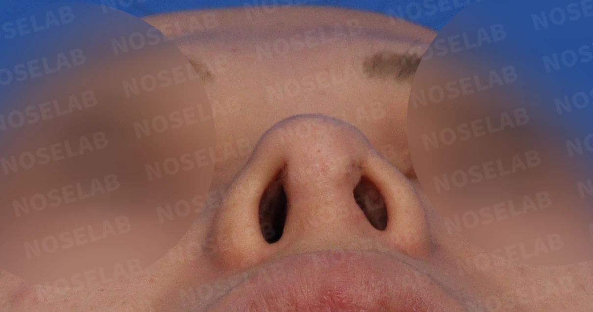
Before surgery: basal (nostril) view
Examining the nostrils, the left nostril is smaller than the right, and scar adhesions from the columella to the alar wall appear to obstruct the upper portion of the nostril.
Surgical plan
We planned to correct the deviated nasal bone using lateral osteotomy.
We planned to reconstruct both the nasal bridge and nasal tip using autologous rib cartilage.
To correct the nostril asymmetry and the upturned nose, we planned septal reconstruction using autologous rib cartilage.
To correct the upturned nose, septal extension was planned, alongside lowering the nostril rims by reconstructing both lower lateral cartilages.
A local flap was planned as needed to widen the left nostril.
Because the skin across the entire nose was expected to have limited elasticity due to contracture, surgical release to expand the dorsal skin was planned.
A dermal graft was planned beneath the depressed skin on the left side.
Before surgery vs. immediately after surgery
Let us compare the preoperative photos with the photos immediately after surgery (Day 0).Immediate postoperative photos show swelling and may differ from the final result.
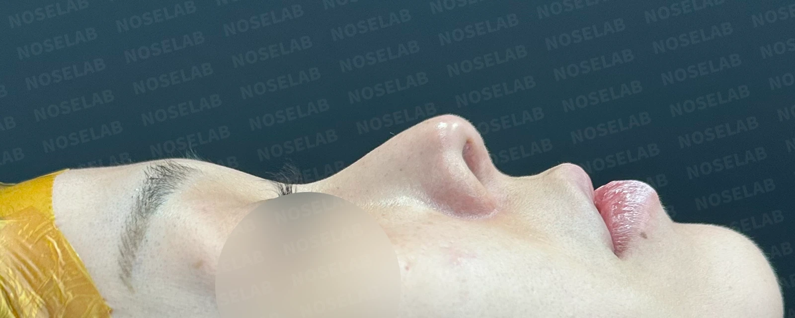
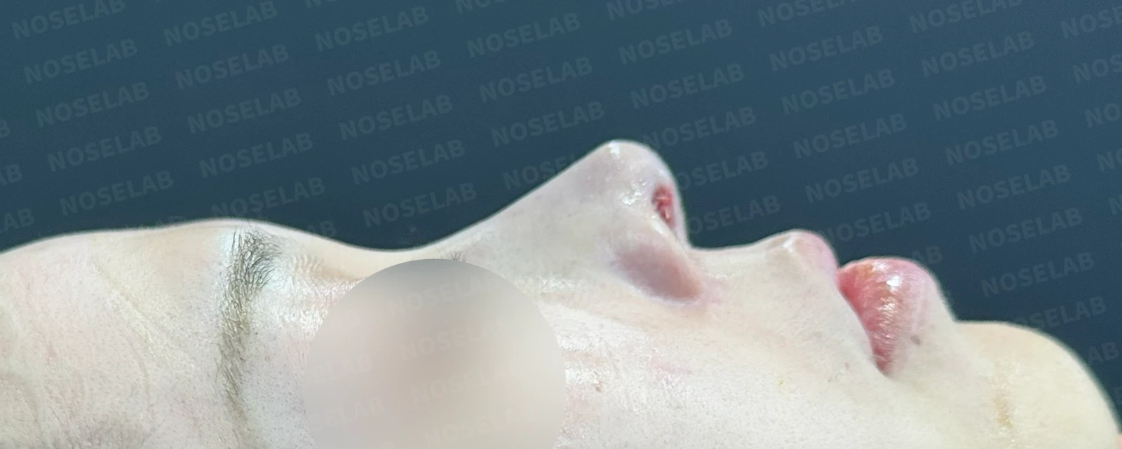
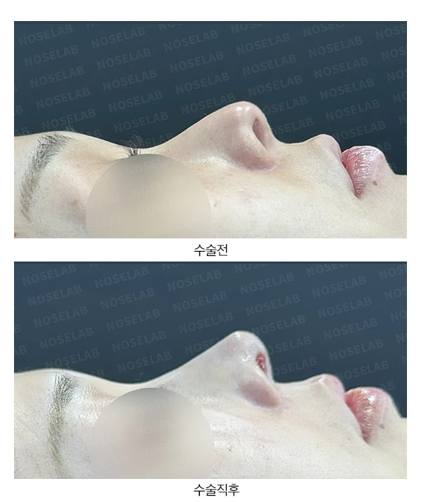
Before surgery / Day 0 immediately after surgery Photos taken immediately after surgery show swelling and may differ from the final result.
On the profile view, the drooping nasal tip has been corrected, the nasolabial angle has decreased, and the bridge line has become much more natural.
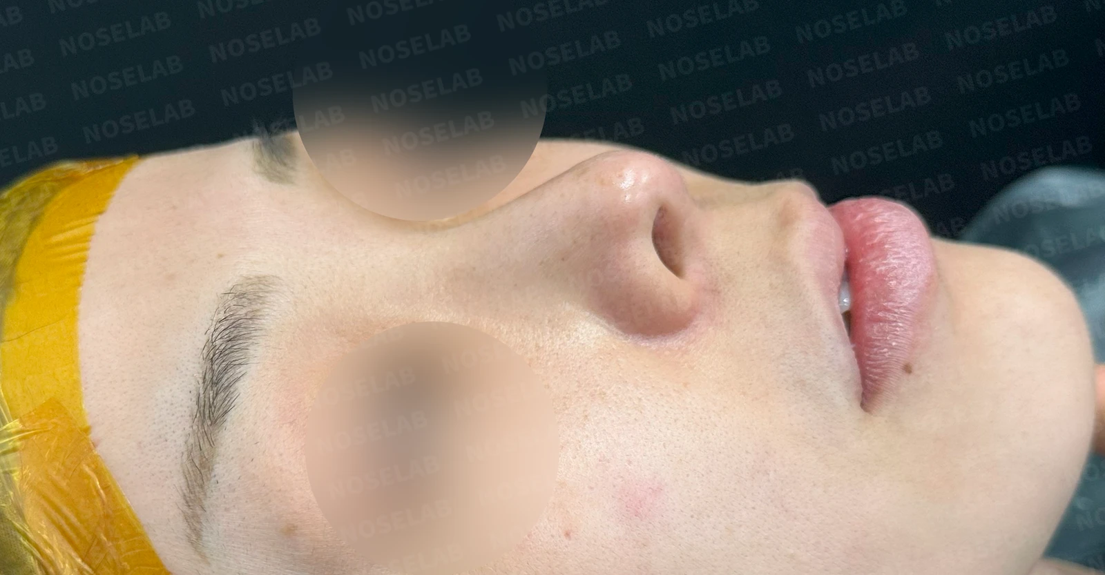
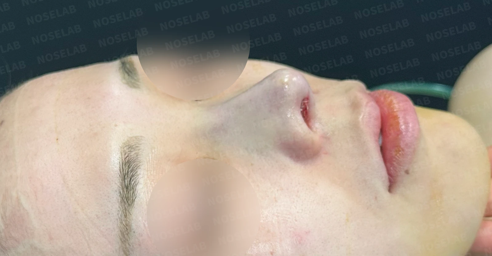
Before surgery / Day 0 immediately after surgery Photos taken immediately after surgery show swelling and may differ from the final result.
On the oblique view, improvement in the nasolabial angle provides the visual effect of improving the appearance of a protruding mouth.
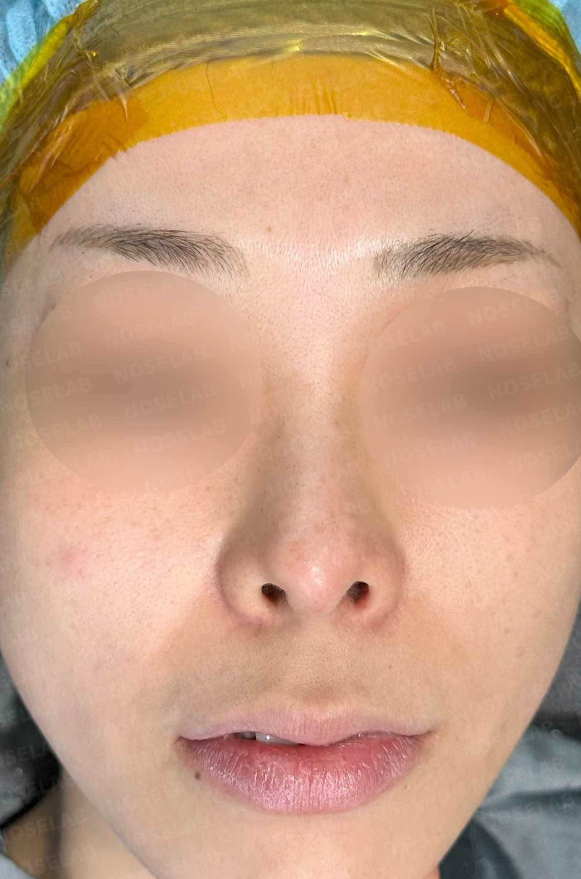
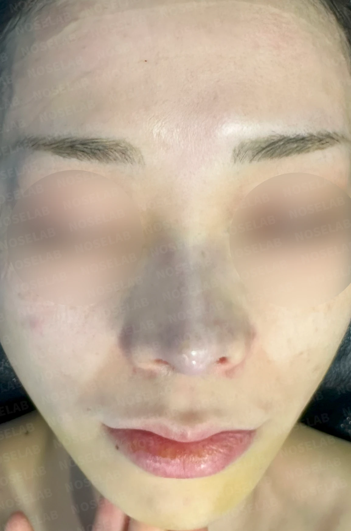
Before surgery / Day 0 immediately after surgery Photos taken immediately after surgery show swelling and may differ from the final result.
The excessive nostril show visible on the front view has significantly improved.
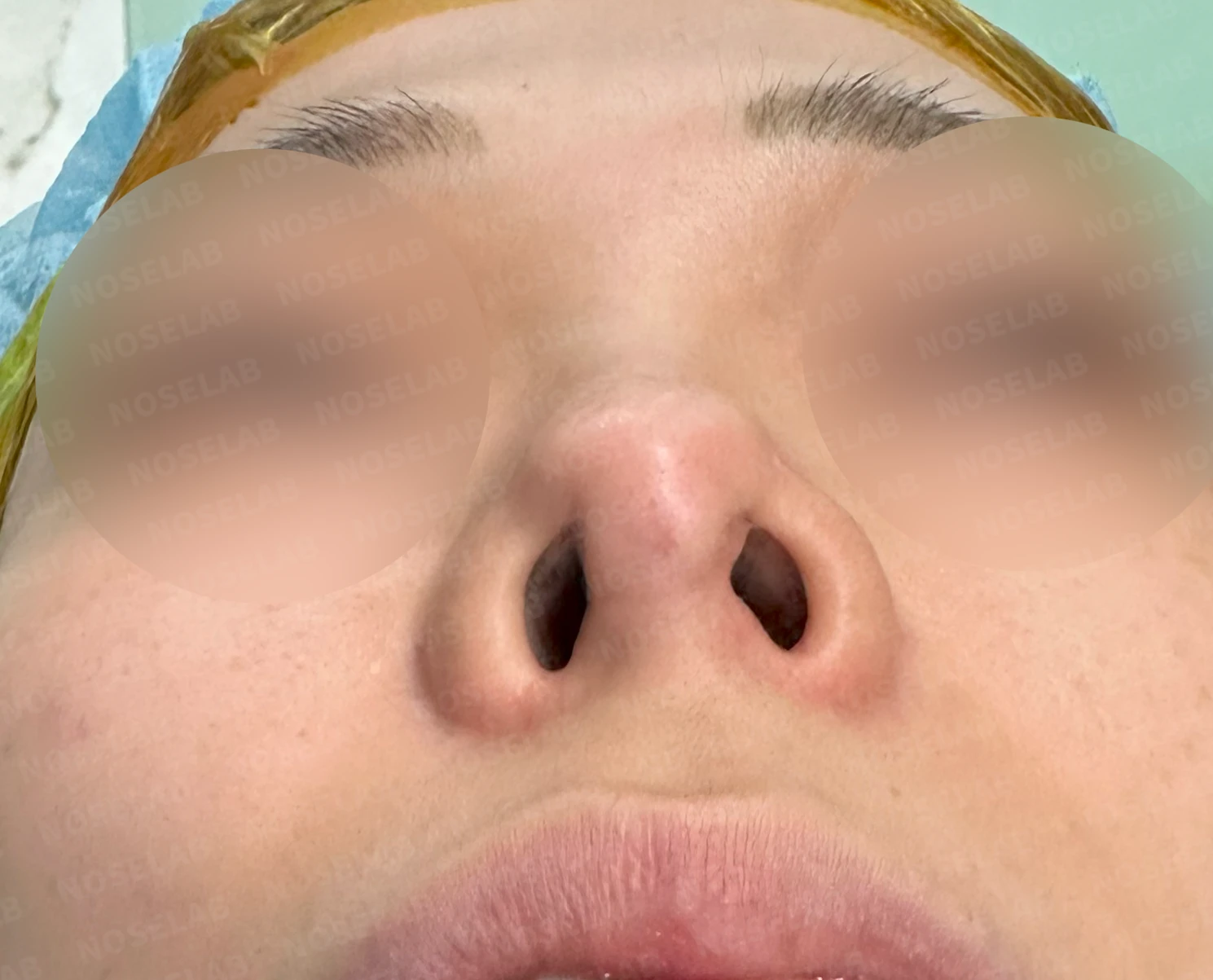
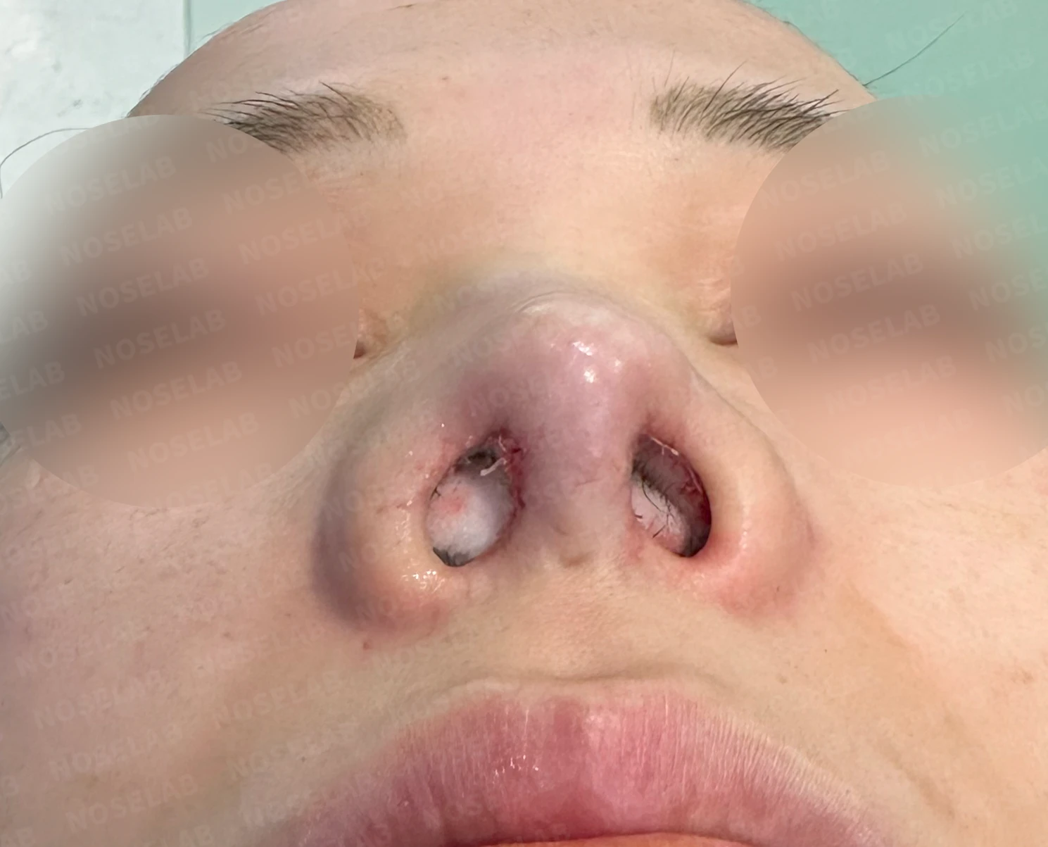
Before surgery / Day 0 immediately after surgery Photos taken immediately after surgery show swelling and may differ from the final result.
The nostril asymmetry has also improved considerably.
Photos immediately after surgery
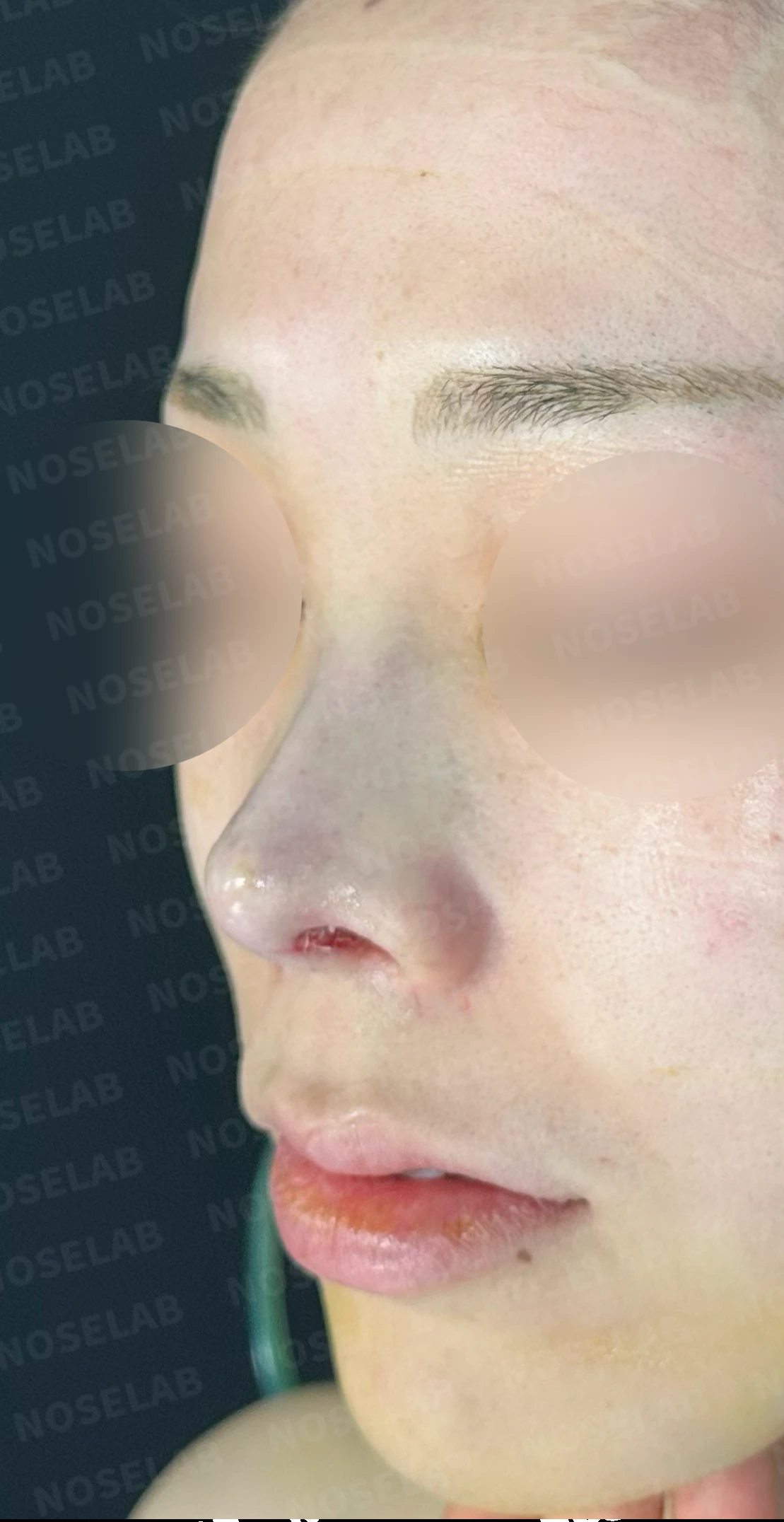
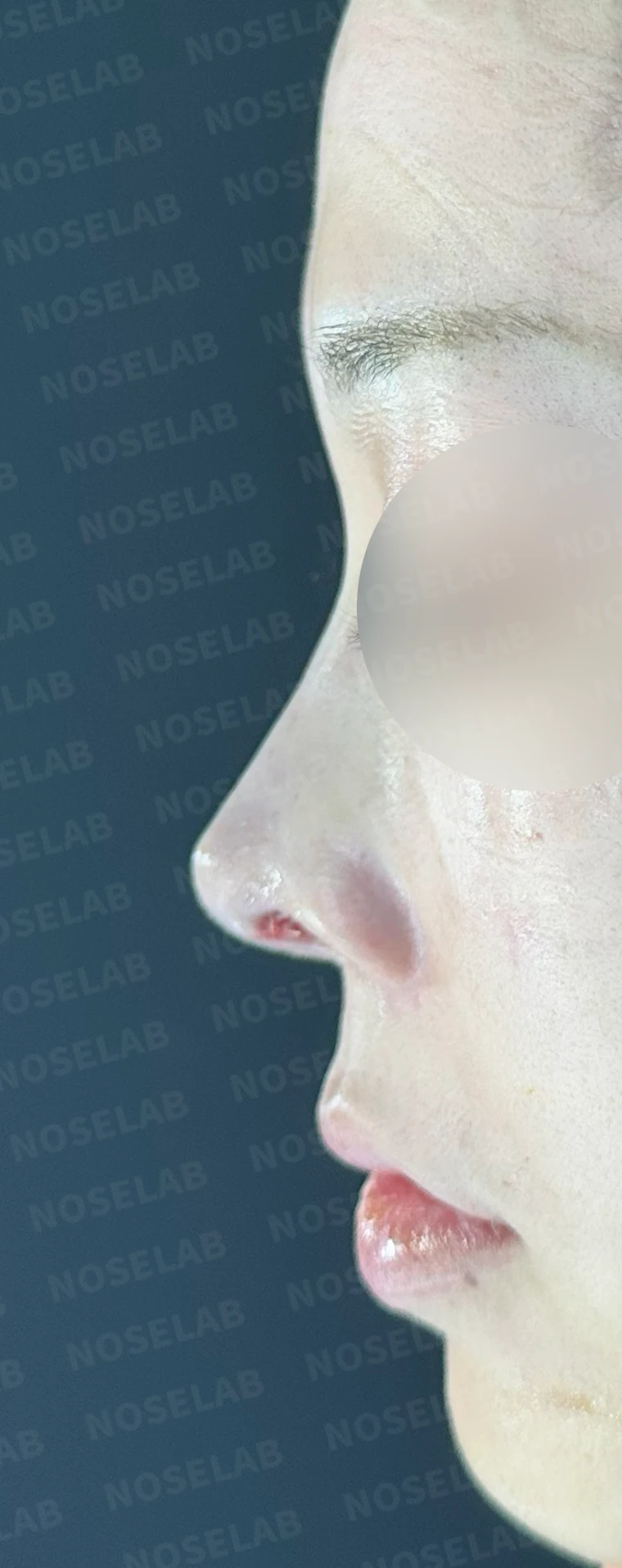
Immediately after surgery (day 0), front, 45-degree, and 90-degree views. Immediate post-operative photos show swelling and may differ from the final result.
Basal (nostril) view immediately after surgery (Day 0)Immediate postoperative photos show swelling and may differ from the final result.
Conclusion
Today, we reviewed a case reconstructing contracture deformity caused by multiple episodes of severe inflammation. After experiencing severe inflammation, damage to normal tissues is inevitable. Inflammation causes the tip cartilages to resorb and soft tissues to transform into firm scar contracture tissue. As this contracts, the nose becomes smaller, distorted, and upturned, resulting in contracture deformity.
In this type of revision surgery, inflammation is the first condition that must be evaluated and treated. In other words, if contracture deformity is actively progressing, residual inflammation is likely present, making active anti-inflammatory treatment essential. Once inflammation is controlled, performing adequate preoperative stem cell therapy to help restore damaged skin tissue is crucial for achieving a favorable final contour.
Additionally, using autologous tissue as much as possible is an approach focused on safety. In particular, using autologous rib cartilage for the nasal tip supports stable results, while autologous rib cartilage or a silicone implant can be considered for the nasal bridge.
In particular, reconstructing the nasal tip cartilages damaged by inflammation as much as possible is the way to prevent future deformities. In addition, because recurrent contracture can occur after contracted nose surgery, follow-up observation is very important, and a second surgery to correct minor deformities may be necessary after 6 months.
Today, we discussed surgery for a contracted nose. I hope this case provides helpful information to those concerned with contracture deformities.
This was Dr. Peter C. Kang. Thank you.

Limitations and risks
When autologous rib cartilage is used, scarring and pain may remain at the donor site on the chest, and pneumothorax (pleural injury) can occur rarely. Over time, the grafted cartilage may warp or undergo partial resorption.
When the septum is corrected at the same time, nasal obstruction may persist or recur, and in rare cases, septal perforation or changes in smell may occur.
Common potential risks include infection, bleeding, asymmetry, scarring, skin necrosis, and insufficient improvement; results vary depending on individual skin condition and previous surgical history.