Hello.
I am Dr. Peter C. Kang, medical director.
The case I would like to discuss today involves a contracted nose. In this case, the patient had undergone 27 formal nasal procedures in total. Having had rhinoplasty surgeries and nasal fat grafting 27 times, the patient visited our clinic because contracture had progressed rapidly over the past 1 year.
Surgical plan
The patient desired a high, dramatic-looking nose, but because the current condition of the nose was expected to be unfavorable, I explained that the shape would be determined in the operating room based on the actual structural findings before proceeding with surgery. In particular, because there was a possibility of acute inflammation, I also carefully discussed the potential need for a second surgery. On the preoperative computed tomography scan, a shape resembling foreign material was visible at the nasal tip, suggesting the possibility of damage to the septal cartilage as well.
Surgical findings
The foreign material seen on the computed tomography scan turned out to be a piece of silicone implant, which appeared to have been placed by a previous surgeon to fill the defect where the septal cartilage at the nasal tip was missing. This implant had migrated toward the nasal tip, leaving the tip skin open by approximately 0.5 mm. Acute inflammation had spread through this opening between the nasal implant and the fat cells previously grafted inside the nose, and the area around the implant was filled with granulation tissue. After completely removing the granulation tissue and inspecting the interior of the nose, 80% of the septal cartilage was gone, and only remnants of the upper lateral cartilages and lower lateral cartilages remained. In addition, the fat cells grafted into the nasal bridge had degenerated into scar tissue and fibrous tissue due to chronic inflammation, so they were thoroughly removed.
The missing septal cartilage was completely reconstructed using autologous rib cartilage, and the upper and lower lateral cartilages were also reconstructed using autologous rib cartilage. On the nasal bridge, the grafted fat tissue was removed, and a silicone implant was placed in that section.
Before surgery vs. immediately after surgery
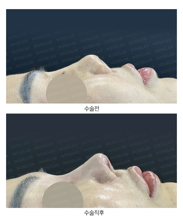
Let us compare the before-and-after photos.
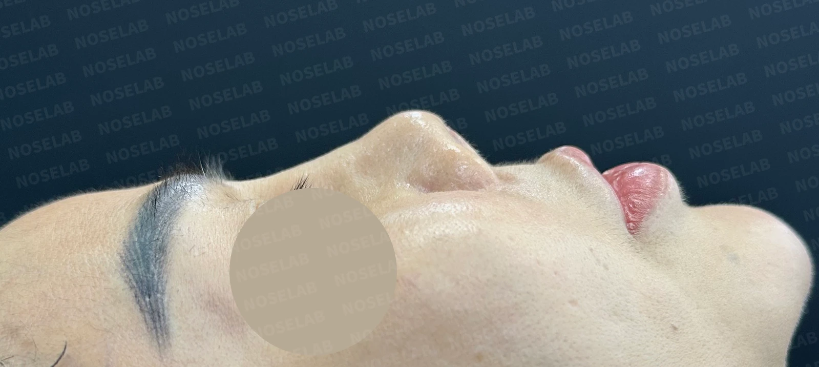
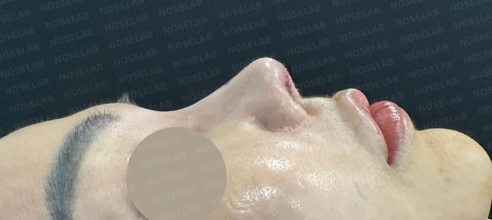
Before surgery / Day 0 immediately after surgery Photos taken immediately after surgery show swelling and may differ from the final result.
The nasal tip, which had been severely upturned before surgery, and the retracted columella showed substantial improvement, resulting in a natural nasolabial angle. In addition, the radix was significantly lowered to maintain a natural line.
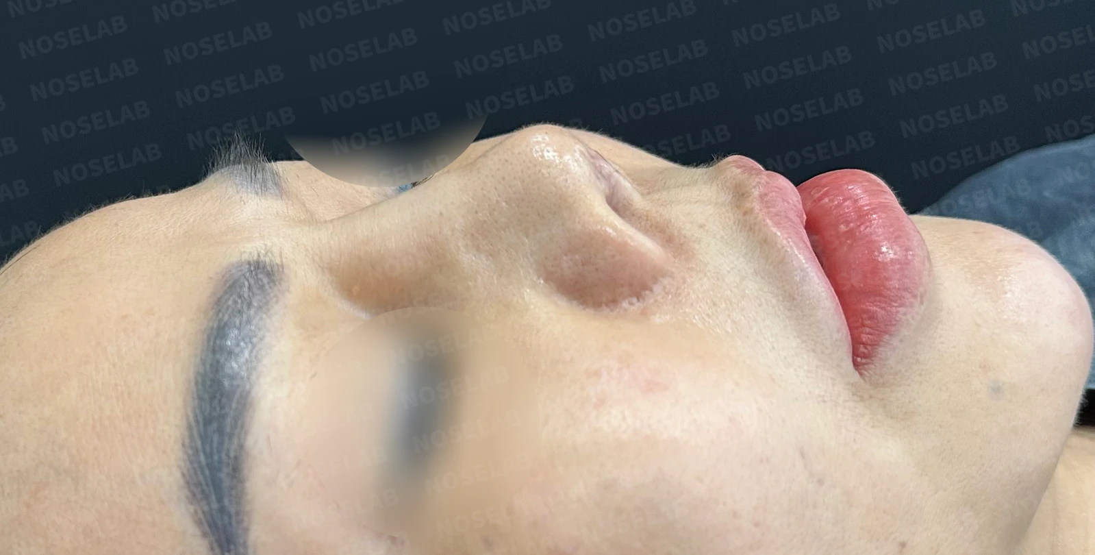
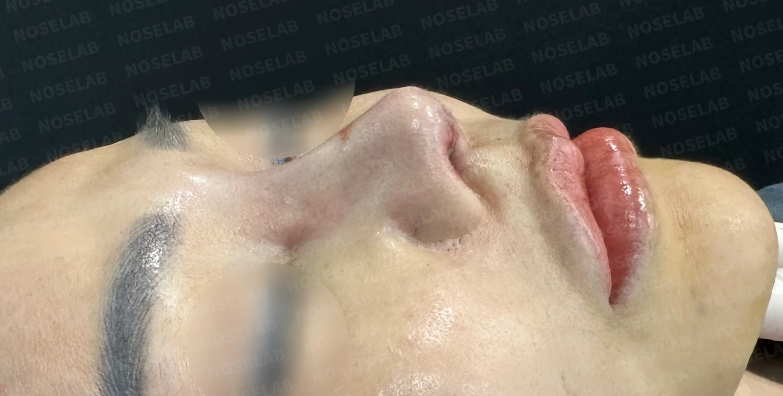
Before surgery / Day 0 immediately after surgery Photos taken immediately after surgery show swelling and may differ from the final result.
The lowering of the radix is the most noticeable change.
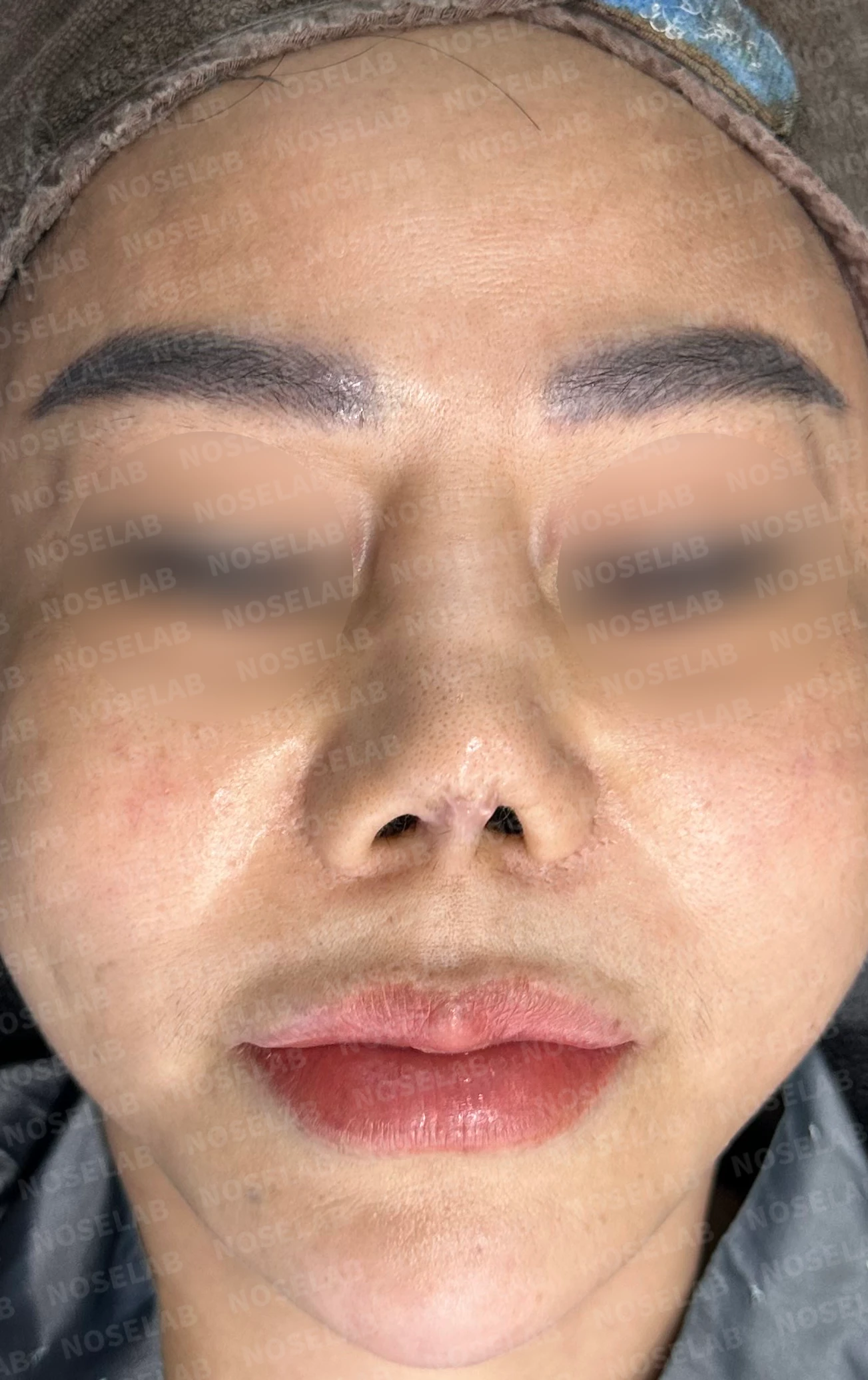
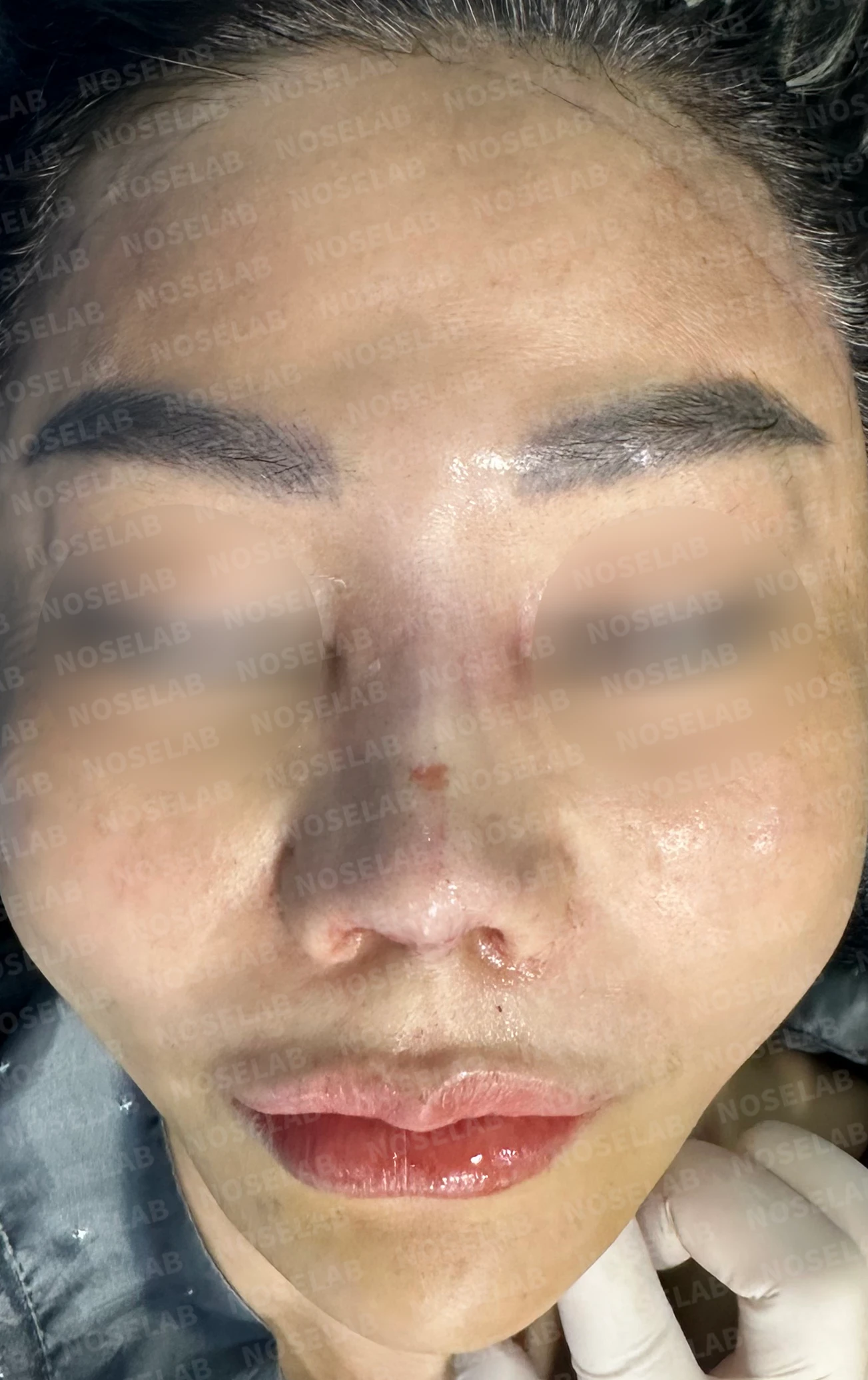
Before surgery / Day 0 immediately after surgery Photos taken immediately after surgery show swelling and may differ from the final result.
From the front view, the previously upturned nose has come down significantly, to the point where the nostrils are barely visible. Swelling and scar tissue during the recovery period may make it appear slightly more upturned than it looks now, but as it settles, it should return close to its current position.
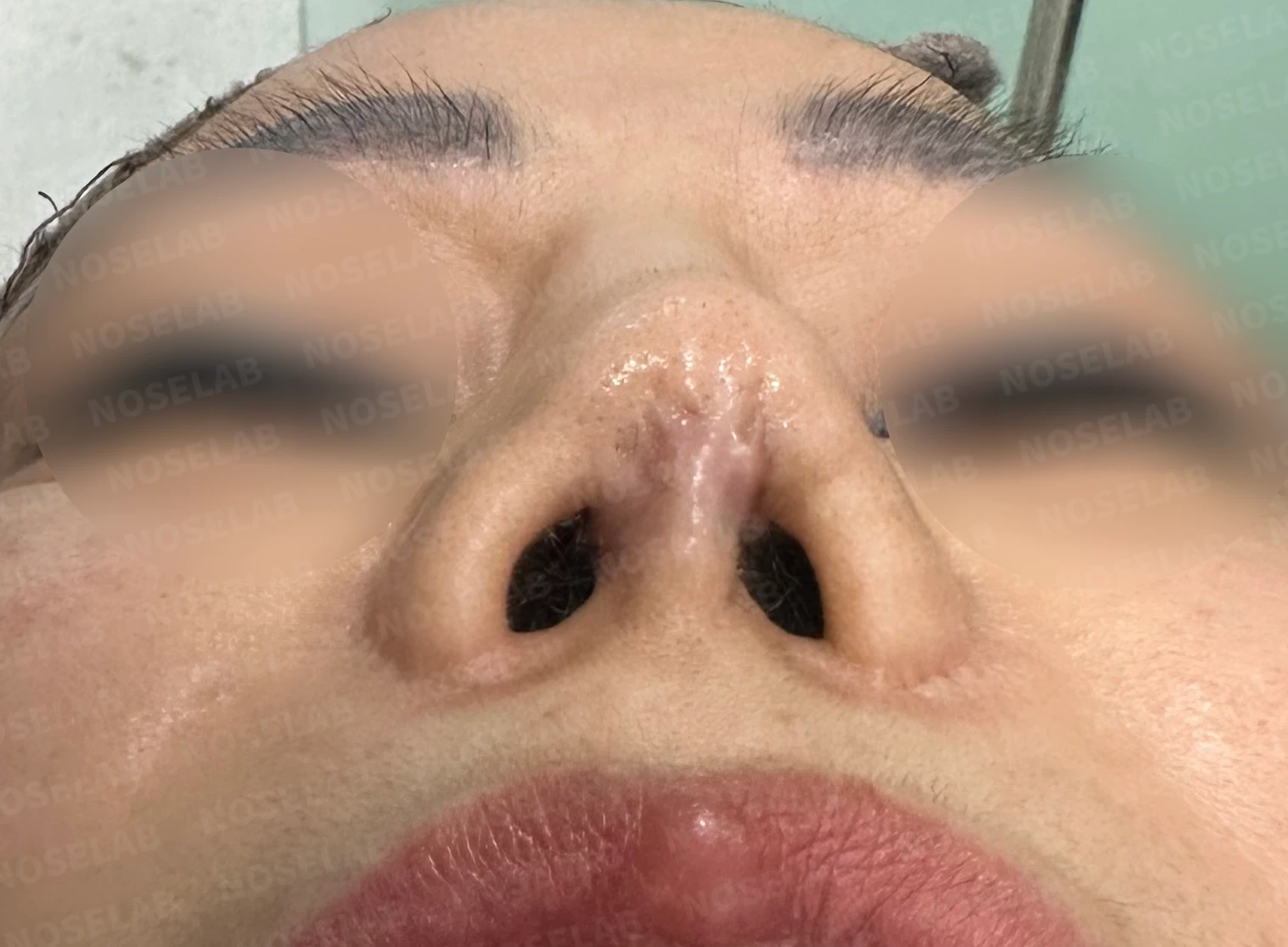
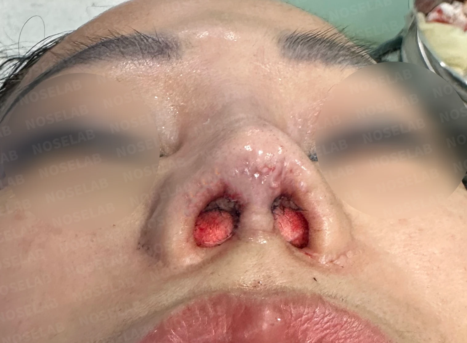
Before surgery / Day 0 immediately after surgery Photos taken immediately after surgery show swelling and may differ from the final result.
The nostrils, which had become severely narrowed due to contracted tissue, have improved considerably.
Photos immediately after surgery
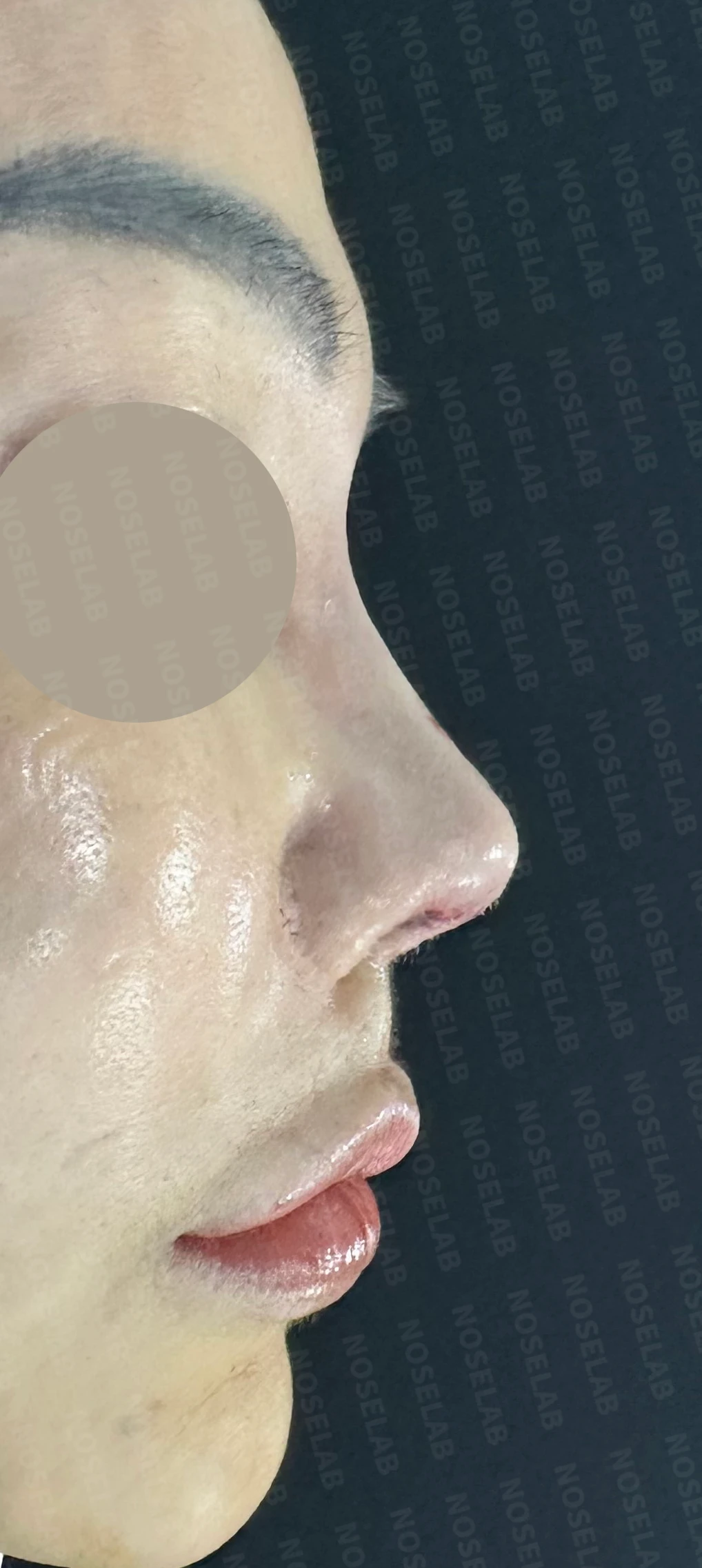
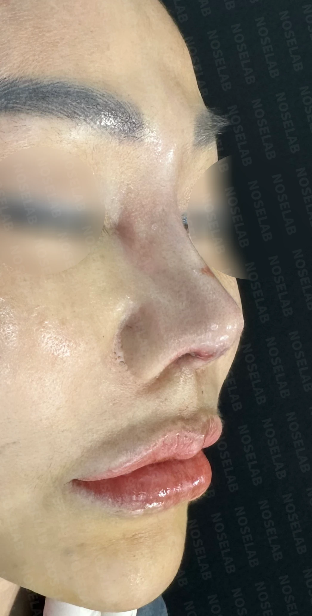
My thoughts on contracted nose deformities
Performing contracture revisions frequently reminds me of how precious the septal cartilage is. When inflammation occurs, nasal tip deformity appears to be less severe when more septal cartilage remains intact. In this case as well, if just a little more septal cartilage had been preserved, the nose would have been less upturned, and lowering the nasal tip again would have been more advantageous. Even so, at our clinic, we reconstruct the septal cartilage firmly to bring the nasal tip down, so it does not present a major issue.
Lowering an upturned nose caused by contracture depends largely on two key factors. The first is how much the skin can stretch, and the second is how sturdy the supporting framework is to bring the tip down. Especially in cases like this, where acute inflammation is present and the structural support of the nose has completely collapsed, the skin does not stretch easily. When there is also no structural support, reconstructing a solid framework first is essential. Once reconstruction is complete, that framework provides the foundation to project the tip slightly higher or lower it a bit further.
Today, we looked at a reconstruction case for a patient with a contracted nose whose septum was almost entirely gone. I hope this information is helpful to those who are concerned about contracted nose deformities.
This was Dr. Peter C. Kang. Thank you.

Limitations and risks
When autologous rib cartilage is used, scarring and pain may remain at the donor site on the chest, and pneumothorax (pleural injury) can occur rarely. Over time, the grafted cartilage may warp or undergo partial resorption.
When the septum is corrected at the same time, nasal obstruction may persist or recur, and in rare cases, septal perforation or changes in smell may occur.
Common potential risks include infection, bleeding, asymmetry, scarring, skin necrosis, and insufficient improvement; results vary depending on individual skin condition and previous surgical history.