Hello.
I am Dr. Peter C. Kang, medical director of the Rhinoplasty Research Center.
Today, I would like to talk about surgery to improve the shape of a small and bulbous nose.
Having focused solely on rhinoplasty, I have performed a wide variety and large number of nasal procedures. However, the cases I find most challenging are those where a nose with minimal skin envelope must be refined aesthetically.
The patient in this case also had a small nose but desired a prominent, defined result. Therefore, during the preoperative consultation, I explained that we could not elevate the nose excessively and promised to do my best within the limits of skin elasticity before proceeding with surgery. When a nose is small, the internal cartilage is usually also small, meaning donor cartilage from the nose itself is often insufficient; thus, we decided to use autologous rib cartilage for the procedure.
Preoperative nasal obstruction examination
The patient had symptoms of nasal obstruction, so diagnostic evaluations related to nasal obstruction were performed at our clinic.
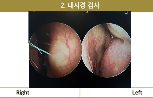
On nasal endoscopy, the septal cartilage was deviated to the right, mucosal blood vessels were dilated, and the left inferior turbinate was hypertrophied. In addition, the internal nasal mucosa appeared generally pale, confirming the presence of concurrent allergic rhinitis.
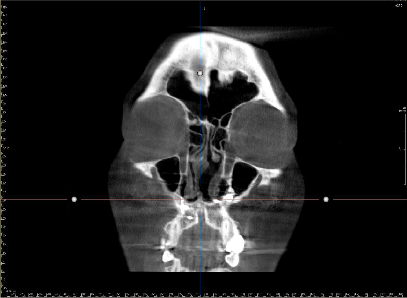
The nasal computed tomography (CT) scan showed severe deviation of the septal cartilage to the right, which significantly narrowed the right nasal airway compared to the left. Considering these objective findings and the patient's symptoms of nasal obstruction, septoplasty to correct the deviated septum and submucosal resection of the inferior turbinate using radiofrequency were planned.
Surgical findings
During surgery, evaluation of the septal cartilage revealed that it was thinner and more deviated than anticipated, leading to partial reconstruction of the septal cartilage using autologous rib cartilage. This significantly corrected the deviation and strengthened the septum, after which septal extension was performed using autologous rib cartilage to elevate the nasal tip with good projection. However, because the tip skin did not stretch as much as expected, a surgical technique was used to gradually mobilize the surrounding skin toward the tip, elevating the nasal tip as much as possible within a safe range.
Before surgery vs. immediately after surgery
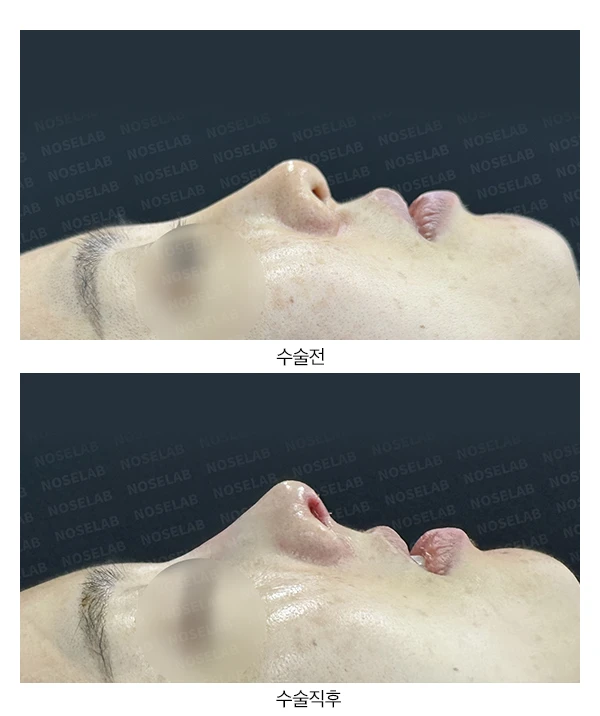
Let us compare the before-and-after photos.
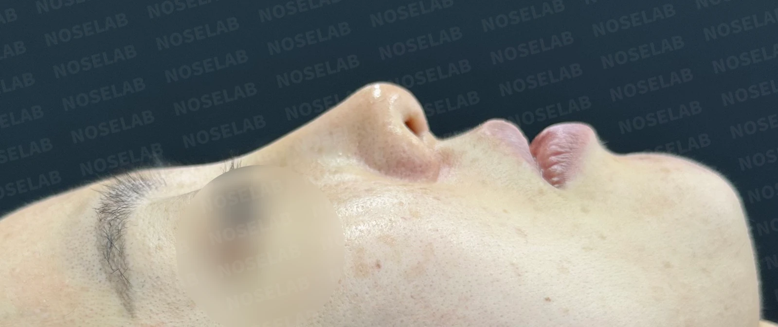
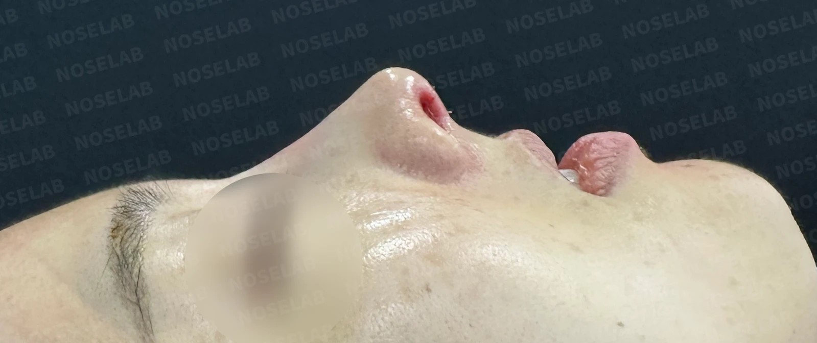
Before surgery / Day 0 immediately after surgery Photos taken immediately after surgery show swelling and may differ from the final result.
One can see that the nasal tip has been noticeably elevated relative to the glabella, and the nasolabial angle has been corrected.
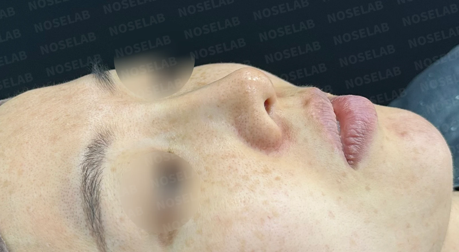
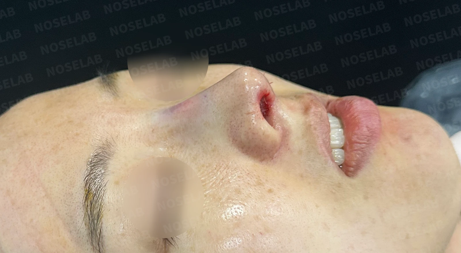
Before surgery / Day 0 immediately after surgery Photos taken immediately after surgery show swelling and may differ from the final result.
The starting point at the glabella can be seen transitioning downward naturally.
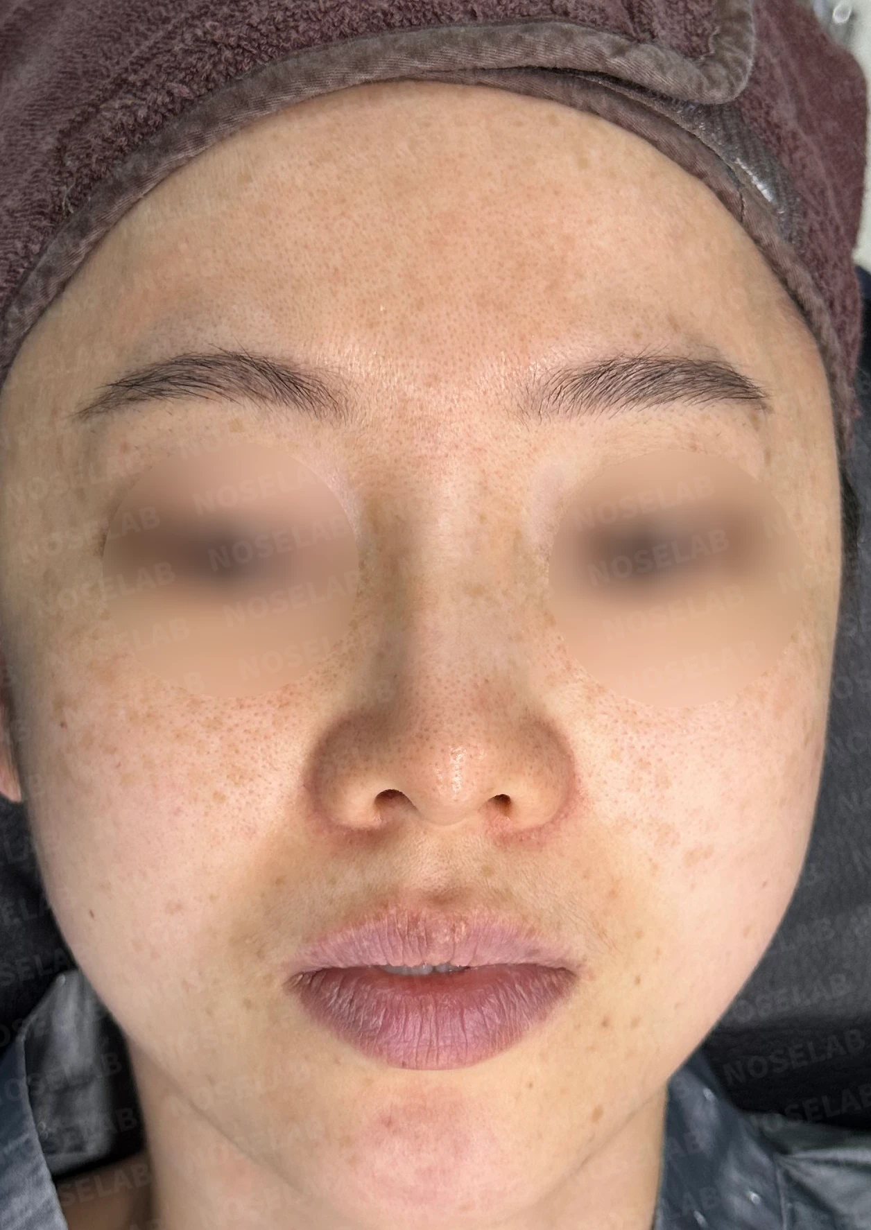
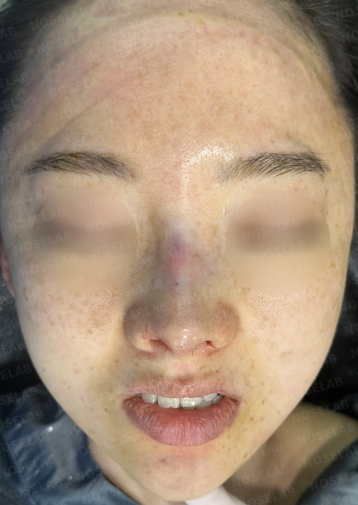
Before surgery / Day 0 immediately after surgery Photos taken immediately after surgery show swelling and may differ from the final result.
The previously upturned nasal tip now sits naturally lower, and the deviation of the nasal bridge to the right has also improved.
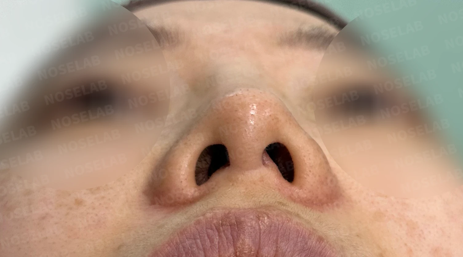
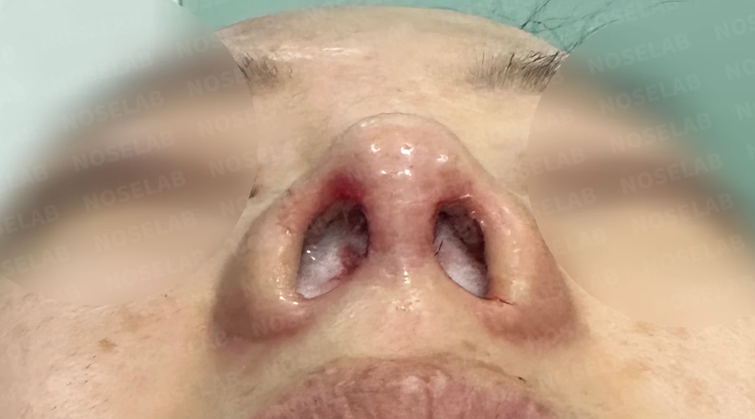
Before surgery / Day 0 immediately after surgery Photos taken immediately after surgery show swelling and may differ from the final result.
Looking at the basal view of the nostrils, the deviation of the columella has been corrected, and the nostrils appear naturally elongated.
Photos immediately after surgery
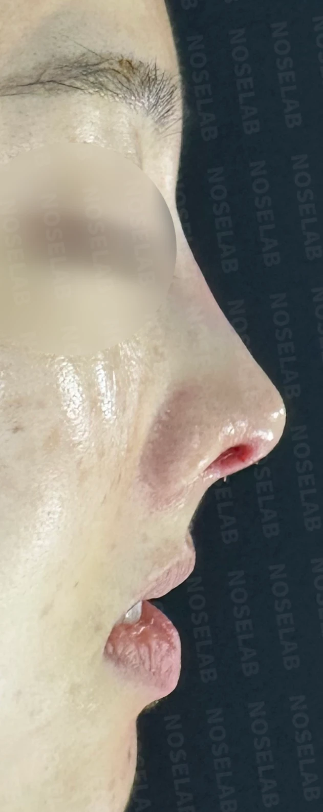
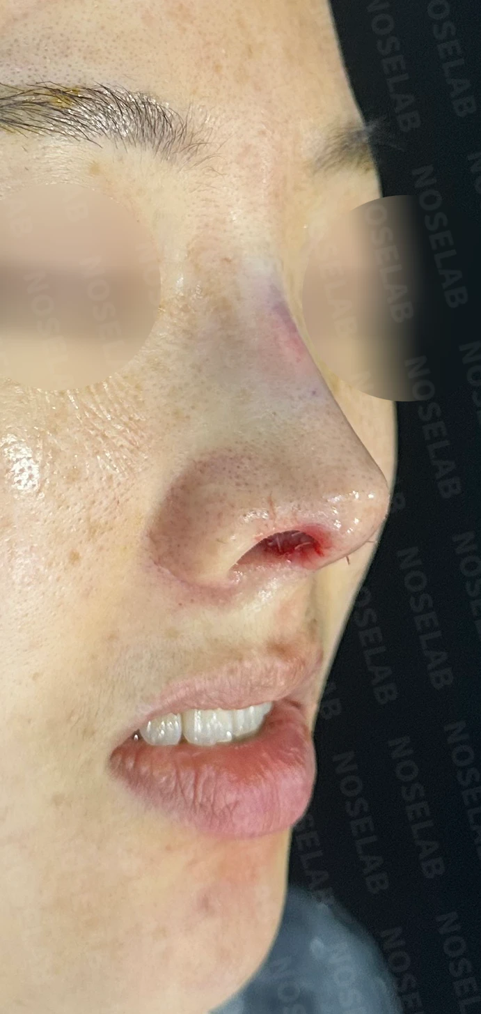
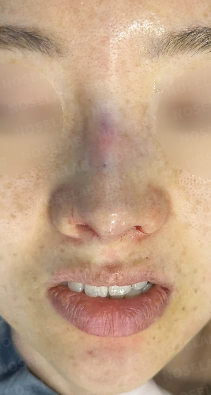
Photos immediately after surgery (Day 0): 90-degree profile, 45-degree oblique, and front viewImmediate postoperative photos show swelling and may differ from the final result.
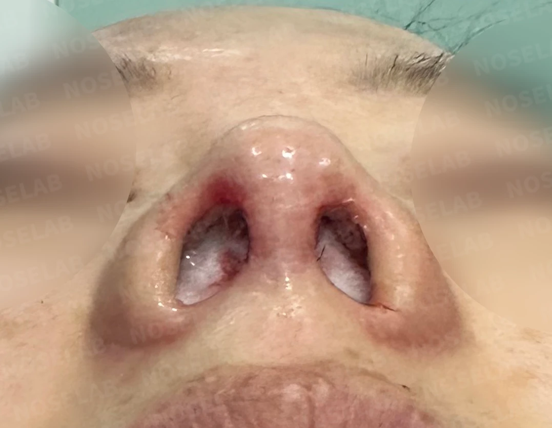
Basal (nostril) view immediately after surgery (Day 0)Immediate postoperative photos show swelling and may differ from the final result.
Conclusion
Surgery to correct a short nose requires continuous focus until the very end. This is especially true for noses with tight, limited skin. Although I frequently utilize techniques to reinforce the tip cartilage as securely as possible and mobilize surrounding skin to elevate the tip, it is easier said than done. Seeing the improvement compared to before surgery toward the end of the procedure is rewarding, but because each patient has different expectations, it can also bring a sense of concern.
For this reason, the promise I can always make is to do my utmost within a safe range. Overly aggressive elevation can lead to cartilage show-through, persistent tip redness, or deformities such as deviation or upward retraction; therefore, avoiding excessive tension is essential. In other words, accepting that there is a limit to how high the nose can be raised and creating the most aesthetically pleasing shape at a safe height is what truly matters.
Today, I shared thoughts on correcting a small and bulbous nose. I hope this helps those who are concerned about similar nasal shapes.
This was Dr. Peter C. Kang. Thank you.

Limitations and risks
When autologous rib cartilage is used, scarring and pain may remain at the donor site on the chest, and pneumothorax (pleural injury) can occur rarely. Over time, the grafted cartilage may warp or undergo partial resorption.
When the septum is corrected at the same time, nasal obstruction may persist or recur, and in rare cases, septal perforation or changes in smell may occur.
Common potential risks include infection, bleeding, asymmetry, scarring, skin necrosis, and insufficient improvement; results vary depending on individual skin condition and previous surgical history.