Hello, I am Dr. Peter C. Kang.
Today, I will introduce a reconstruction case of a patient who suffered from breathing difficulties because an implant used in previous surgery obstructed the nasal cavity.
📍 Surgical history
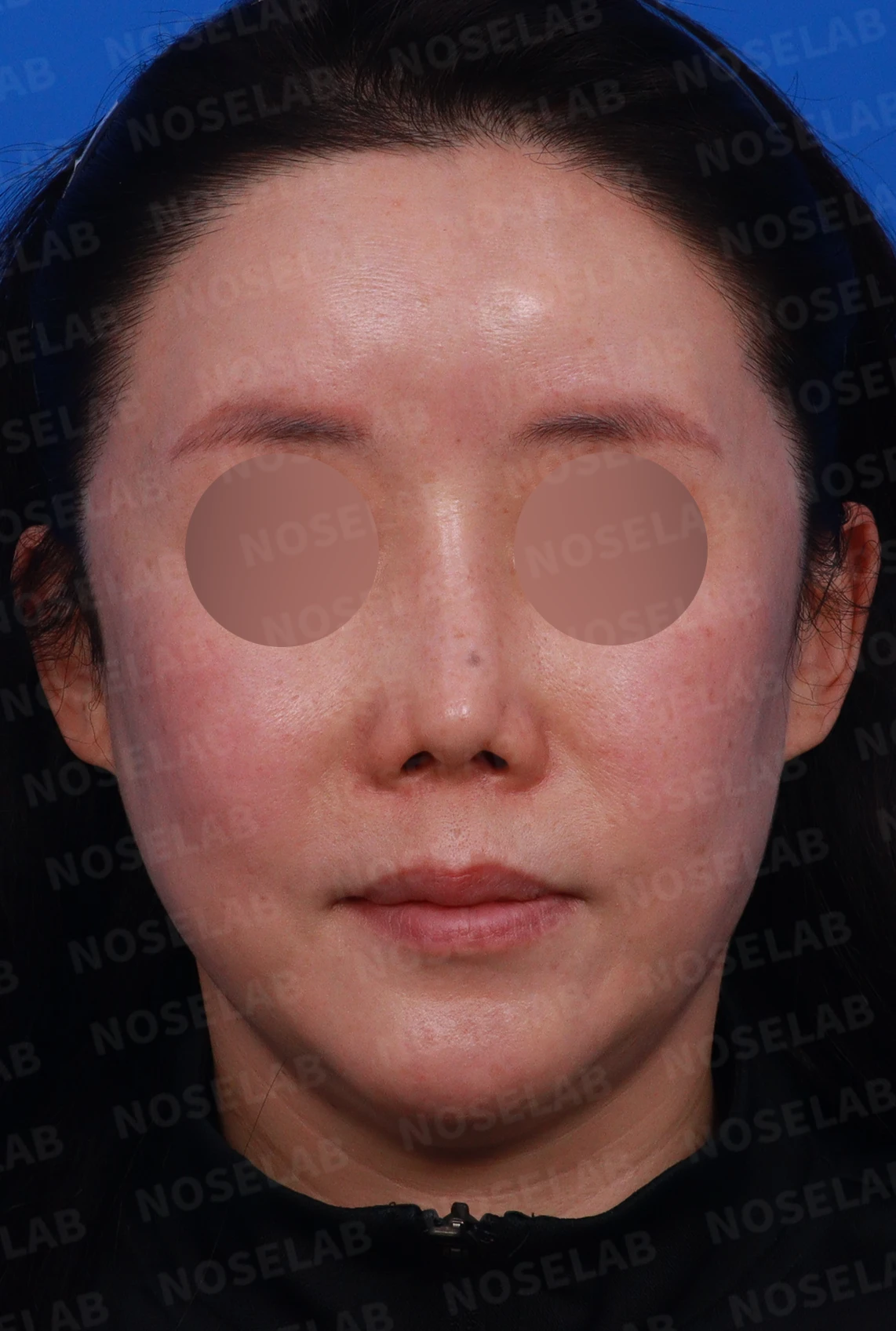
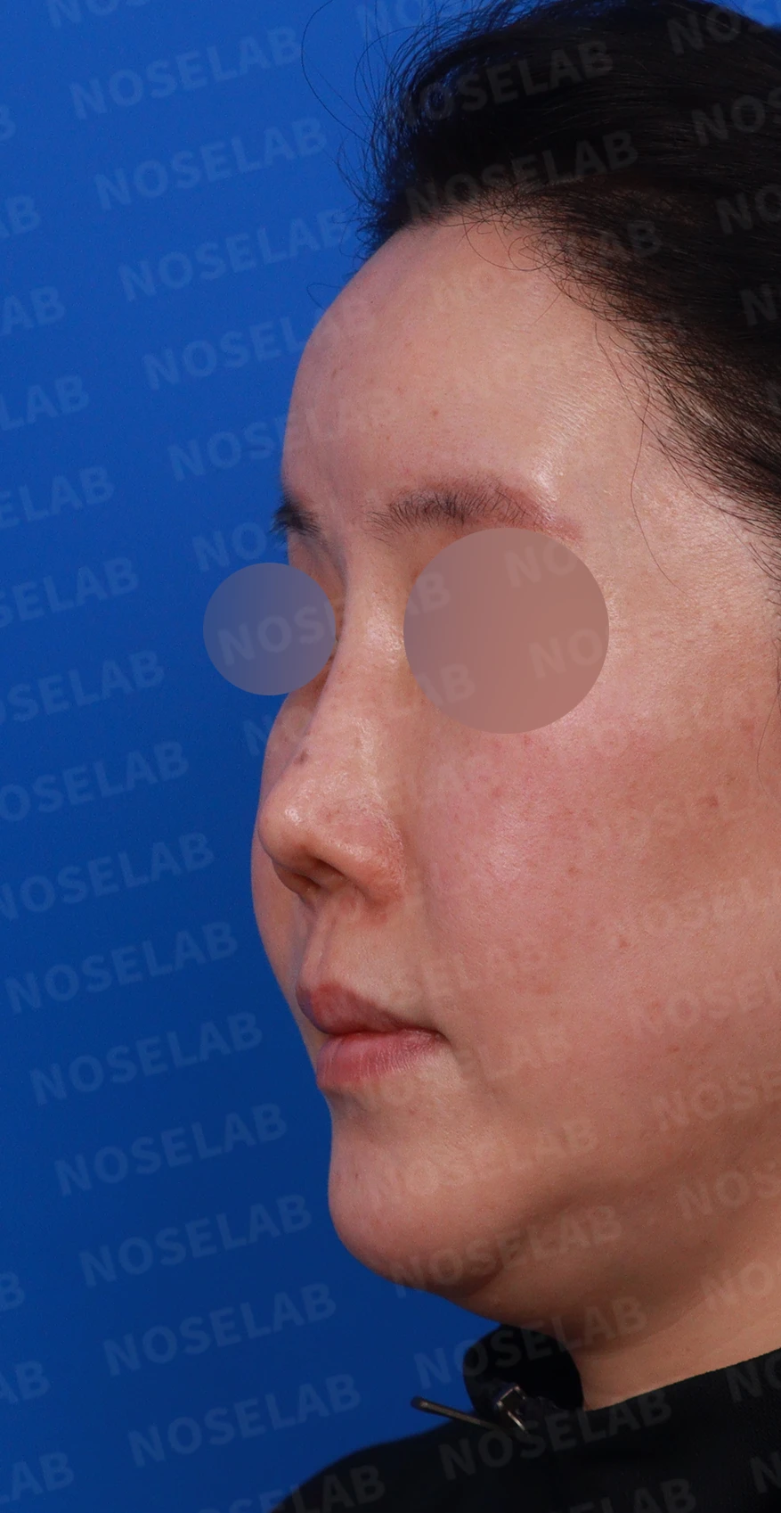
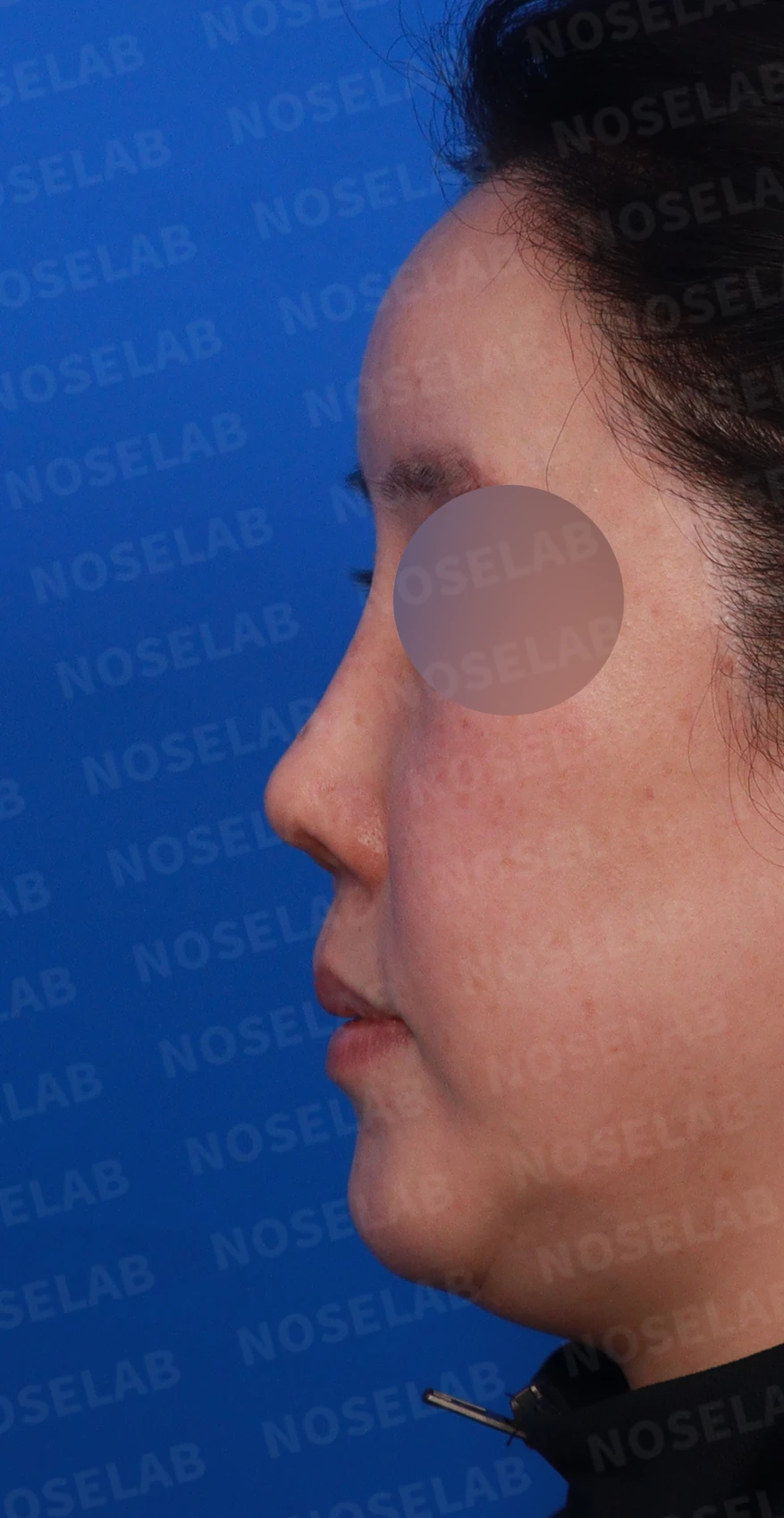
These are design consultation photos: front, left oblique, and profile views.
2010: Rhinoplasty + alar reduction + paranasal augmentation
2012: Septum + silicone implant + donor rib cartilage (DR) surgery
2013: Nasal valve + 3 functional nasal procedures
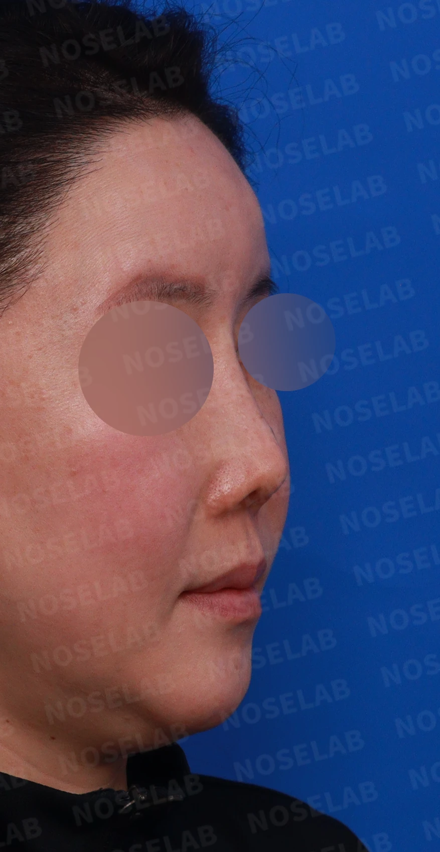
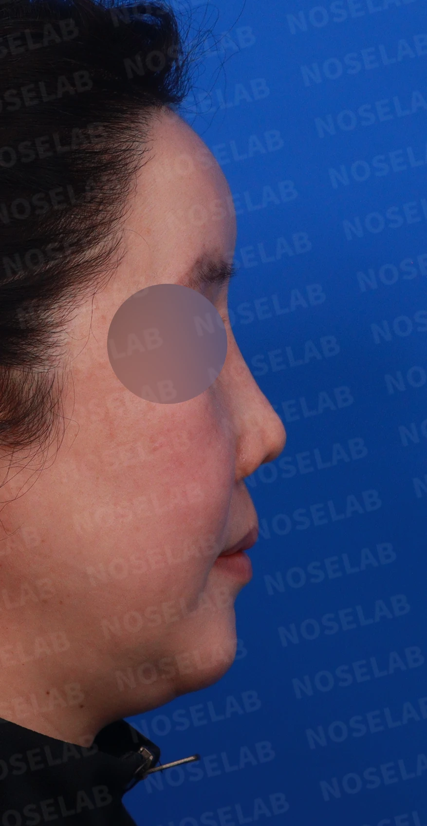
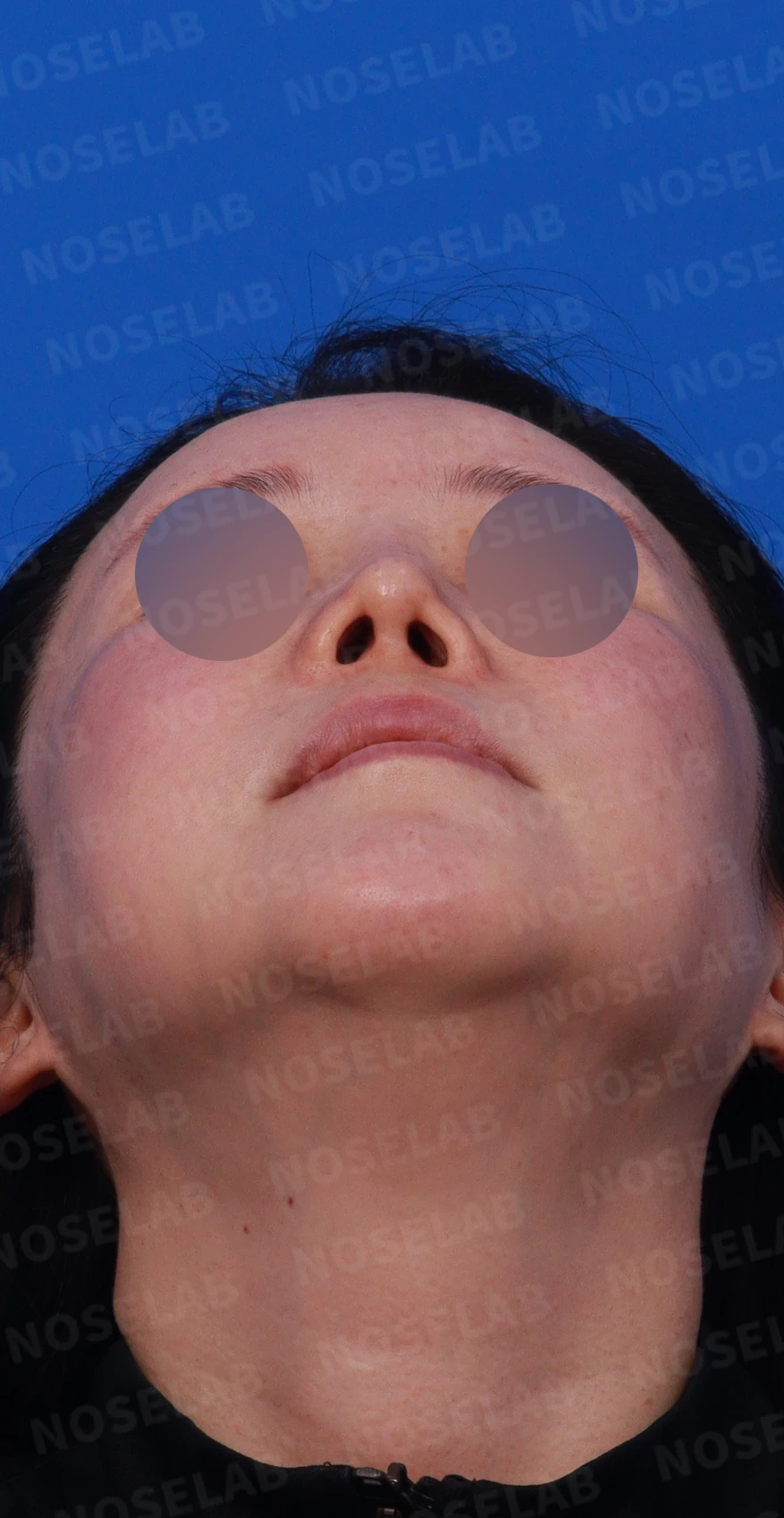
Design consultation: right oblique, profile, and basal (nostril) views.
🔎 Initial consultation findings
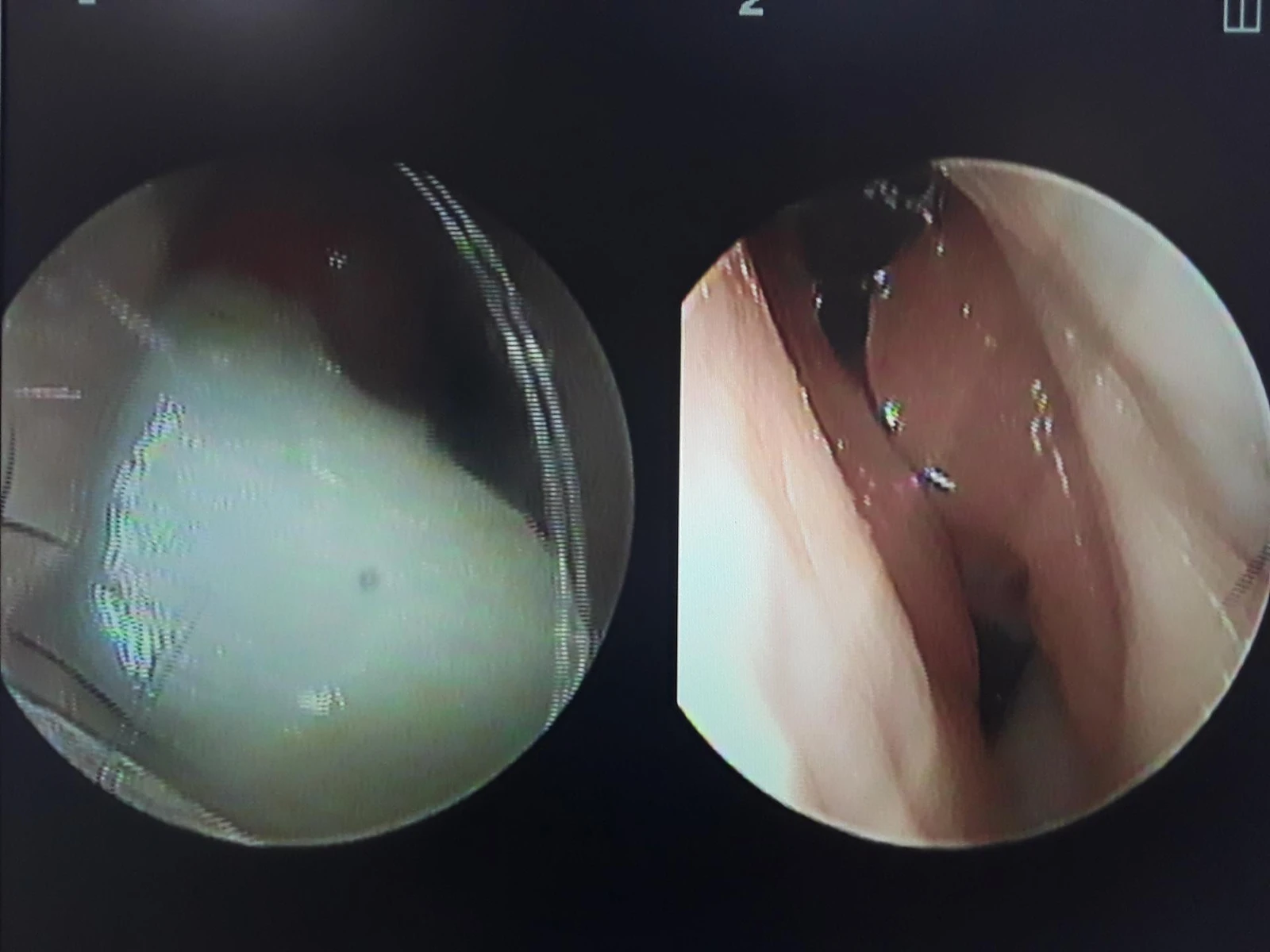
Nasal endoscopy photo
Severe functional issues
State where the implant protruded into the right nasal cavity, blocking 2/3 of the airway
Unable to breathe through the right side
Rhinitis symptoms
Structural damage
The septum was in very poor condition
Retracted columella
Severe damage to both lower lateral cartilages
Pinched appearance of the alae
Aesthetic concerns
Deviated nose
Drooping tip
✨ Patient's requests
Correction of deviated nose
Desire to elevate the nasal tip
Relief of internal nasal obstruction
💉 Surgical procedure details
Functional recovery
Removal of the implant obstructing the nasal cavity
Removal of the contralateral paranasal implant
Structural reconstruction
Septal reconstruction using autologous rib cartilage
Reconstruction of both lower lateral cartilages using autologous rib cartilage
Restoration of retracted columella
Morphological improvement
Correction of crooked nose
Elevation of the drooping tip
Improvement of the pinched appearance of the alae
Correction of the nasolabial angle
Functional improvement
Improvement of rhinitis through septoplasty and radiofrequency submucosal turbinoplasty of the inferior turbinates
✅ Surgical results
Functional recovery
The obstructed right nasal cavity was cleared
Restoration of normal bilateral breathing function
Alleviation of rhinitis symptoms
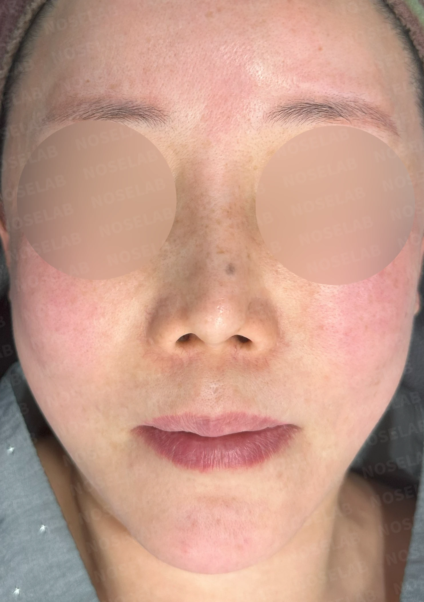
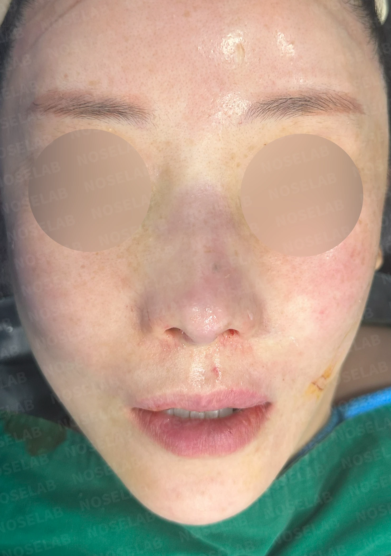
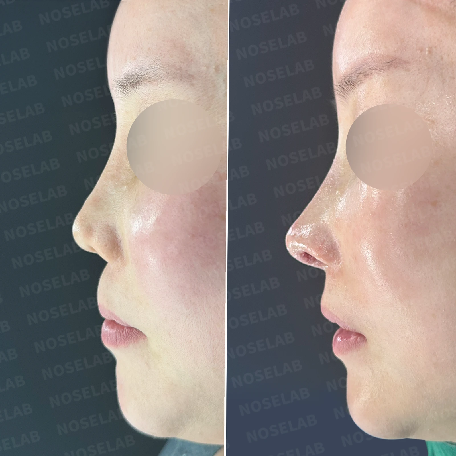
Before surgery / Day 0 immediately after surgery Photos taken immediately after surgery show swelling and may differ from the final result.
Front view
Correction of crooked nose
Naturally restored alae
Improvement of the pinched areas
Balanced nasal shape
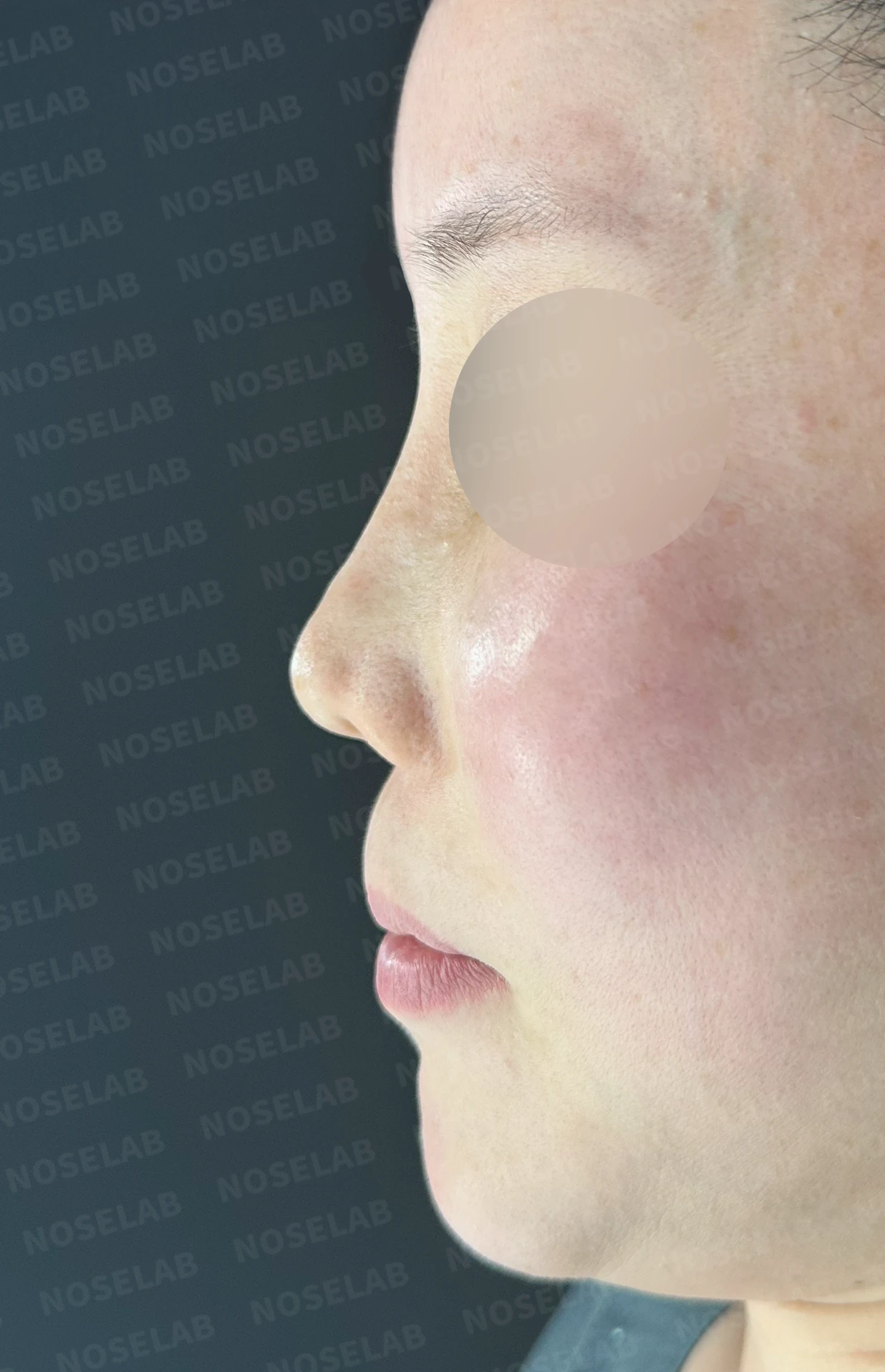
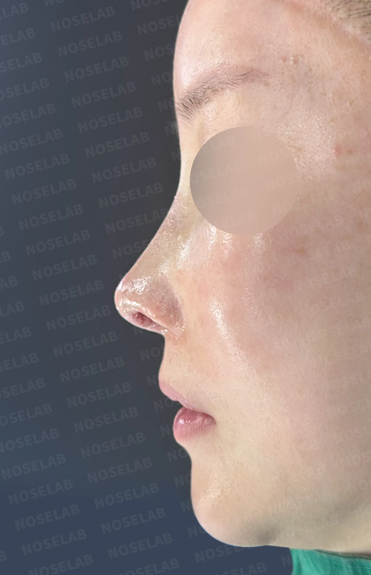
Before surgery / Day 0 immediately after surgery Photos taken immediately after surgery show swelling and may differ from the final result.
Profile view
Naturally elevated nasal tip
Restored columella
Improved nasolabial angle
Harmonious profile line
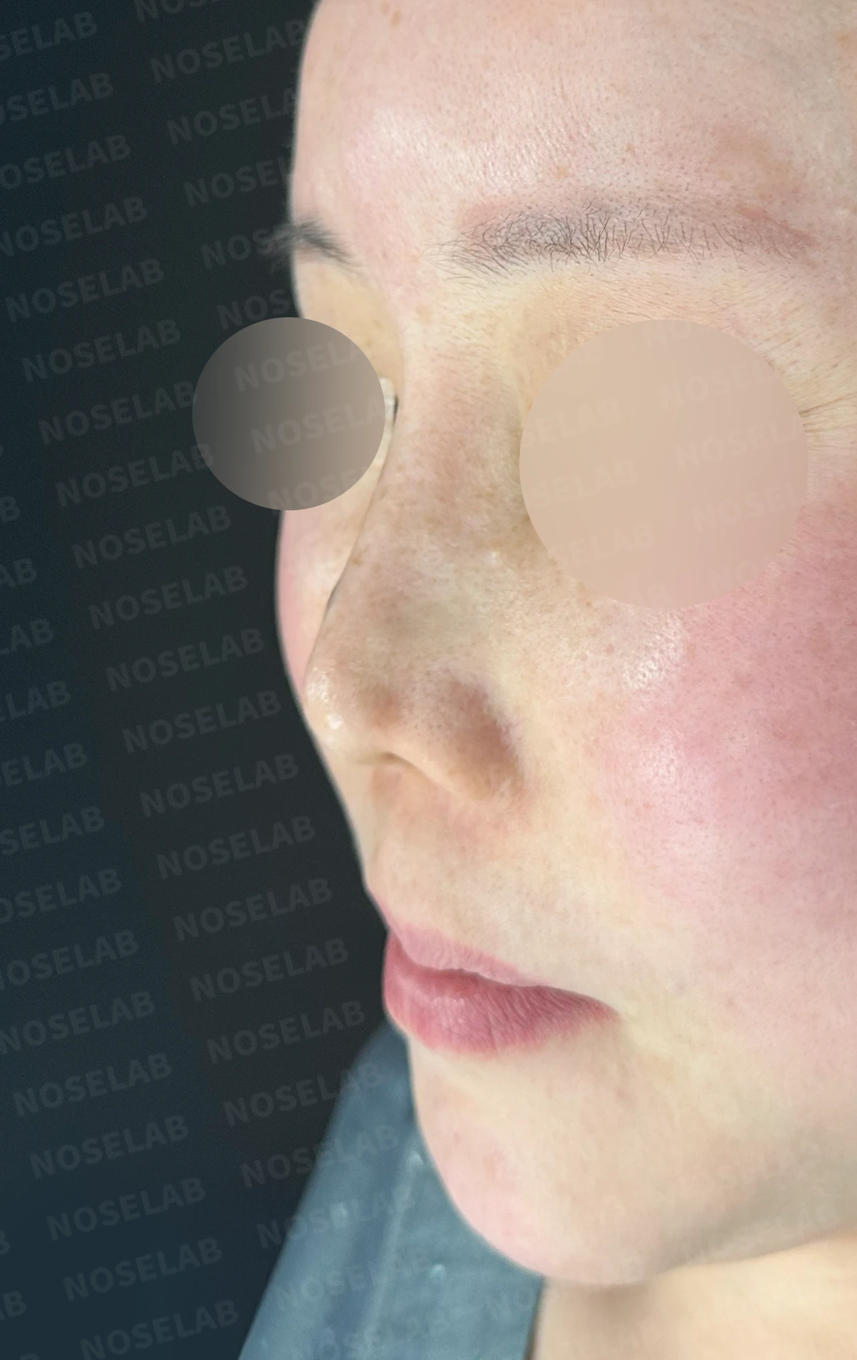
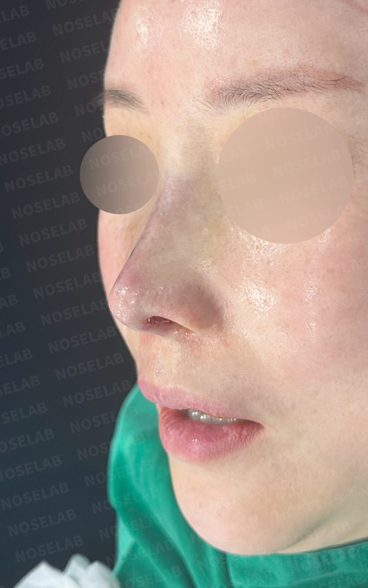
Before surgery / Day 0 immediately after surgery Photos taken immediately after surgery show swelling and may differ from the final result.
Structural stability
Sturdy septal reconstruction
Stable lower lateral cartilage structure
Long-term stable outcome
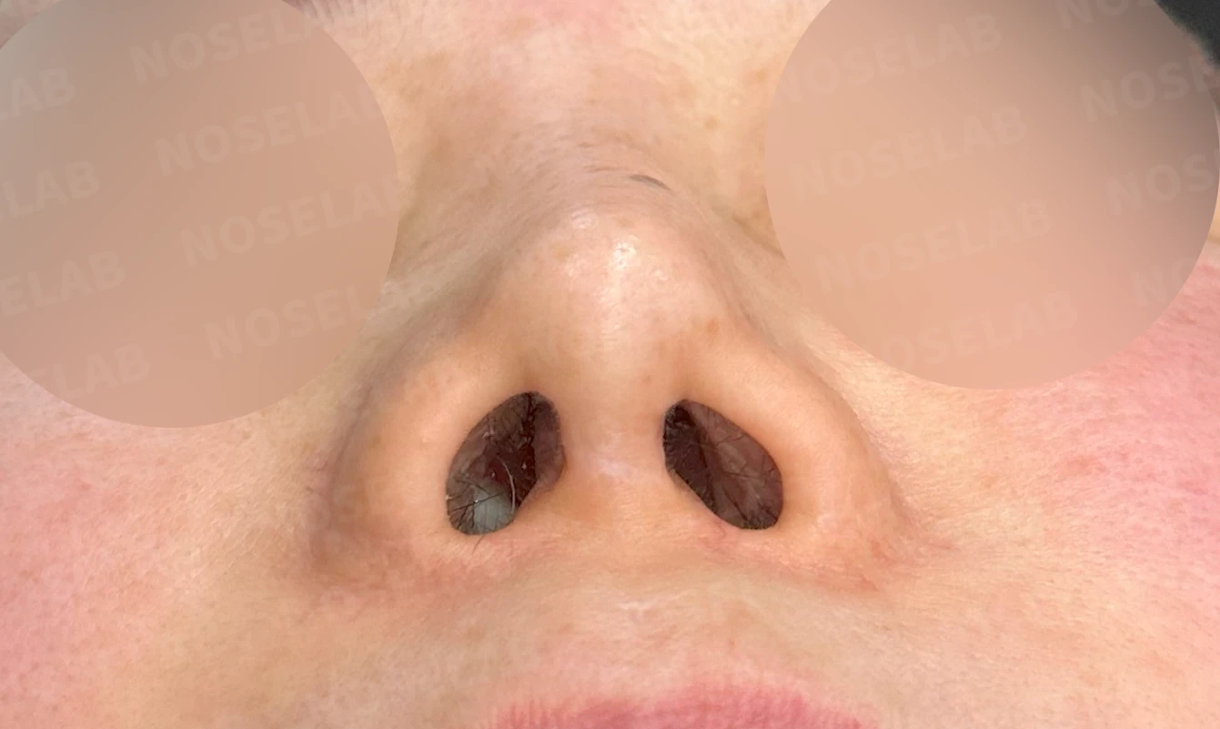
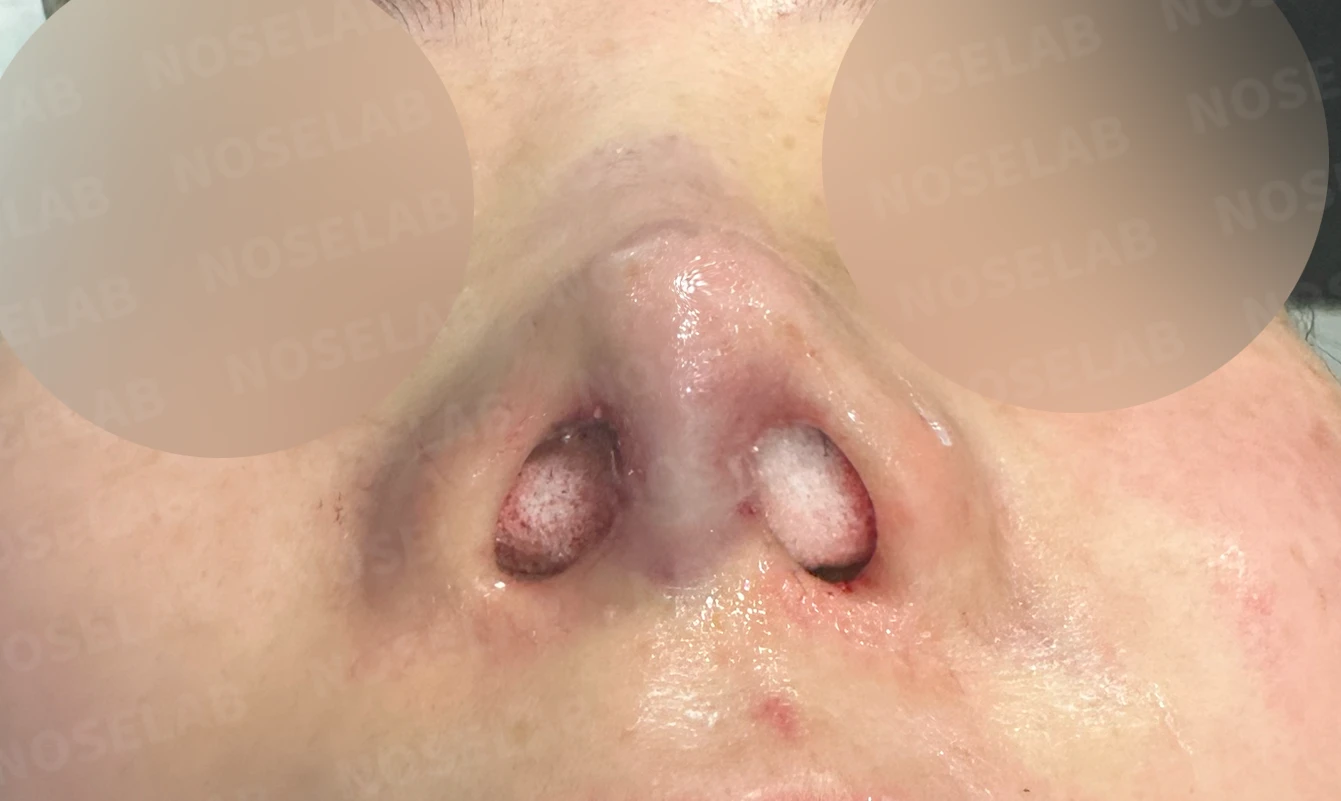
Before surgery / Day 0 immediately after surgery Photos taken immediately after surgery show swelling and may differ from the final result.
👨⚕️ Specialist's assessment
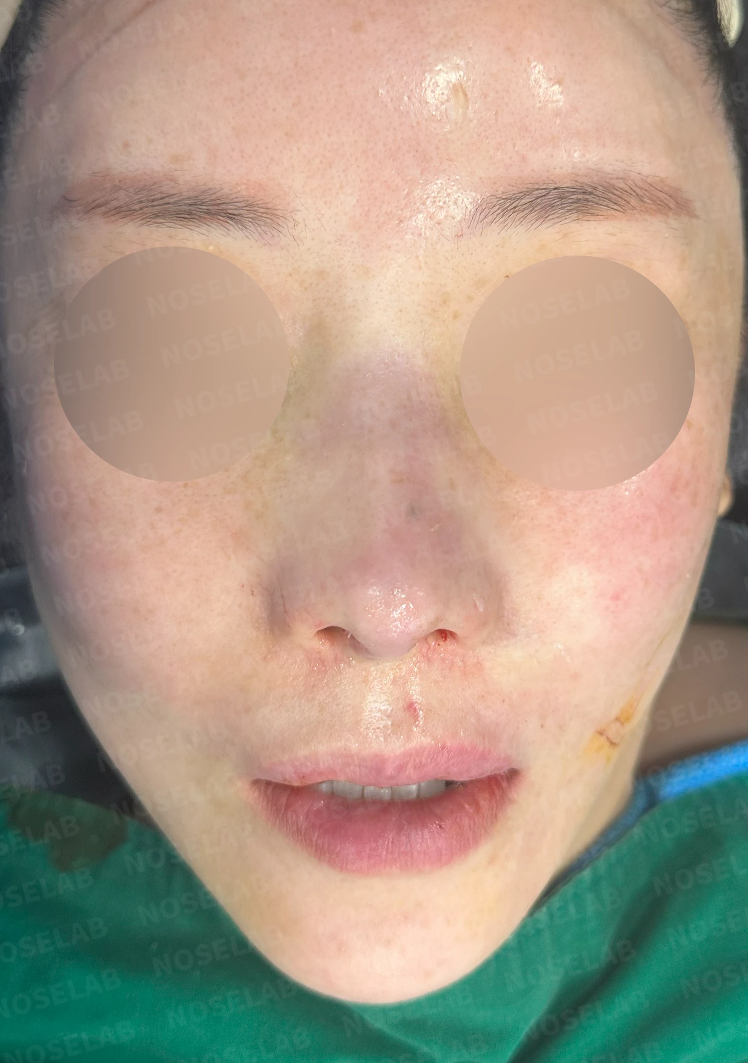
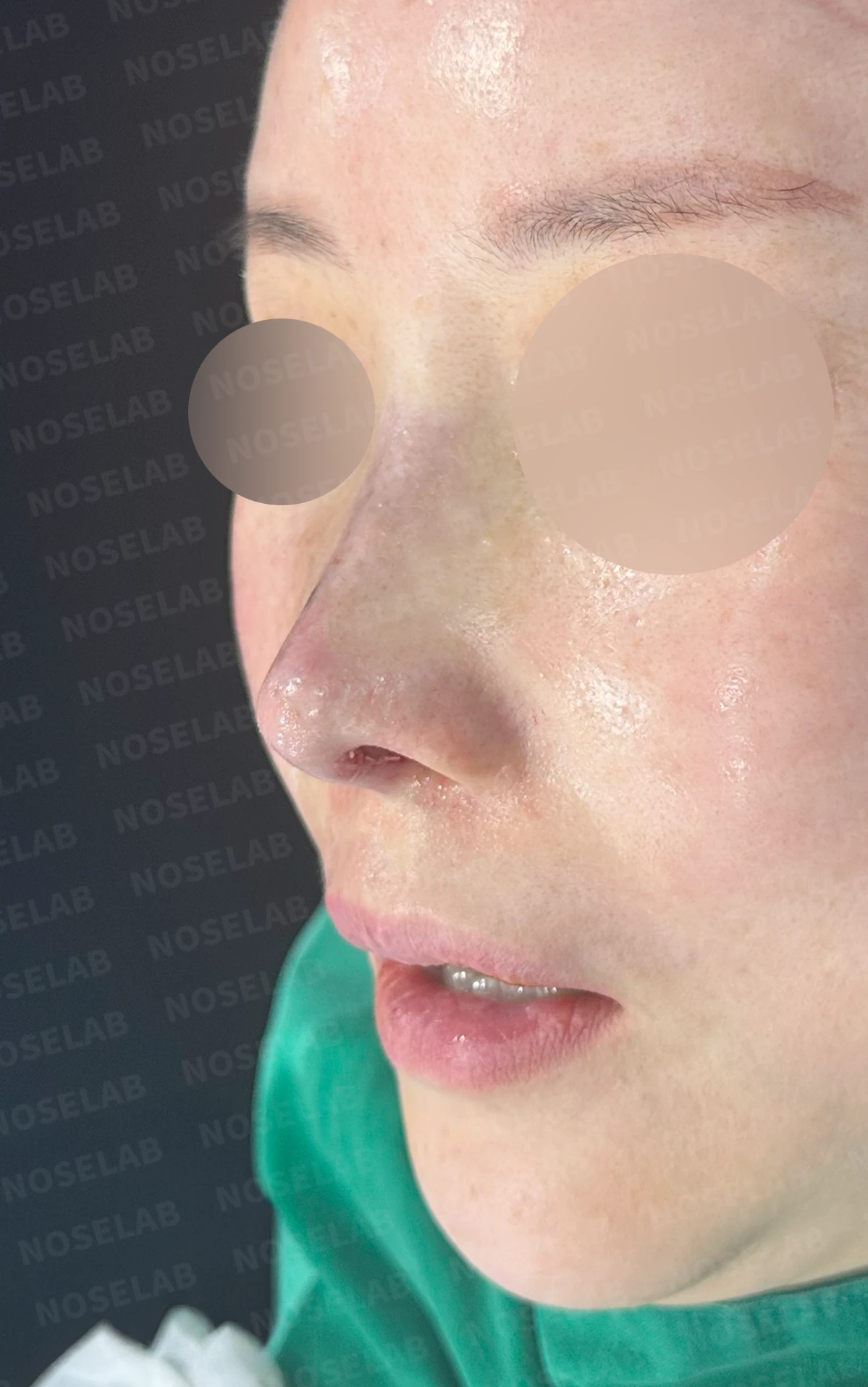
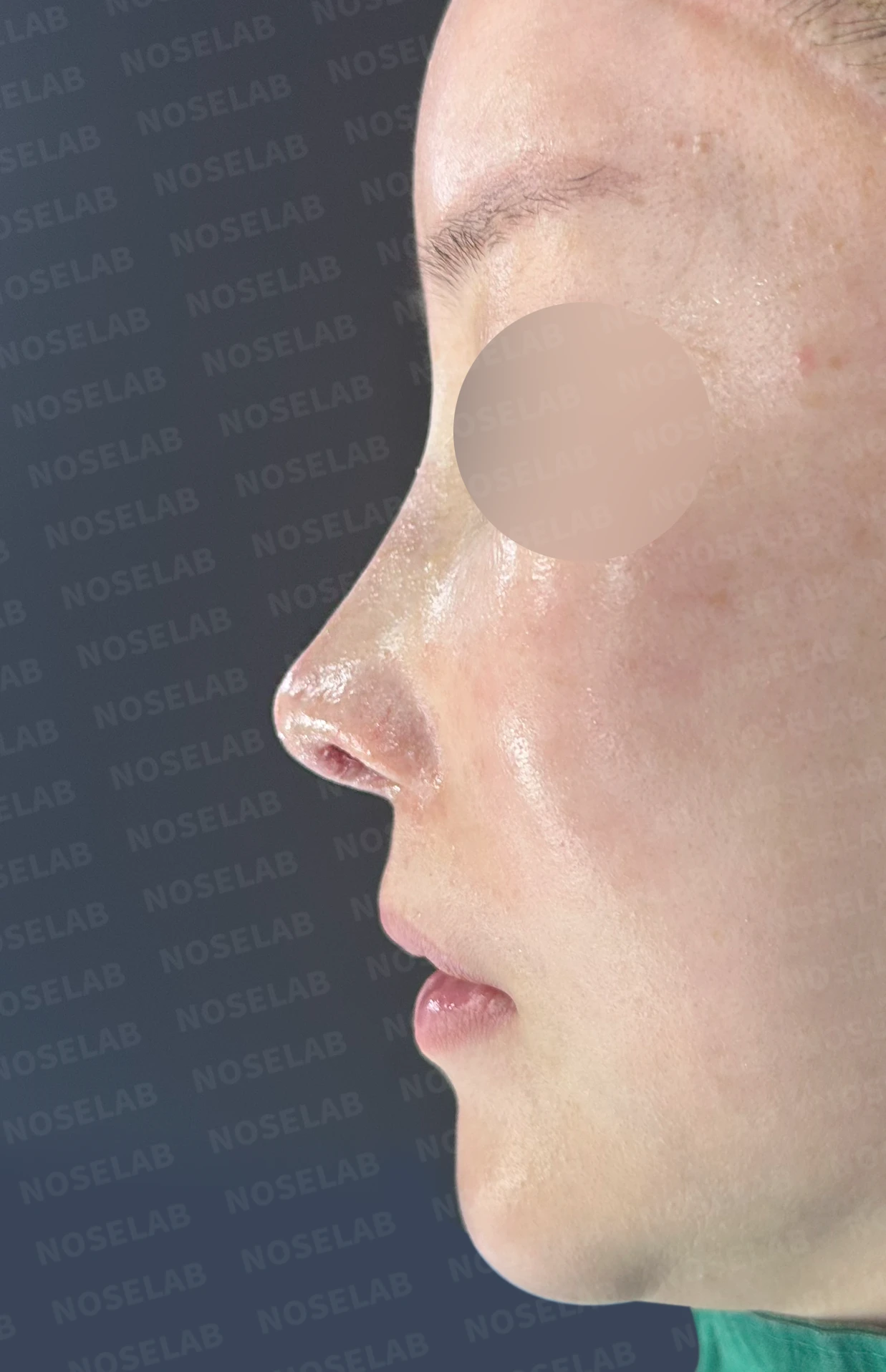
Front, profile, and oblique views immediately after surgery on day 0 Photos taken immediately after surgery show swelling and may differ from the final result.
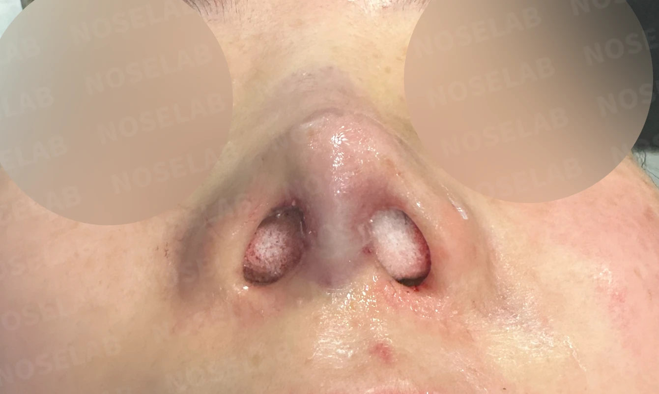
Nostril view on day 0 immediately after surgery Photos taken immediately after surgery show swelling and may differ from the final result.
This patient visited our clinic with a very unusual condition. Over time, an alar base implant placed in 2010 had migrated from its original position and protruded into the right nasal cavity, obstructing 2/3 of the nasal cavity. As a result, the patient was completely unable to breathe through the right side of the nose, experiencing significant daily discomfort and a severely reduced quality of life.
Migration of a paranasal implant is an uncommon complication, but it can occur over time due to changes in surrounding tissues or weakening of the implant's fixation. In this case, the displaced implant pushed into the nasal cavity, causing severe breathing obstruction.
At the same time, due to the nasal surgeries performed in 2012 and 2013, the septum was in very poor condition, and both lower lateral cartilages were severely damaged. The columella was retracted, and the alae appeared pinched, presenting multiple complex structural issues.
First and foremost, to restore breathing function, we removed the paranasal implant obstructing the nasal cavity. At the same time, we also removed the other paranasal implant to prevent similar issues from occurring in the future.
Next, to fundamentally rebuild the damaged nasal structure, we performed reconstruction using autologous rib cartilage. We completely reconstructed the septum to restore the central support structure of the nose, and also rebuilt the severely damaged bilateral lower lateral cartilages using autologous rib cartilage to correct the pinched alar appearance naturally. The retracted columella was restored as well.
Correction of the crooked nose and elevation of the nasal tip, which the patient desired, were also achieved during the structural reconstruction process. Correcting the nasolabial angle improved the harmony of the profile line as well.
As a result, we resolved the severe breathing obstruction caused by the migrated paranasal implant, and simultaneously corrected the structural damage from previous nasal surgeries through autologous rib cartilage reconstruction. In particular, using autologous tissue supports a stable long-term result.
This was Dr. Peter C. Kang. Thank you.
Thank you.
#closedrhinoplasty #implantremoval #nasalobstruction #autologousribcartilagereconstruction #alarreconstruction #functionalrecovery

Limitations and risks
When autologous rib cartilage is used, scarring and pain may remain at the donor site on the chest, and pneumothorax (pleural injury) can occur rarely. Over time, the grafted cartilage may warp or undergo partial resorption.
When using donor rib cartilage, long-term follow-up is necessary because the grafted cartilage may resorb or warp.
When revising after implant removal, contracture may progress again after removal, the nasal height and length may decrease, and staged surgery may be necessary depending on the skin condition.
When the septum is corrected at the same time, nasal obstruction may persist or recur, and in rare cases, septal perforation or changes in smell may occur.
Common potential risks include infection, bleeding, asymmetry, scarring, skin necrosis, and insufficient improvement; results vary depending on individual skin condition and previous surgical history.