Hello, I am Dr. Peter C. Kang.
Today, I would like to share a structural reconstruction case of a patient who had undergone surgery where the implant was unexpectedly replaced with an L-shaped silicone implant following a previous rhinoplasty.
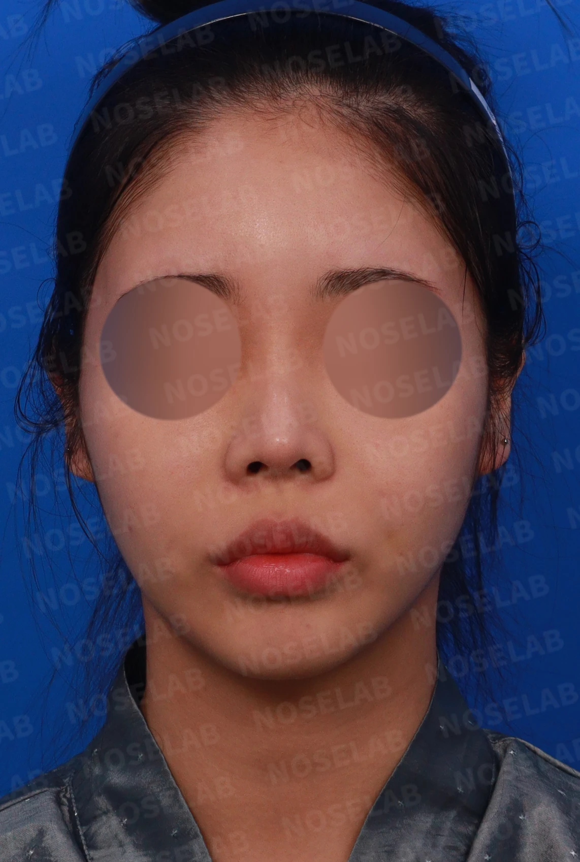
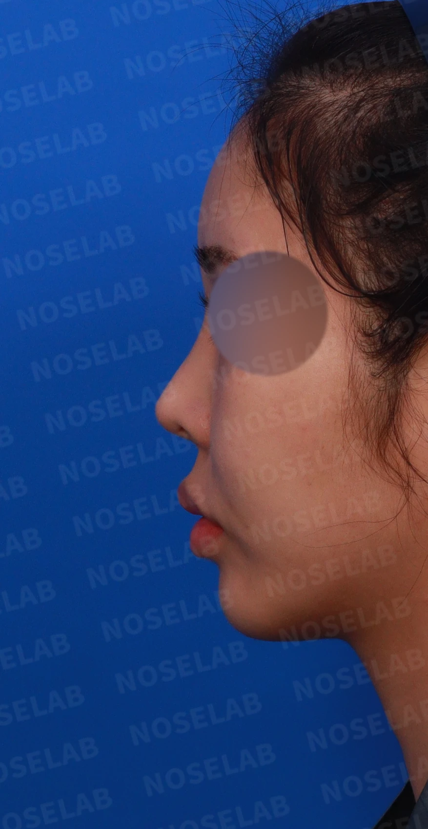
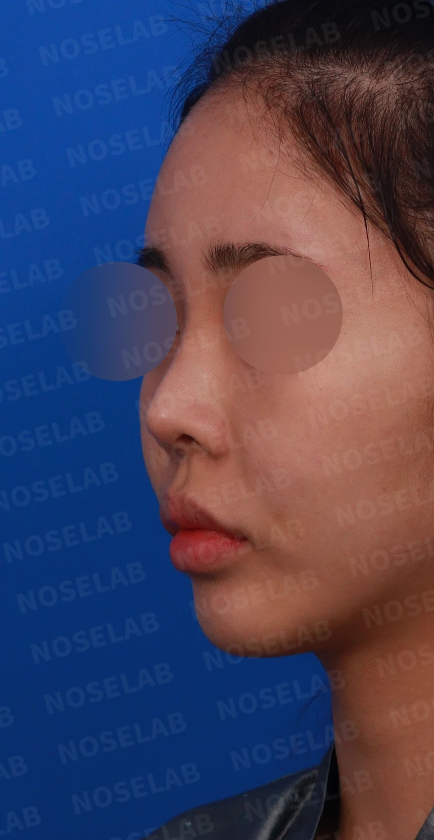
Front, left profile, and oblique views during design consultation
📍 Surgical history
10 years ago: Septal cartilage and ear cartilage were used for the nasal tip, and an I-shaped implant was used for the nasal bridge
6 months later: During a revision procedure for nasal deviation, it was unexpectedly replaced with an L-shaped silicone implant
🔎 Initial consultation findings
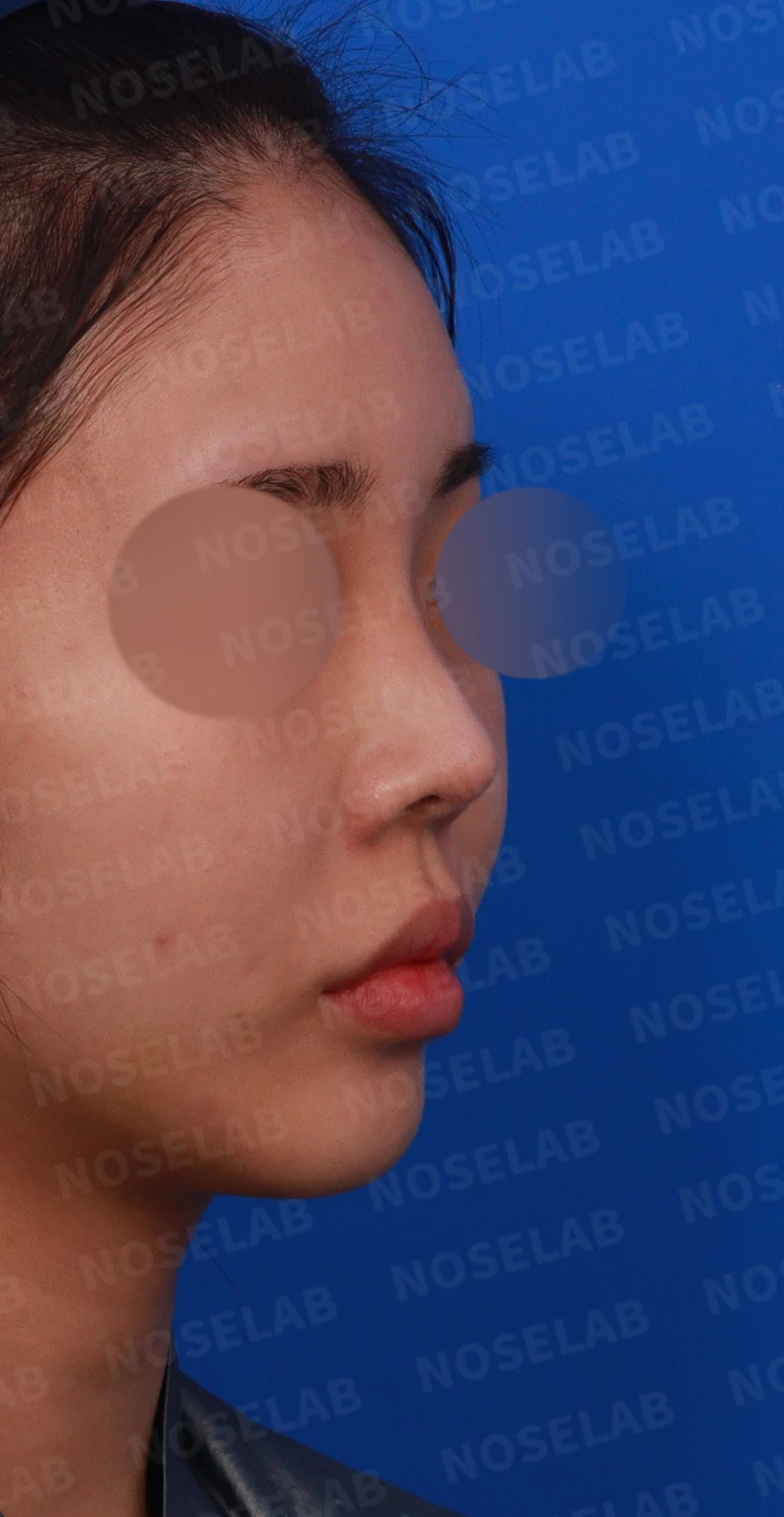
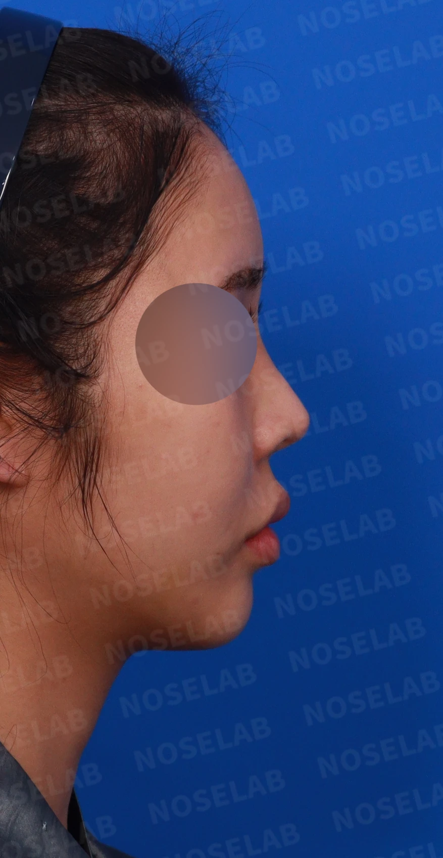
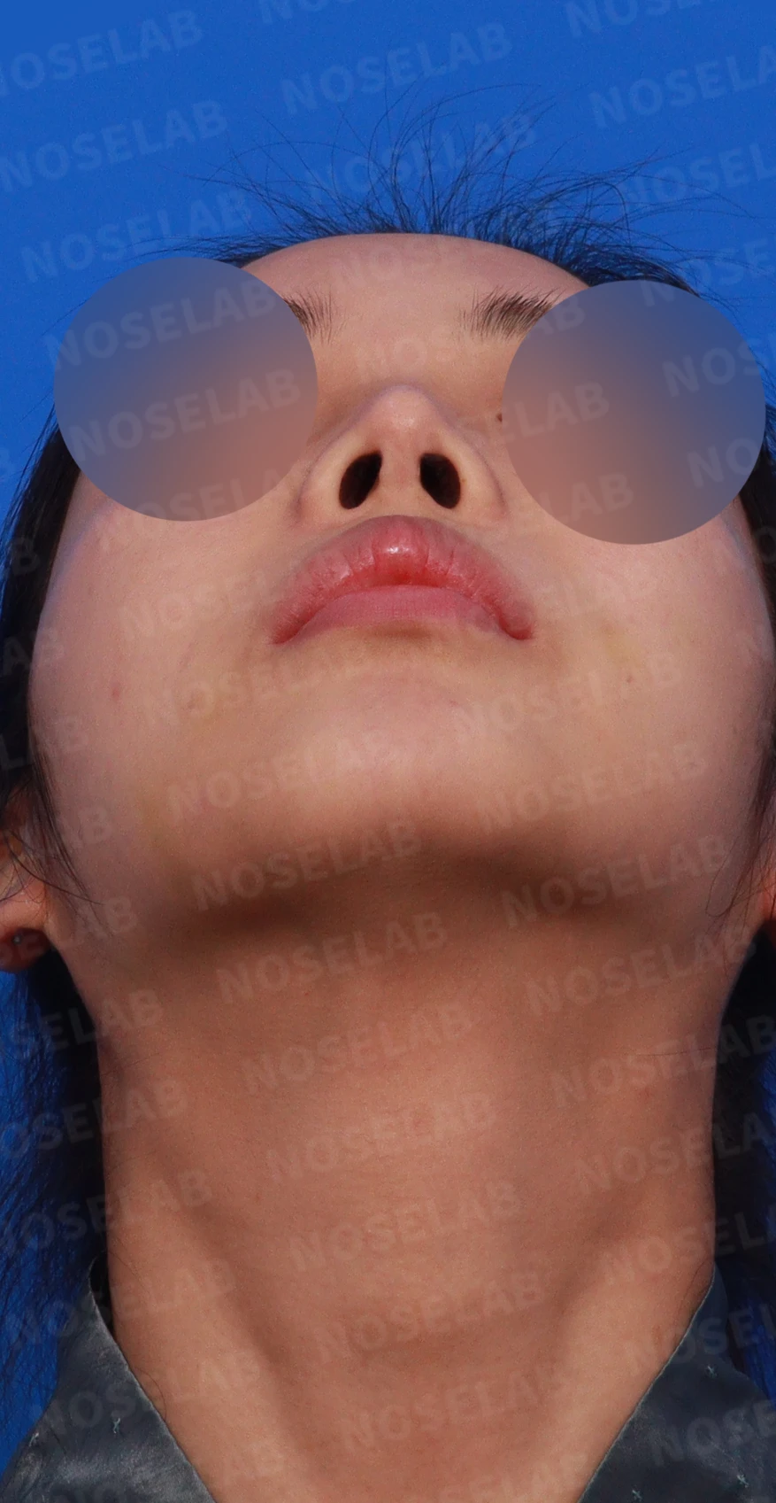
Design consultation: right oblique, profile, and basal (nostril) views
Front view
Upturned nose
Crooked nose
Narrow nasal bridge
2. Profile view
High radix
Acute nasolabial angle
Drooping nasal tip appearance
3. Nostrils
Nostril asymmetry
Columella deviated to the right
✨ Patient's requests


Correction of upturned nose
Creating a straight bridge line
Lowering the radix
Improvement of the nasolabial angle
Improvement of nostril asymmetry
💉 Surgical procedure details
Reconstruction with a stable structure
Removal of the L-shaped silicone implant
Reconstruction of the nasal tip using only autologous rib cartilage with safety in mind
Nasal bridge changed to an I-shaped implant
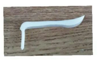
Removed L-shaped implant
2. Structural improvement
Naturally lowering the radix
Correction of upturned nose
Correcting the columella tilted to the right
3. Shape improvement
Creating a straight bridge line
Correction of the nasolabial angle
Improvement of nostril asymmetry
✅ Surgical results
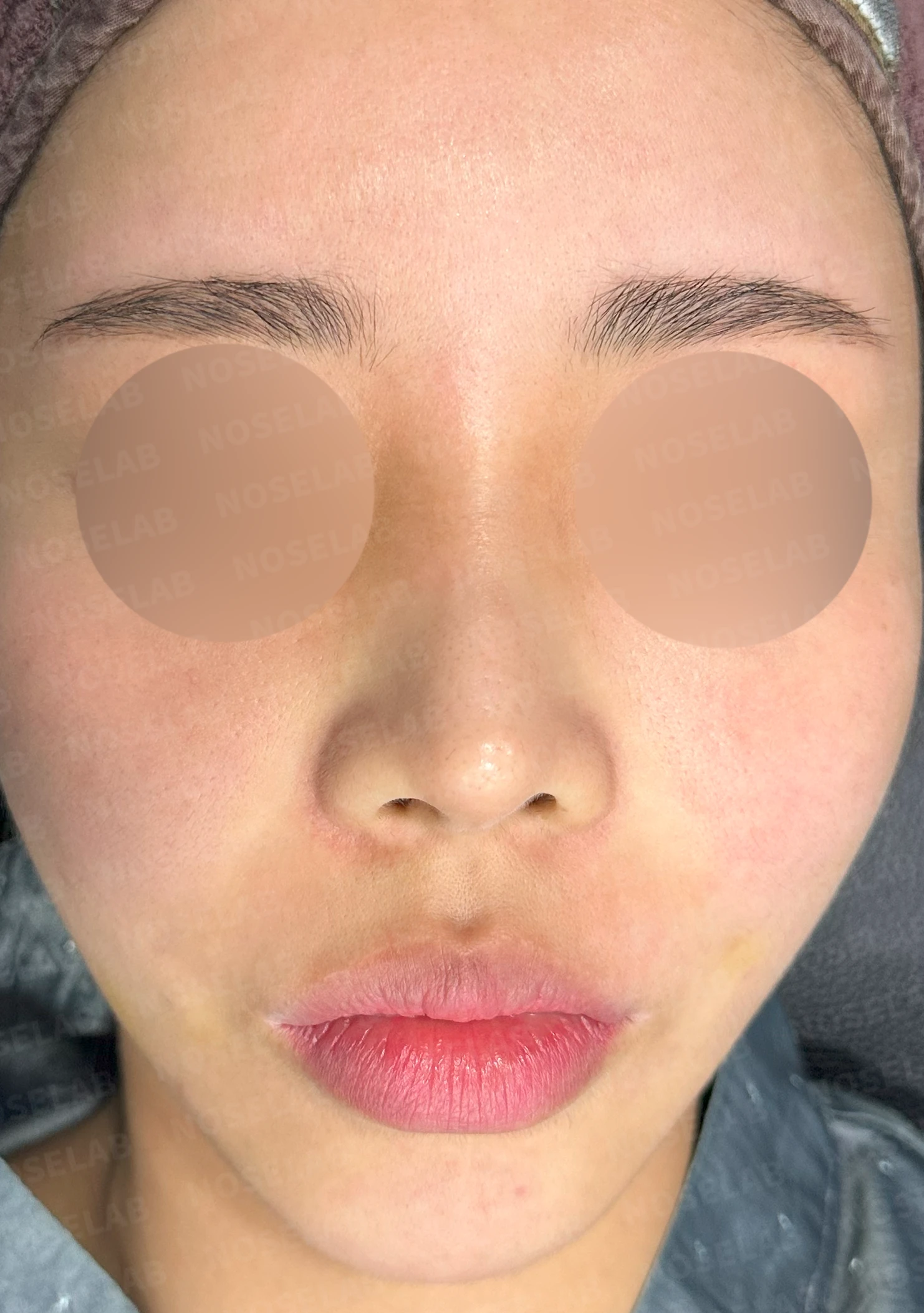
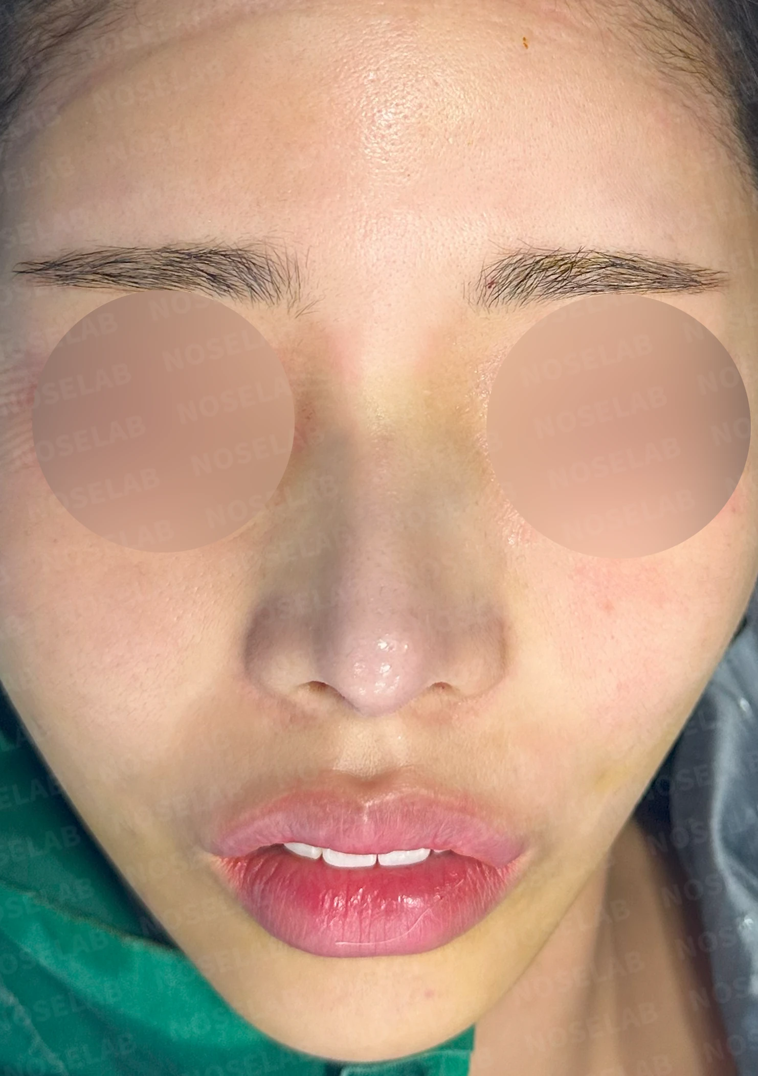
Before surgery / Day 0 immediately after surgery Photos taken immediately after surgery show swelling and may differ from the final result.
Front view
Radix lowered to create a natural appearance between the eyebrows
Significant nostril asymmetry and nostril show improved
Crooked areas improved
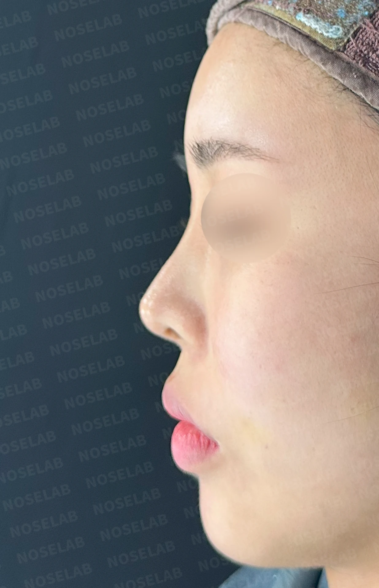
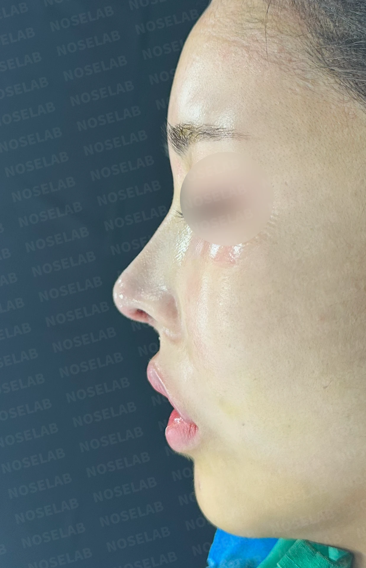
Before surgery / Day 0 immediately after surgery Photos taken immediately after surgery show swelling and may differ from the final result.
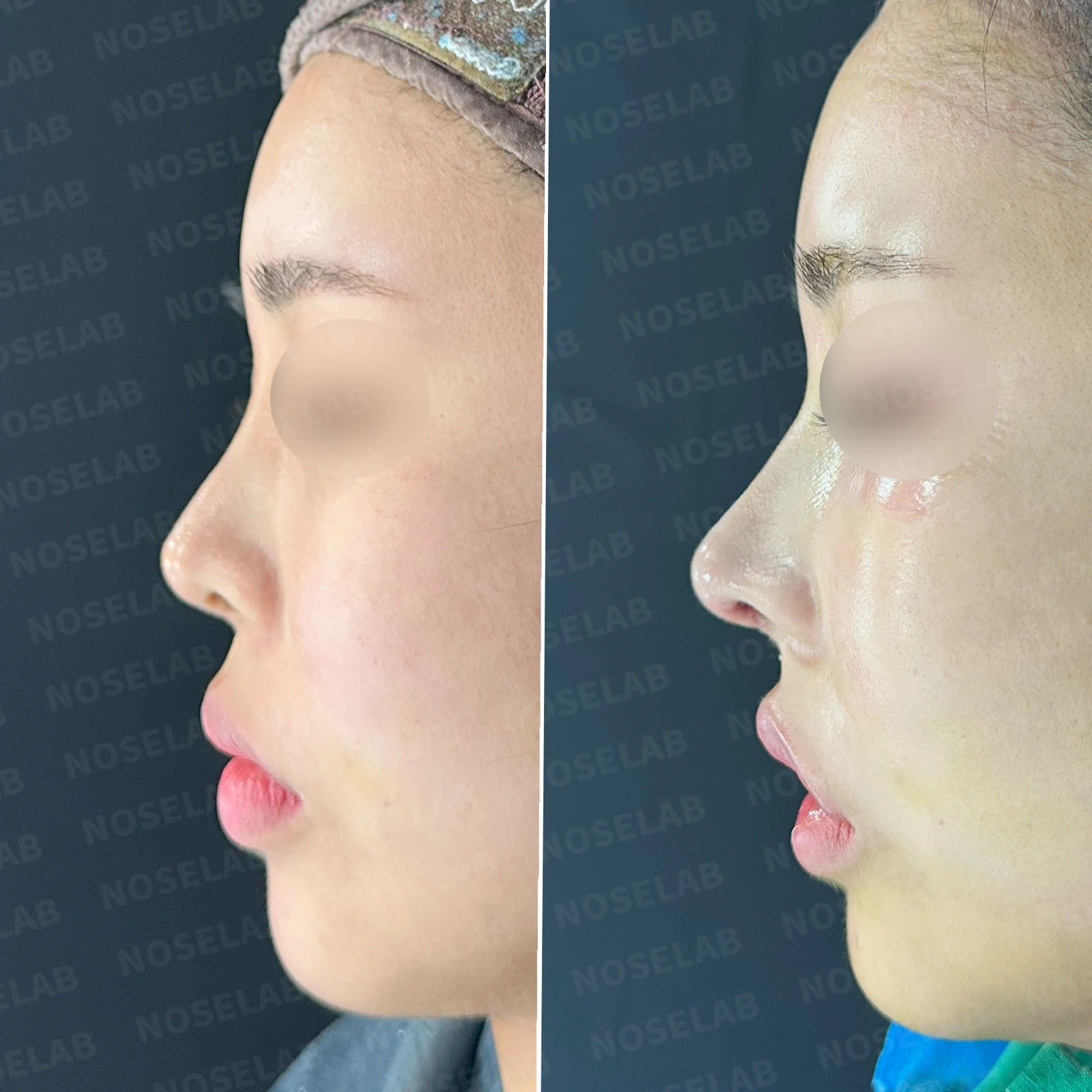
2. Profile view
Natural radix position
Improvement of upturned nose
Natural nasolabial angle
Straight bridge line
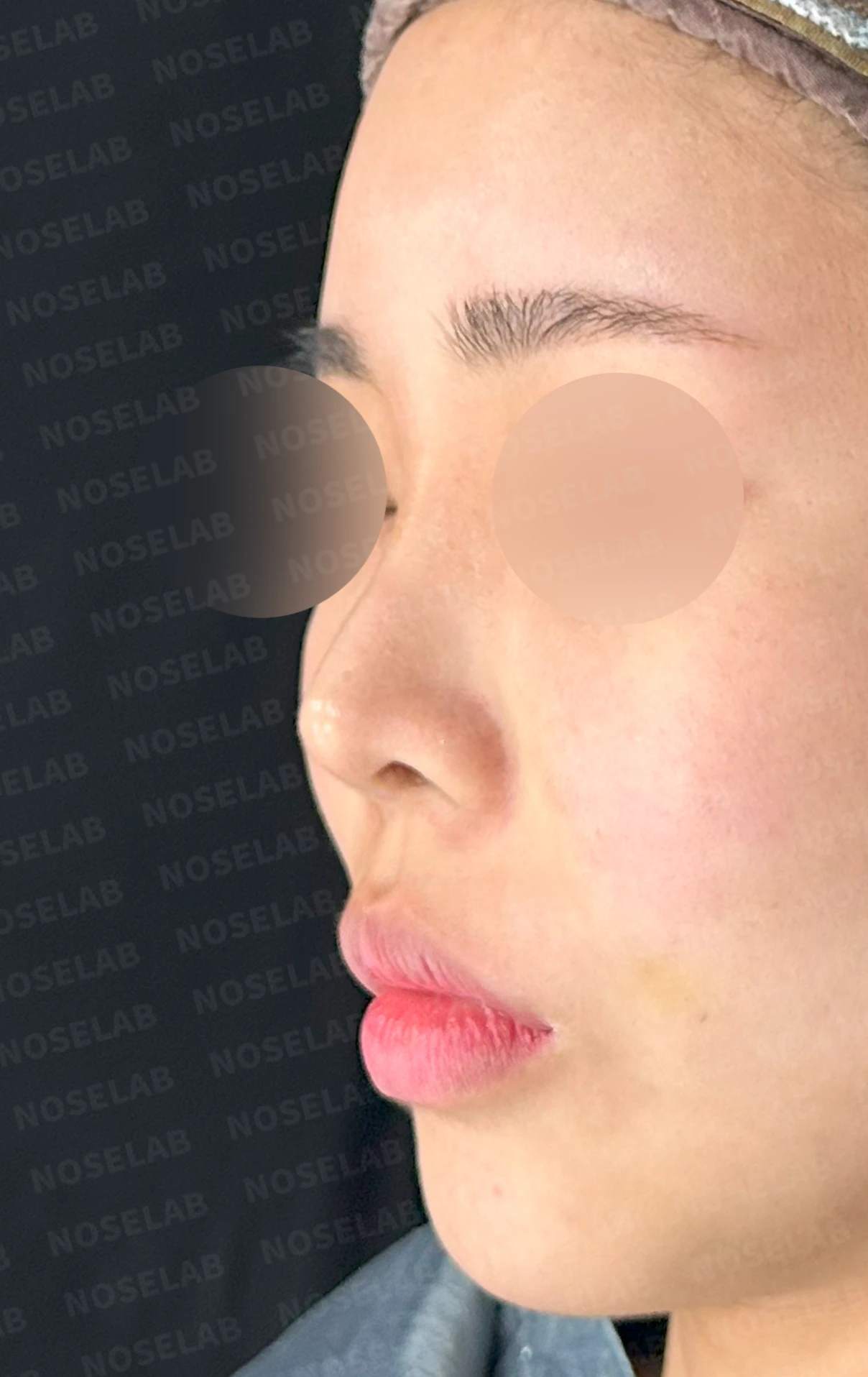
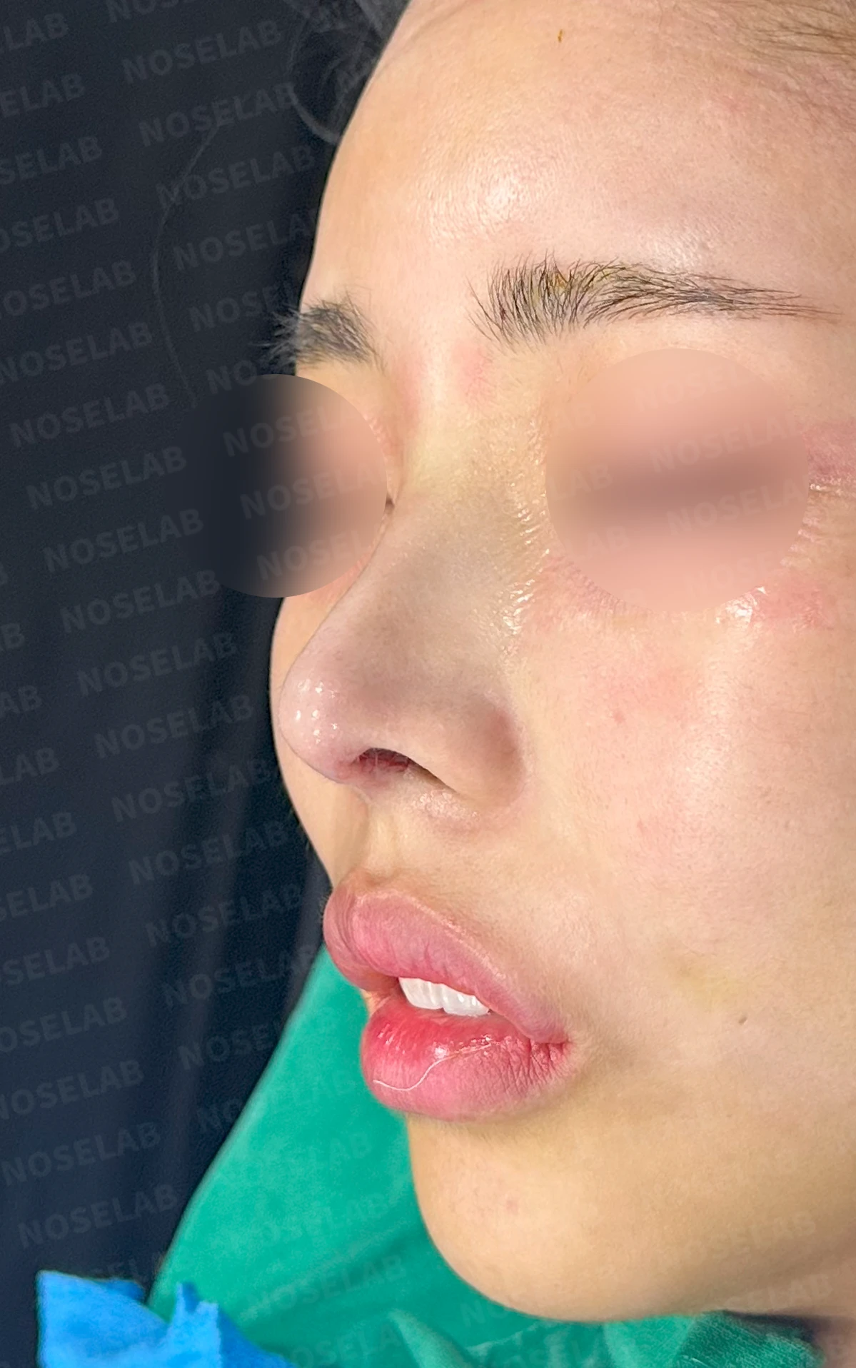
Before surgery / Day 0 immediately after surgery Photos taken immediately after surgery show swelling and may differ from the final result.
3. Nostrils
Improvement in nostril symmetry
Correction of the tilted columella
Softened nasal tip appearance
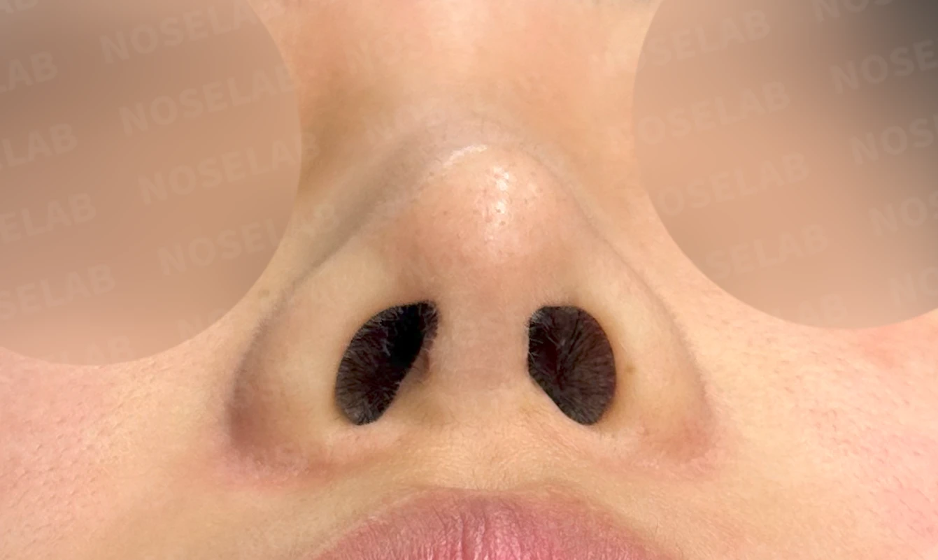
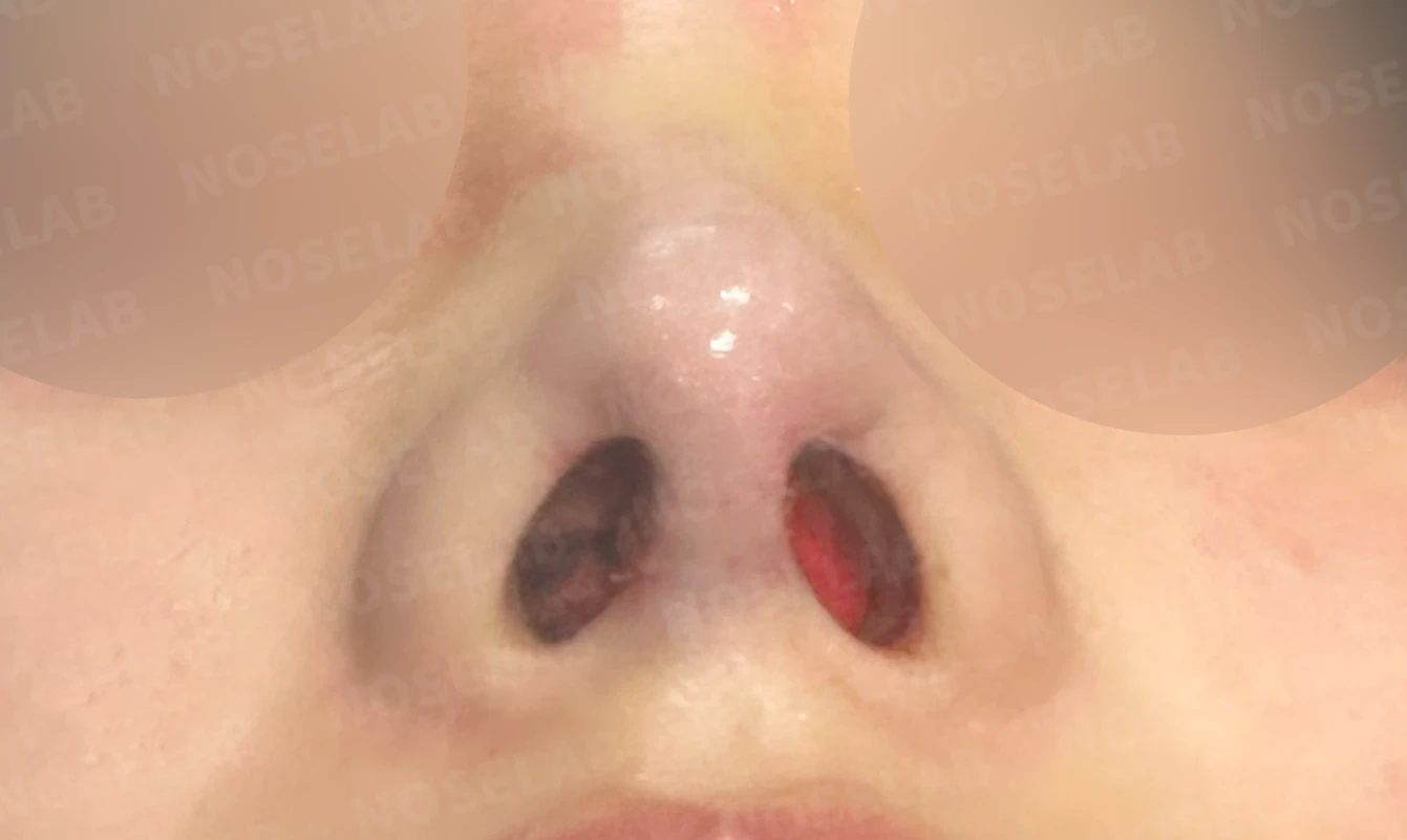
Before surgery / Day 0 immediately after surgery Photos taken immediately after surgery show swelling and may differ from the final result.
👨⚕️ Specialist's assessment
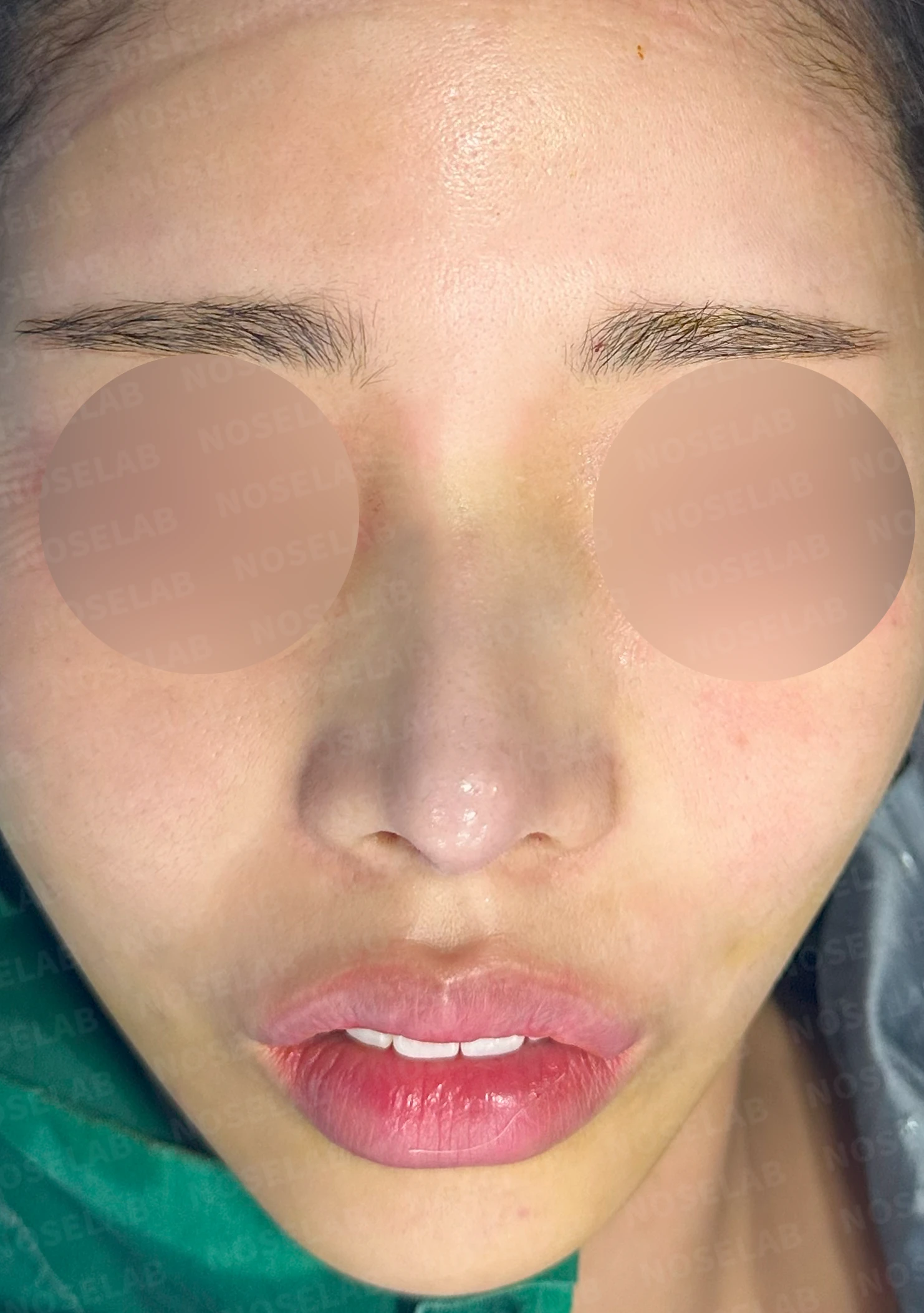
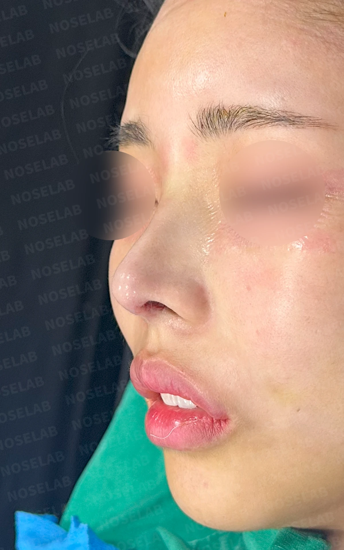
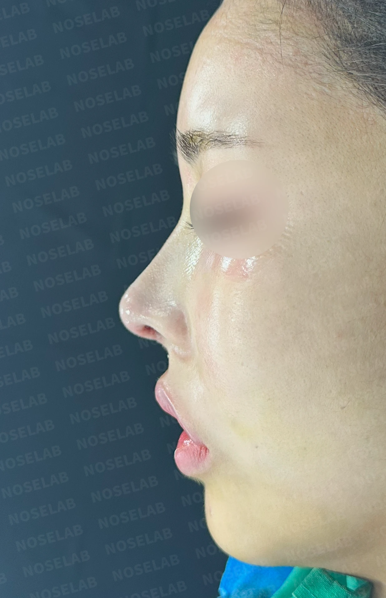
Front, oblique, and profile views on day 0 immediately after surgery Photos taken immediately after surgery show swelling and may differ from the final result.
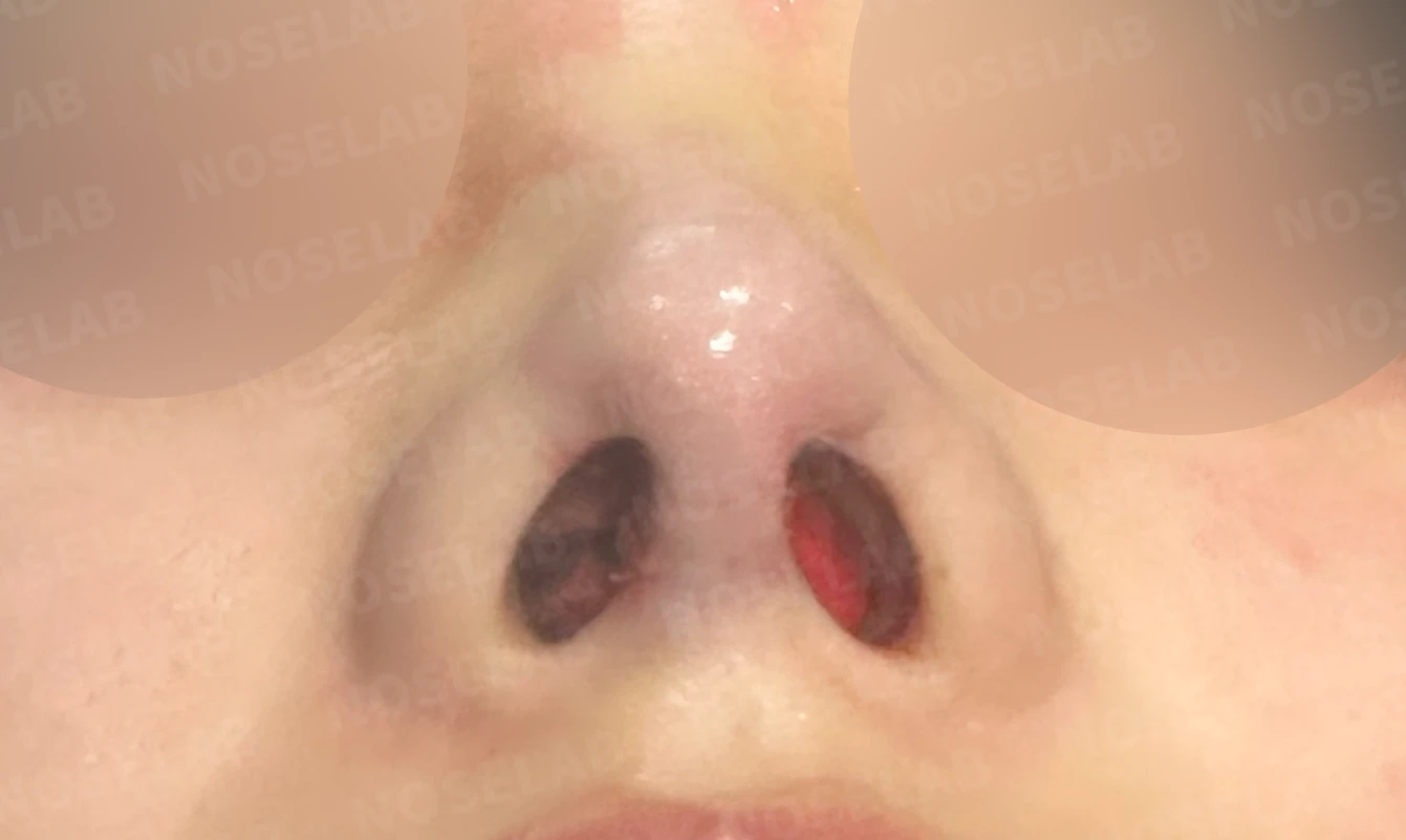
Nostril view on day 0 immediately after surgery Photos taken immediately after surgery show swelling and may differ from the final result.
This case clearly illustrates the importance of material selection in rhinoplasty. In the patient's primary surgery 10 years ago, a structurally stable technique was used: septal and ear cartilage for the nasal tip, and an I-shaped implant for the nasal bridge. However, during a subsequent revision to correct deviation, the cartilaginous support structure of the nasal tip had inexplicably been replaced with an L-shaped silicone implant.
In rhinoplasty, the nasal tip requires the most delicate approach, and using cartilage is generally natural and stable. In particular, replacing a nasal tip that was already well-supported by cartilage with an L-shaped silicone implant departs from sound rhinoplasty principles. Because an L-shaped silicone implant extends in a single piece into the nasal tip, long-term use carries a relatively high risk of foreign body sensation, show-through, and structural deformation.
In this surgery, we focused on removing the L-shaped silicone implant, reconstructing the nasal tip using only autologous rib cartilage, and switching the nasal bridge to a reliable I-shaped implant. At the same time, we lowered the radix, corrected the nasolabial angle, and addressed the nostril asymmetry and right-tilted columella.
This case reaffirms that selecting the appropriate materials and surgical methods tailored to each patient is a vital factor in achieving long-term stability and satisfaction in rhinoplasty.
This was Dr. Peter C. Kang. Thank you.
Thank you.
#closedrhinoplasty #revisionrhinoplasty #Lshapedimplant #Ishapedimplant #customrhinoplasty

Limitations and risks
When autologous rib cartilage is used, scarring and pain may remain at the donor site on the chest, and pneumothorax (pleural injury) can occur rarely. Over time, the grafted cartilage may warp or undergo partial resorption.
When revising after implant removal, contracture may progress again after removal, the nasal height and length may decrease, and staged surgery may be necessary depending on the skin condition.
When the septum is corrected at the same time, nasal obstruction may persist or recur, and in rare cases, septal perforation or changes in smell may occur.
Common potential risks include infection, bleeding, asymmetry, scarring, skin necrosis, and insufficient improvement; results vary depending on individual skin condition and previous surgical history.