Hello, I am Dr. Peter C. Kang.
Today, I would like to introduce a surgical case of a patient who was troubled by nasal bridge pain, headaches, and an unstable, shifting implant.
This patient had undergone surgery with an I-shaped silicone implant 20 years ago and had recently been taking long-term medication for severe rhinitis.
Preoperative consultation
The patient's main symptoms:
- Pain in the glabella and nasal bridge (particularly severe when wearing glasses)
- Mobility of the silicone implant
- Intermittent pain along the nasal dorsum
- Severe rhinitis
Analysis of preoperative condition:
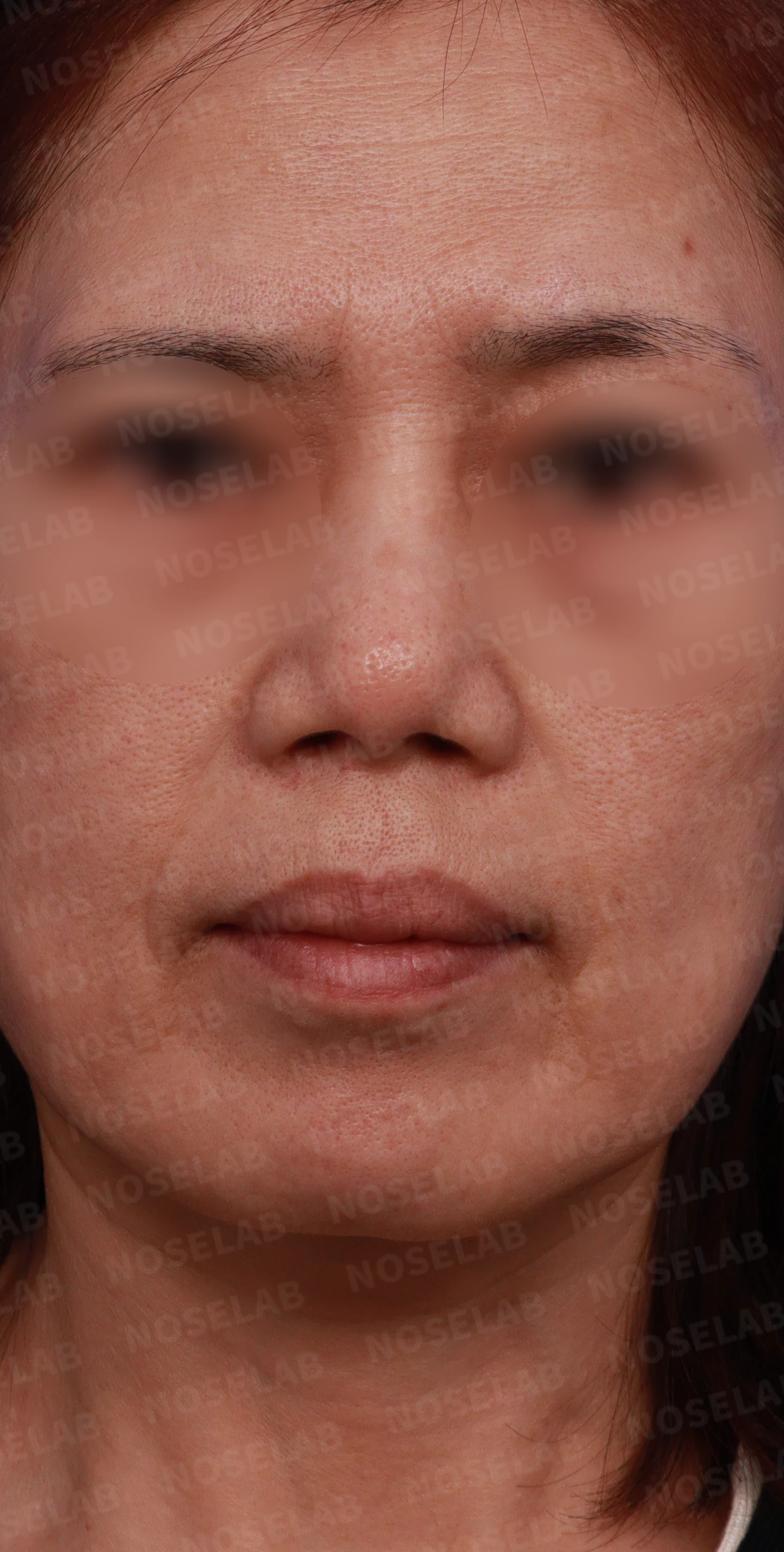
Front view from the design consultation.
Front view:
- The nasal bridge appears swollen and widened, with a drooping tip
- The nasal tip is blunt and bulbous
- The alae are splayed and drooping
- The philtrum appears long
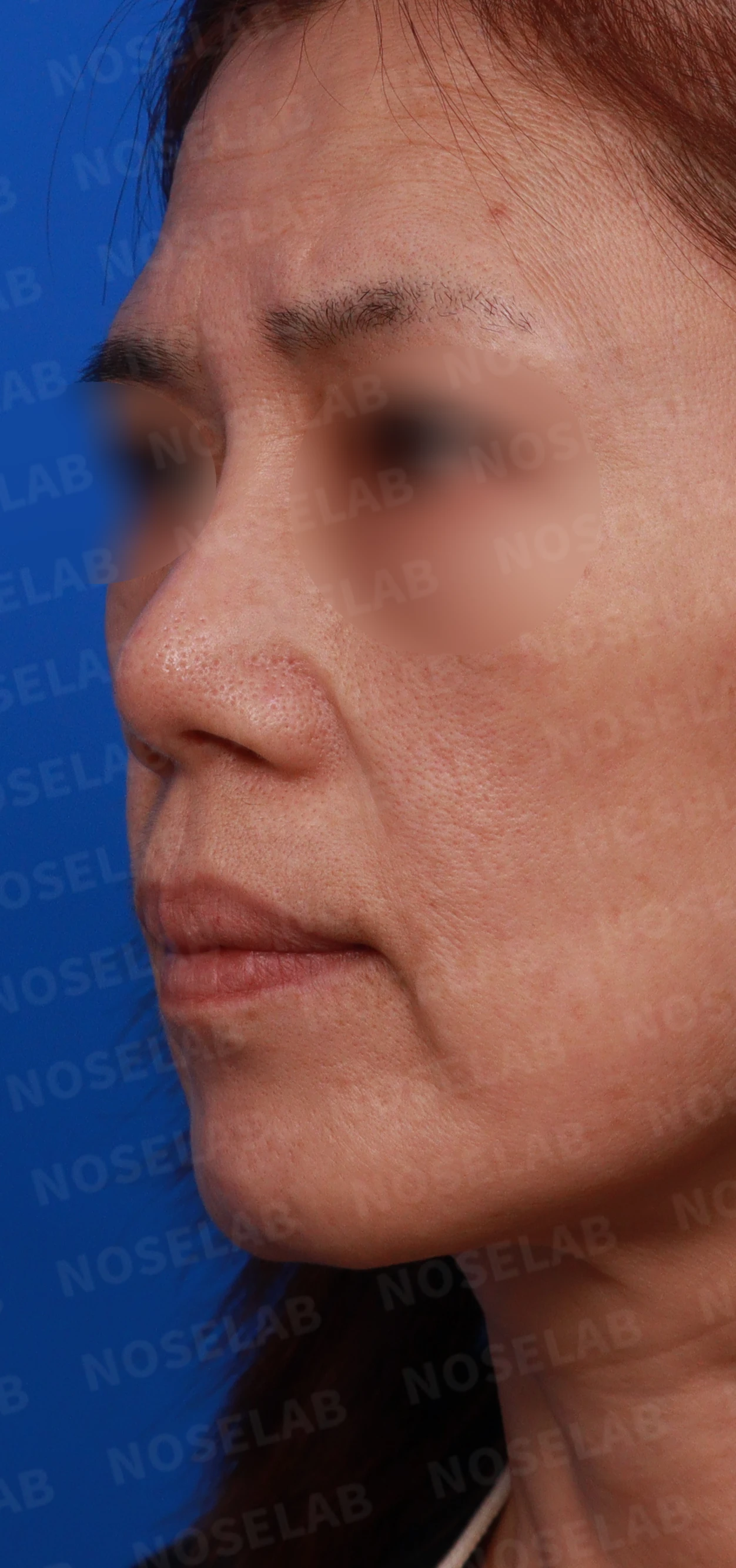
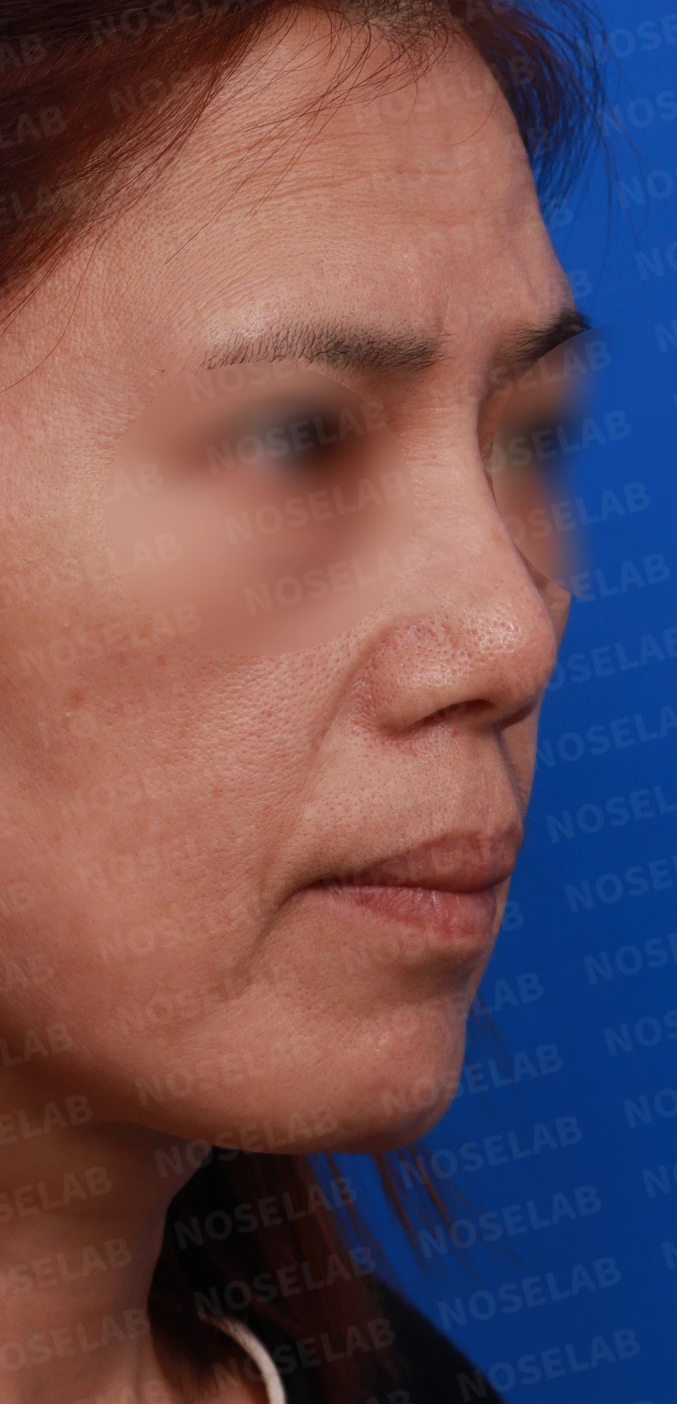
Oblique view from the design consultation.
Oblique view:
- The nasal tip appears bulbous and drooping
- Findings of a protruding mouth are observed
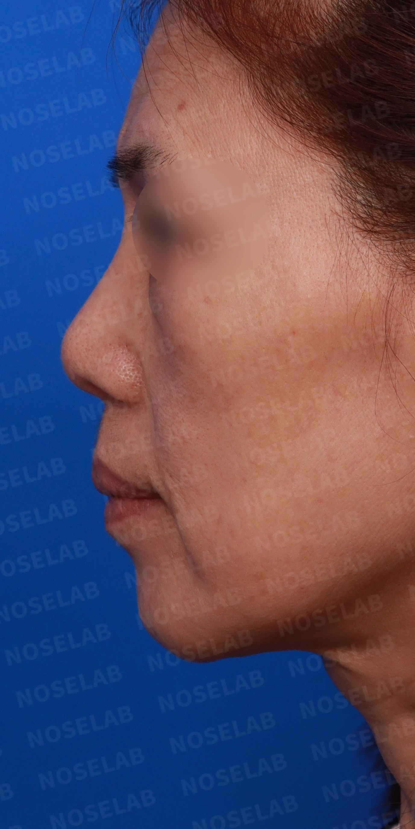
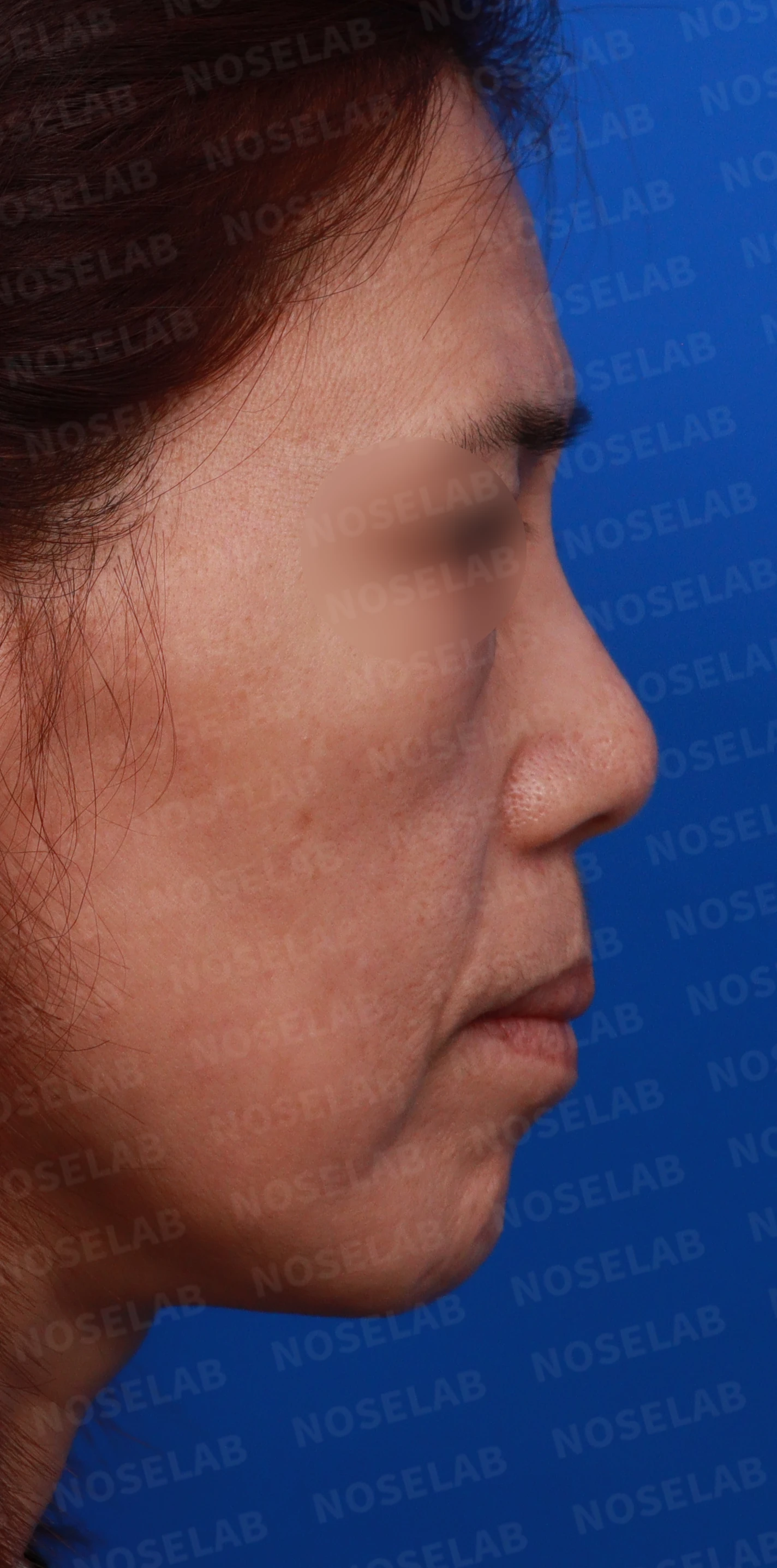
Profile view from the design consultation.
Profile view:
- The nasal tip looks compressed and bulbous
- Retracted columella and the appearance of a protruding mouth are observed
- Receding chin is observed
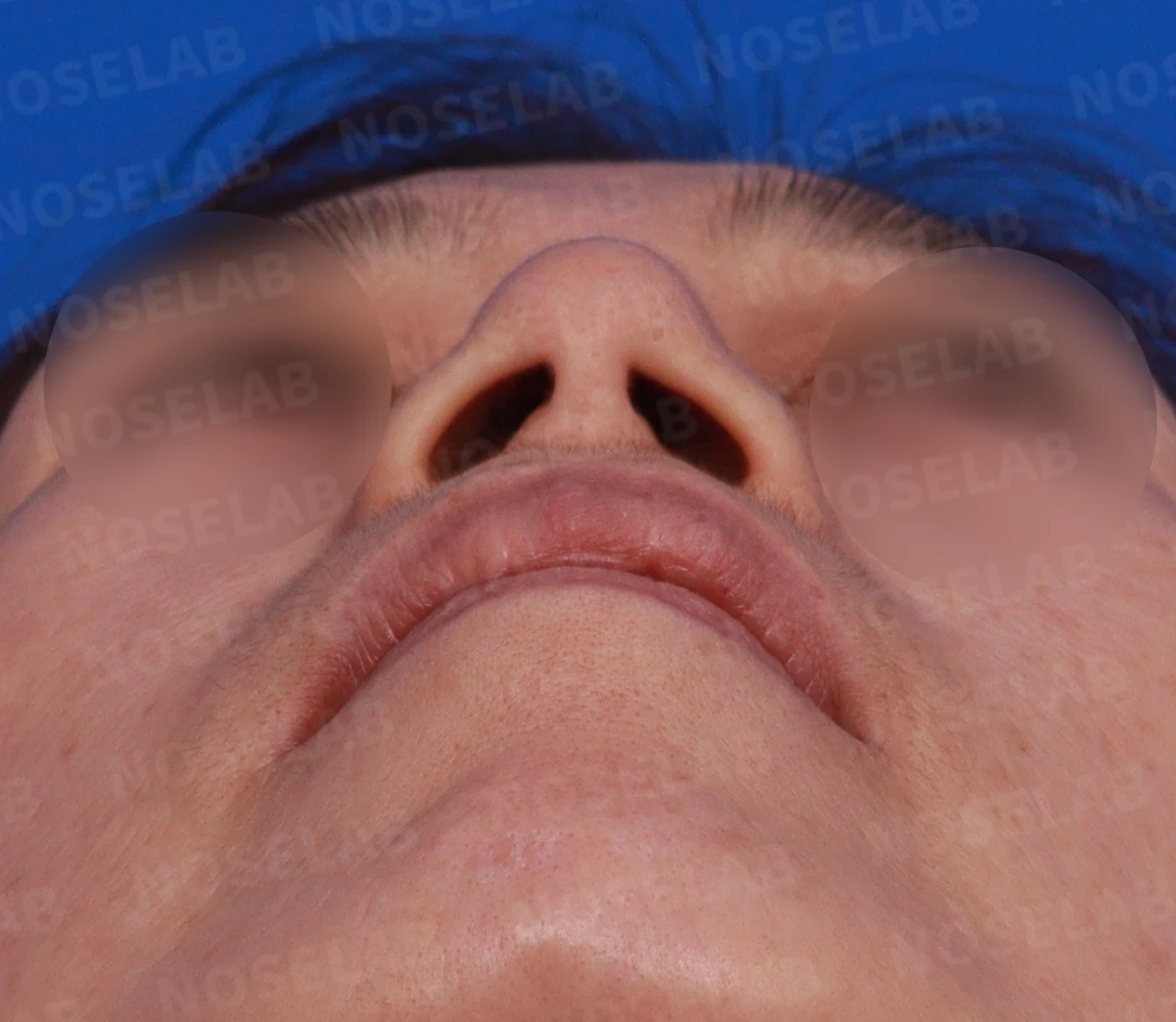
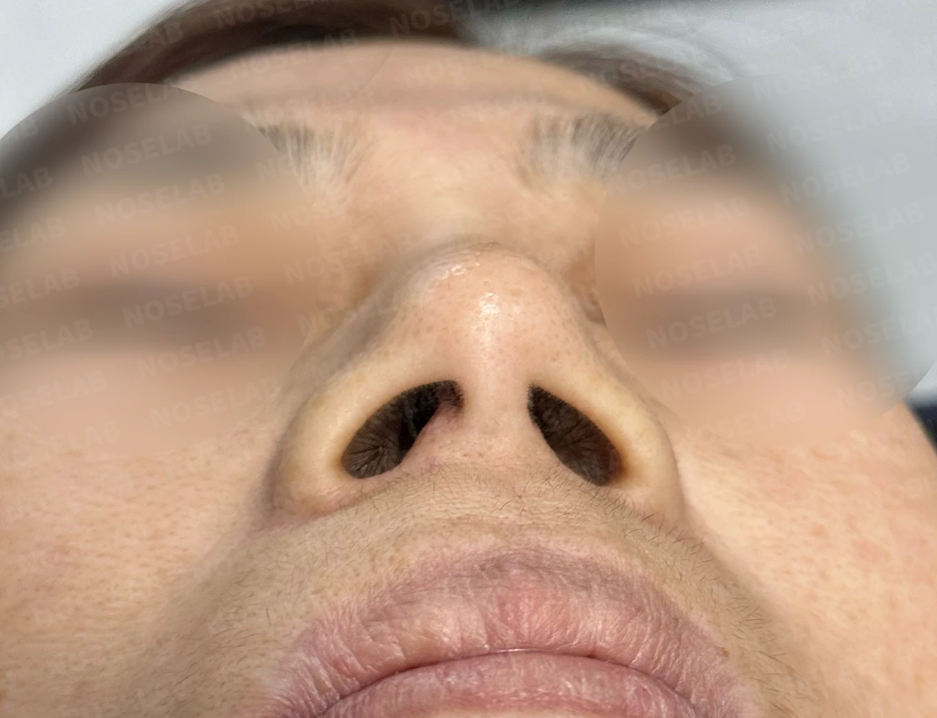
Base view (nostril) photo
Nostrils:
- Slight nostril asymmetry is visible
- The nostrils appear compressed and splayed
Surgical plan
Removal of existing silicone implant
Partial removal of tissue around the implant
Placement of a new implant (thinner and wider than the previous one)
Tip elevation and nasolabial angle correction
Septoplasty and septal extension
Surgical procedure
1. Removal of the existing implant:
- Removal of a rigid silicone implant measuring 8 mm in width and 3 to 4 mm in thickness
- Removal of granulation tissue around the implant and irrigation with saline
2. Nasal tip reconstruction:
- Removal of scar tissue around the lower lateral cartilages
- Septal extension performed using autologous rib cartilage
- Reshaping and elevating the nasal tip
3. Nasolabial angle correction:
- Lowering the columella and correcting the nasolabial angle
- Expected improvement in protruding mouth appearance
4. Placement of the new implant:
- Placement of an implant designed with minimal height and thickness
- Positioned beneath the periosteum to ensure stability
5. Septoplasty:
- Corrected only the deviated portion minimally to preserve cartilage
Before-and-after photo comparison
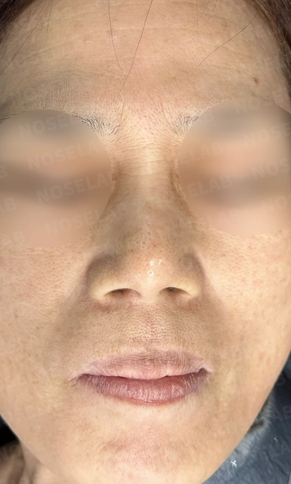
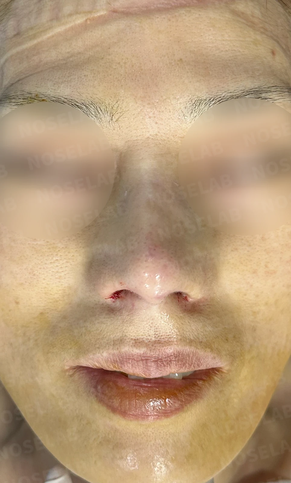
Before surgery / Day 0 immediately after surgery Photos taken immediately after surgery show swelling and may differ from the final result.
Front view:
- The previously widened nasal bridge is slimmer, with swelling reduced
- The bulbous nasal tip has been naturally elevated
- Alar spreading has improved
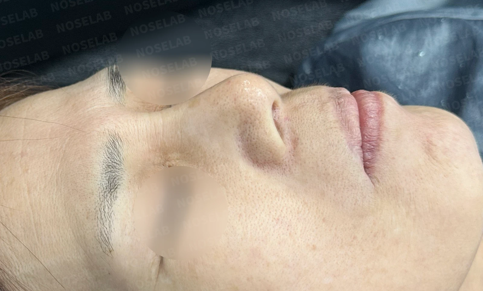
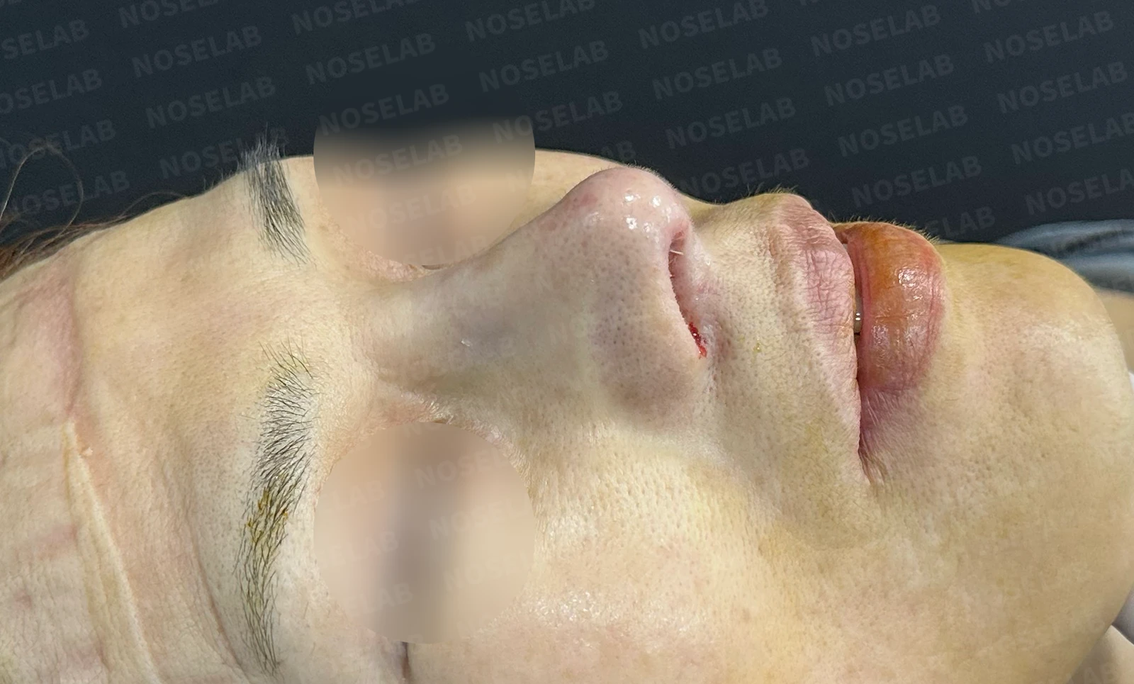
Before surgery / Day 0 immediately after surgery Photos taken immediately after surgery show swelling and may differ from the final result.
Oblique view:
- Drooping of the nasal tip is corrected, achieving a natural height
- The appearance of a protruding mouth has been softened
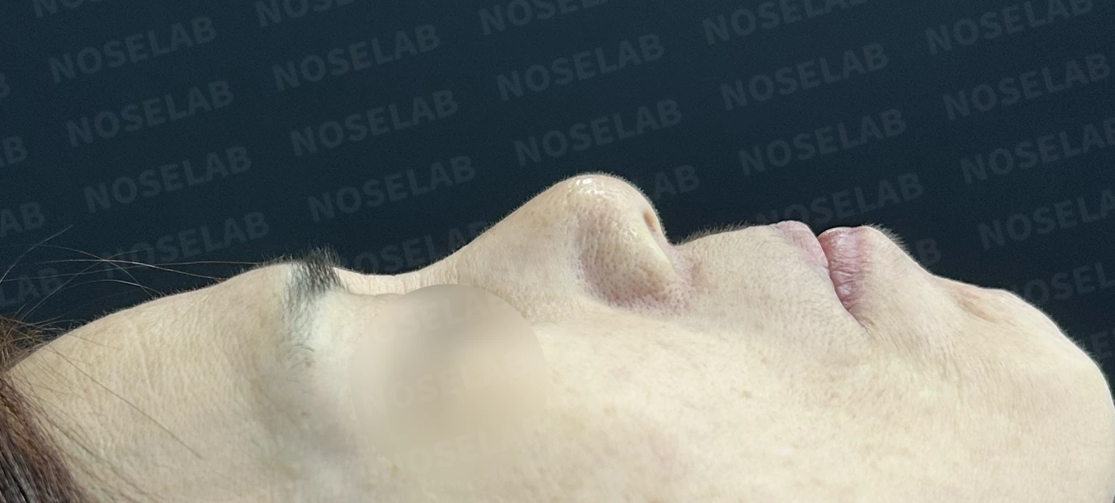
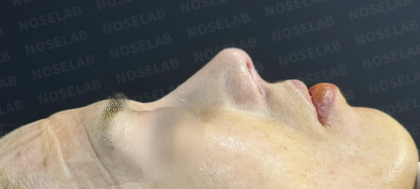
Before surgery / Day 0 immediately after surgery Photos taken immediately after surgery show swelling and may differ from the final result.
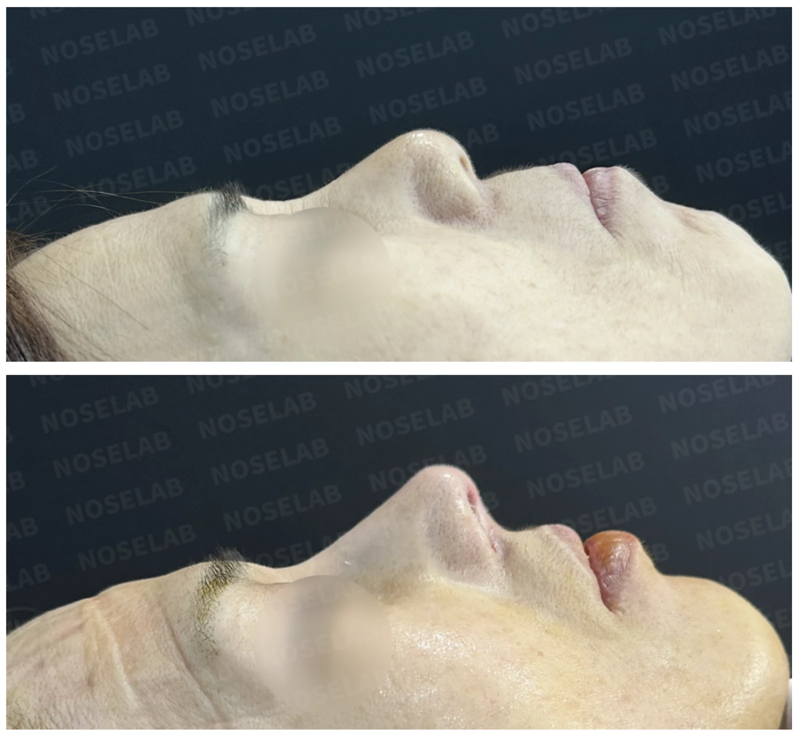
Profile view:
- The nasal tip is naturally lifted, and bulbousness is improved
- Columellar retraction is corrected, creating a natural nasolabial angle
- Visible improvement in protruding mouth appearance
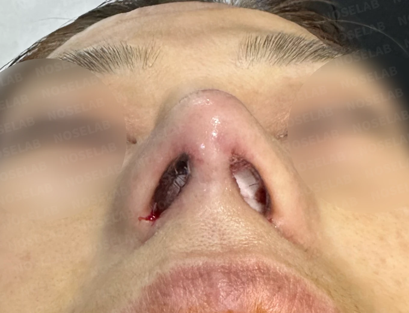
Before surgery / Day 0 immediately after surgery Photos taken immediately after surgery show swelling and may differ from the final result.
Nostrils:
- Nostril asymmetry has improved
- The previously compressed nostril shape looks natural
Photos immediately after surgery
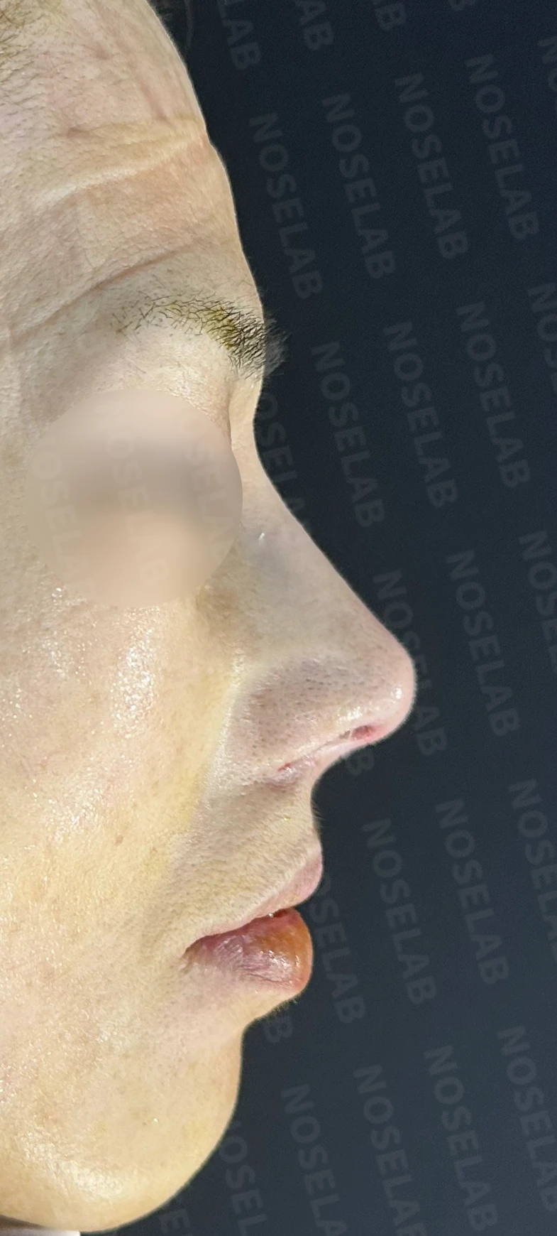
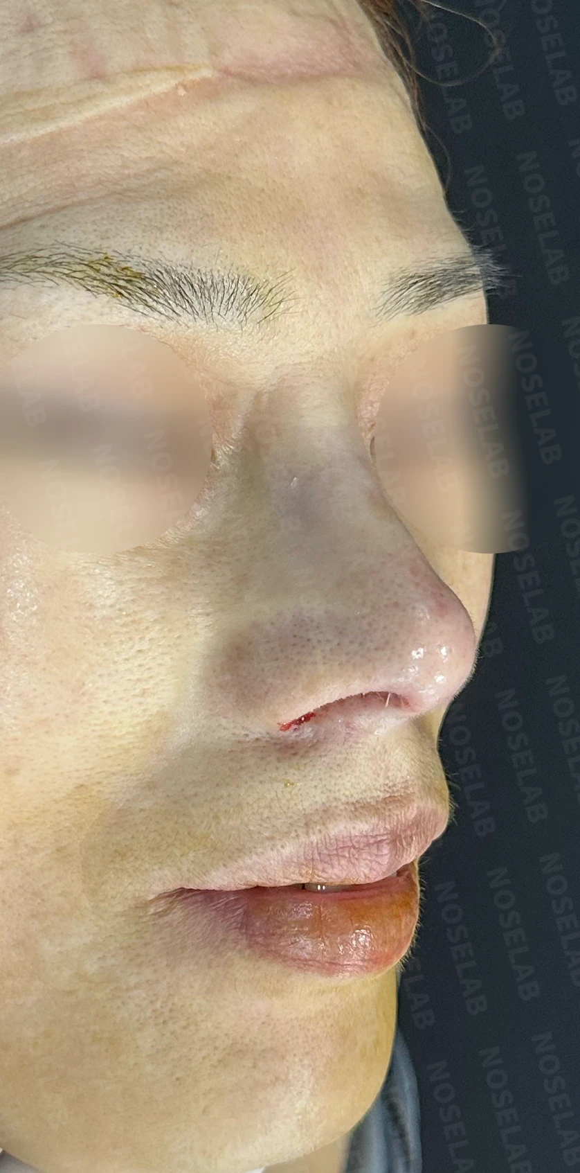
Front, profile, and oblique views taken immediately after surgery (day 0).Immediate postoperative photos show swelling and may differ from the final result.
Nostril view on day 0 immediately after surgery. Photos taken immediately after surgery show swelling and may differ from the final result.
Conclusion
This was a complex case requiring aesthetic improvement alongside the resolution of complications from an old rhinoplasty (pain, implant mobility).
By combining multiple techniques—including removal of the old implant and replacement with a new one, nasal tip reconstruction using autologous rib cartilage, and nasolabial angle correction—both functional and aesthetic improvements were achieved.
In this particular case, neuralgia due to a chronic foreign body reaction was suspected, so the focus was placed on eliminating the foreign body reaction and relieving pressure.
Symptom changes will be closely monitored through follow-up visits over a 6-month period after surgery, and additional measures will be taken if necessary.
Closing remarks
Following rhinoplasty, some patients occasionally experience a feeling of pressure or pain along the nasal bridge. Most of these cases arise within 6 months after surgery and naturally improve over time as scar tissue diminishes. In some patients, the thickness of the implant exceeds what the nasal bridge can comfortably accommodate, making adaptation difficult. In such cases, lowering the implant height can resolve the issue.
When dorsal pain develops several years or decades later after being asymptomatic initially, a chronic foreign body reaction to the implant or acute or chronic inflammation should be considered. In most of these cases, the issue can be resolved simply by surgically evaluating the condition of the dorsum, correcting the implant position, and performing thorough irrigation.
If you are considering rhinoplasty or experiencing discomfort following a past surgery, I recommend consulting an ENT specialist to identify the precise cause and determine the appropriate solution.
I always prioritize patient safety and satisfaction, striving to achieve the intended results.
This was Dr. Peter C. Kang. Thank you.
Thank you.
#rhinoplasty #implantremoval #bridgepain #headacherelief #rhinitistreatment #nasalobstructionrelief #widenednosecorrection #bulboustipcorrection #protrudingmouthcorrection #closedrhinoplasty #autologousribcartilagegraft #ENTspecialistrhinoplasty #bulbousnose #widenednose

Limitations and risks
When autologous rib cartilage is used, scarring and pain may remain at the donor site on the chest, and pneumothorax (pleural injury) can occur rarely. Over time, the grafted cartilage may warp or undergo partial resorption.
When revising after implant removal, contracture may progress again after removal, the nasal height and length may decrease, and staged surgery may be necessary depending on the skin condition.
When the septum is corrected at the same time, nasal obstruction may persist or recur, and in rare cases, septal perforation or changes in smell may occur.
Common potential risks include infection, bleeding, asymmetry, scarring, skin necrosis, and insufficient improvement; results vary depending on individual skin condition and previous surgical history.