Hello.
I am Dr. Peter C. Kang, medical director.
Today, I will share the story of a patient who received nasal filler 14 years ago, later underwent filler removal surgery and rhinoplasty using septal and ear cartilage, but visited our clinic because the nose still appeared long.
Because this patient also had a history of two-jaw surgery, the septal cartilage was expected to be significantly weakened.
Preoperative consultation
When I first met the patient in the consultation room, my initial impression was that the glabella was high and the nose was long. From the front, the nose appeared generally bulbous and widened, and its length made the patient look slightly older than their actual age.
On the profile view, the nasal tip appeared long and drooping, and the mouth looked protruding. Because the nasolabial angle was acute, the area connecting the nasal tip to the philtrum and mouth appeared unnatural.
On the nostril view, the noticeable issues were nostril size discrepancy and asymmetry, and the widening of the nose seemed to have become more pronounced following the two-jaw surgery.
What was the surgical plan?
Therefore, we needed to focus on reinforcing the septal cartilage. The history of two-jaw surgery and the long, drooping nasal appearance were clues pointing to a weakened septum. We planned to perform septal cartilage reconstruction using autologous rib cartilage.
The overall shape of the nose strongly resembled a nose with filler. Although the patient was told that the filler was removed during previous surgery, a considerable amount was suspected to remain, making filler removal a crucial component of this operation.
The bulbous appearance of the nasal tip was thought to be due to substantial scar tissue, so we planned to reduce the skin thickness by partially removing subcutaneous tissue.
On the nasal bridge, we planned to remove foreign material and augment the dorsum using a silicone implant.
Before surgery vs. immediately after surgery
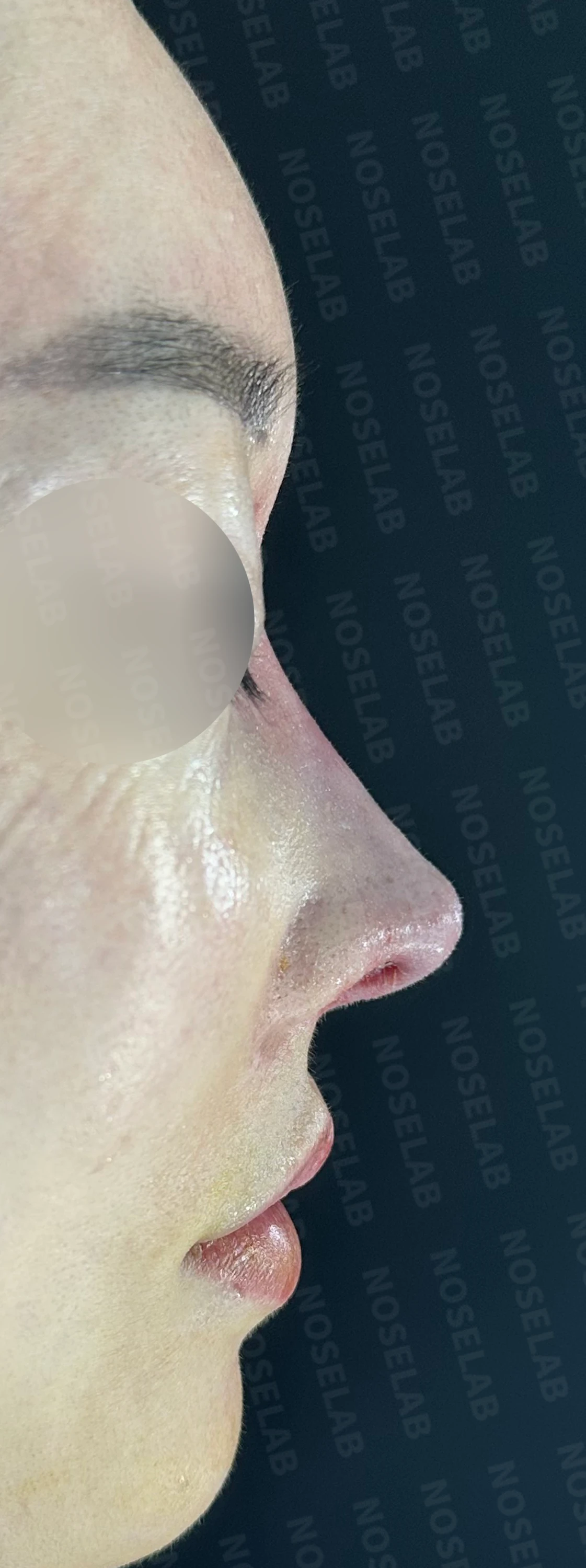
Looking at the profile photo, you can see that the radix has been lowered significantly and the nasolabial angle has changed considerably. The bulbous look of the nasal tip has also improved.
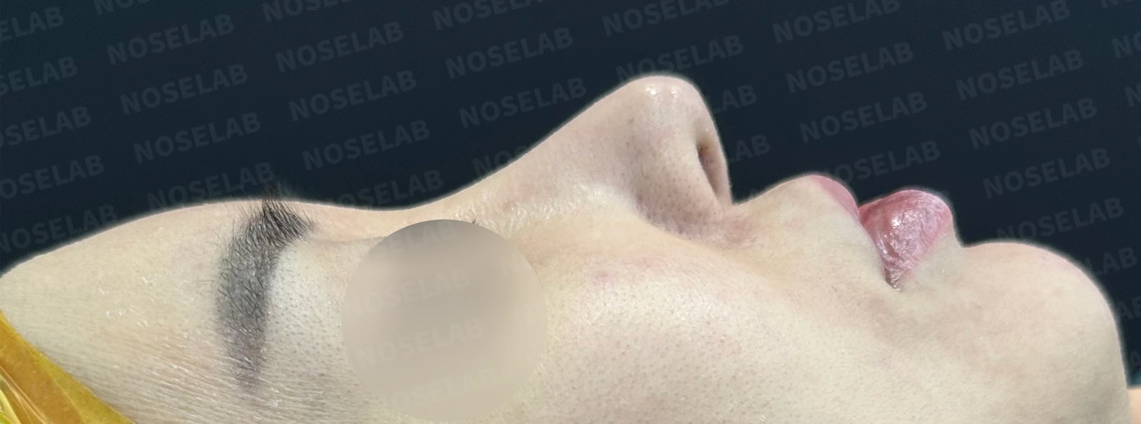
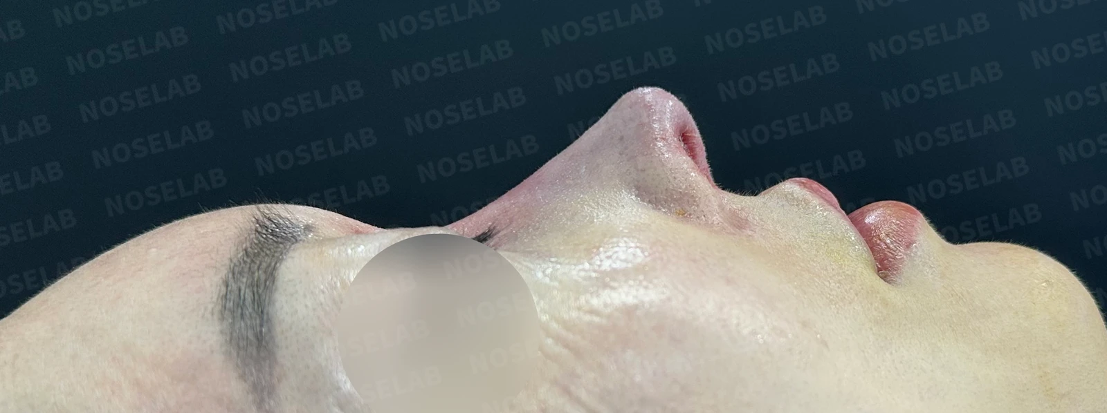
Before surgery / Day 0 immediately after surgery Photos taken immediately after surgery show swelling and may differ from the final result.
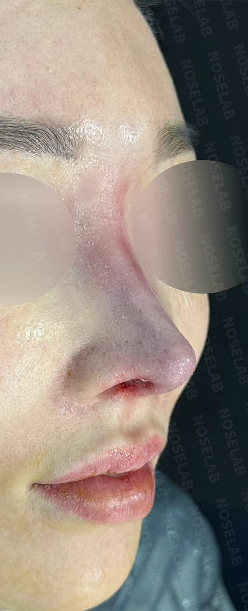
In the 45-degree oblique view, you can see that the nasal tip has become significantly slimmer.
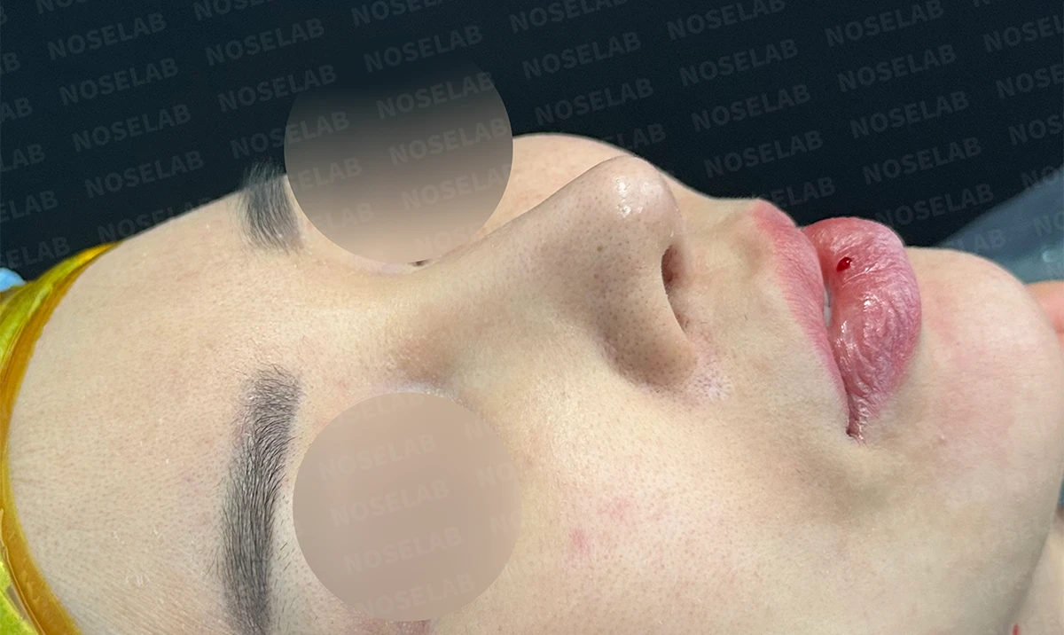
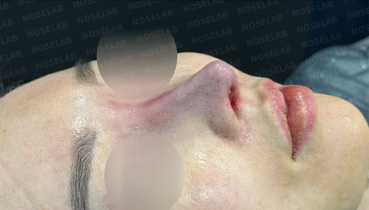
Before surgery / Day 0 immediately after surgery Photos taken immediately after surgery show swelling and may differ from the final result.
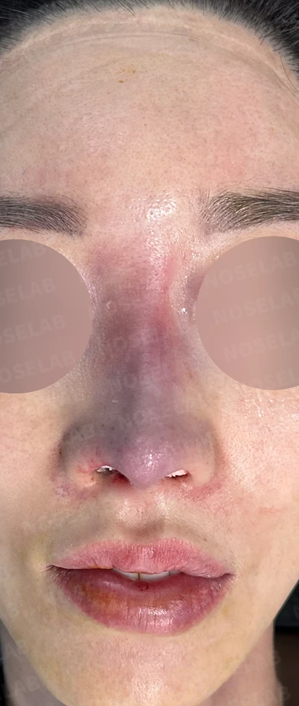
The overall bulbous and deviated appearance of the nose has improved. While the nasal dorsum shows redness due to bruising from the foreign material removal, it is expected to recover quickly. The nostril asymmetry has also improved significantly.
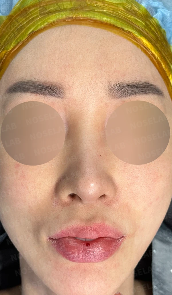
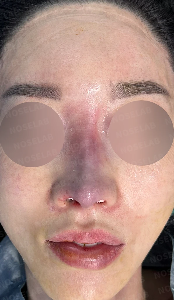
Before surgery / Day 0 immediately after surgery Photos taken immediately after surgery show swelling and may differ from the final result.
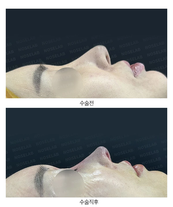
Looking at the nostril shape, the right nostril had appeared noticeably larger with severe asymmetry, but the shapes have become more alike, reducing the difference.
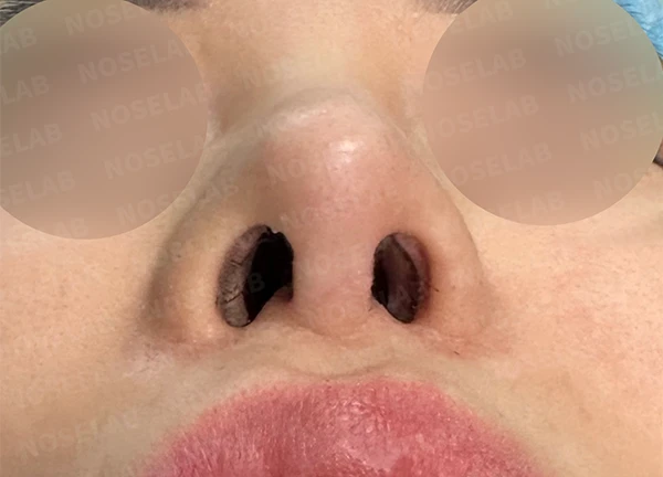
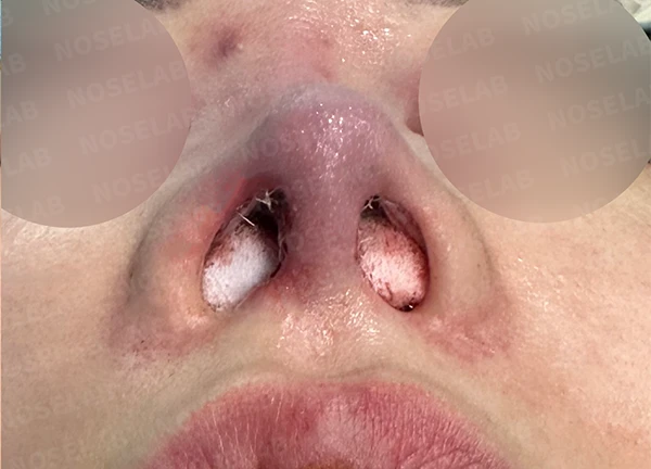
Before surgery / Day 0 immediately after surgery Photos taken immediately after surgery show swelling and may differ from the final result.
Photos taken immediately after surgery
The relationship between two-jaw surgery and rhinoplasty
Today, let us look at the relationship between facial contouring surgery and rhinoplasty. The contouring procedure that can affect nasal surgery is two-jaw surgery. Two-jaw surgery separates, trims, and refixes the upper and lower jawbones. The area where the upper jawbone separates from the facial skeleton is the lower portion of the septal cartilage. Therefore, undergoing two-jaw surgery can damage or weaken the septal cartilage. If septal extension is performed during rhinoplasty, prior two-jaw surgery can influence the nasal procedure.
First, because the septal cartilage is weakened, harvesting septal cartilage as a rhinoplasty graft material can be difficult. Stronger cartilage, such as donor rib cartilage or autologous rib cartilage, may need to be used instead.
Second, if the anterior part of the septal cartilage is weak, performing a septal extension can lead to issues such as nasal tip deviation or drooping. Therefore, septal cartilage reconstruction must be performed to reinforce the septum alongside the rhinoplasty to prevent nasal tip deformities.
Today, we covered rhinoplasty for patients who have undergone two-jaw surgery. I hope this is helpful for those who have had two-jaw surgery and are considering rhinoplasty.
This was Dr. Peter C. Kang. Thank you.

Limitations and risks
When autologous rib cartilage is used, scarring and pain may remain at the donor site on the chest, and pneumothorax (pleural injury) can occur rarely. Over time, the grafted cartilage may warp or undergo partial resorption.
When using donor rib cartilage, long-term follow-up is necessary because the grafted cartilage may resorb or warp.
When foreign materials such as injected fillers are removed and reconstructed, contracture may recur, nasal height and length may decrease, and staged surgery may be necessary depending on the skin condition.
When the septum is corrected at the same time, nasal obstruction may persist or recur, and in rare cases, septal perforation or changes in smell may occur.
Common potential risks include infection, bleeding, asymmetry, scarring, skin necrosis, and insufficient improvement; results vary depending on individual skin condition and previous surgical history.