Hello.
I am Dr. Peter C. Kang, medical director.
The nose is aesthetically important, but it is also a vital organ for breathing. While breathing through the nose comes naturally to most people, for some it can be a constant struggle.
Today, I would like to discuss functional rhinoplasty—a procedure that aims to enhance appearance while improving breathing.
This patient visited our clinic complaining of nasal obstruction. A CT scan taken 1 year prior showed a deviated septum and hypertrophic rhinitis, for which septoplasty and radiofrequency submucosal resection of the inferior turbinates were performed. Although the hypertrophic inferior turbinates were reduced and overall congestion improved, the septal cartilage was very thin and severely deviated, eventually reverting to its original crooked state. Therefore, we planned a procedure combining septal cartilage reconstruction with cosmetic refinement (functional rhinoplasty).
Preoperative consultation details
On the front view, the nasal tip deviated to the right, nostril asymmetry was noticeable, and the area above the left nostril wing was depressed.
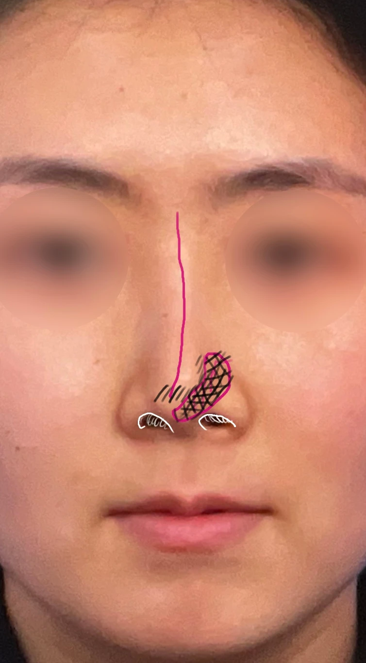
Preoperative design, front view
On the profile view, the nasal tip appeared drooping, and the border between the nasal tip and the implant showed through.
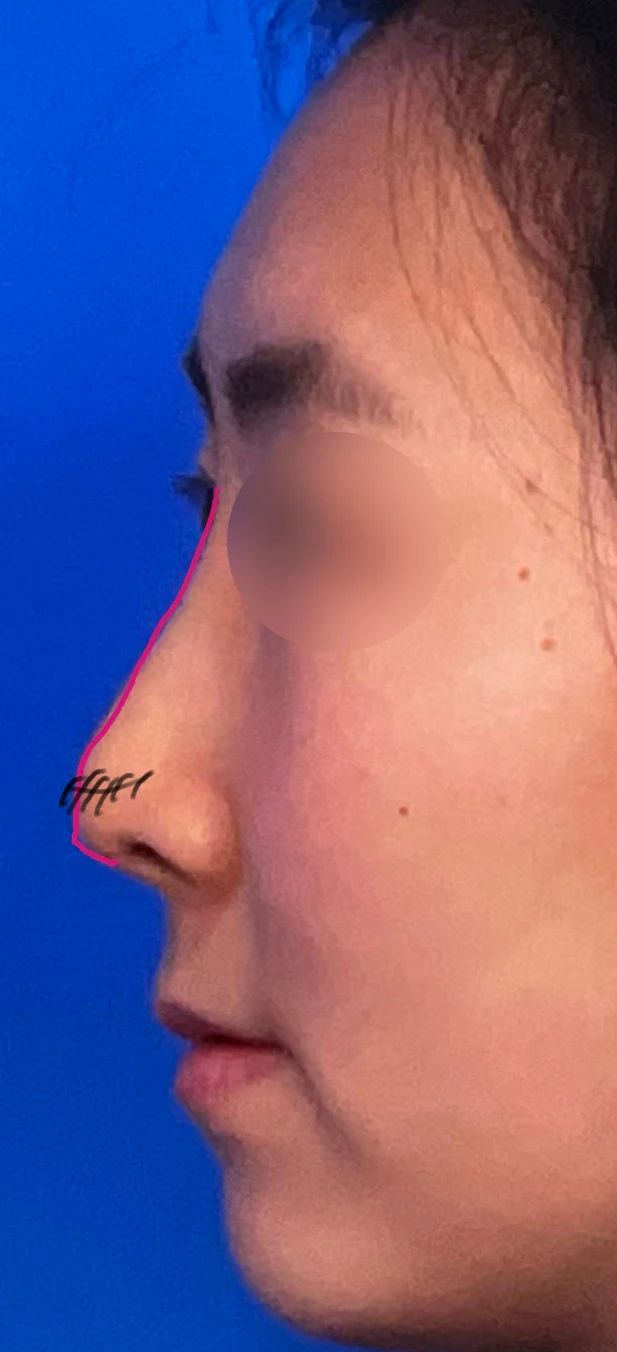
Preoperative design profile view
The columella leaned to the right, and the septum inside the left nostril was severely deviated, severely blocking the internal nasal passage.
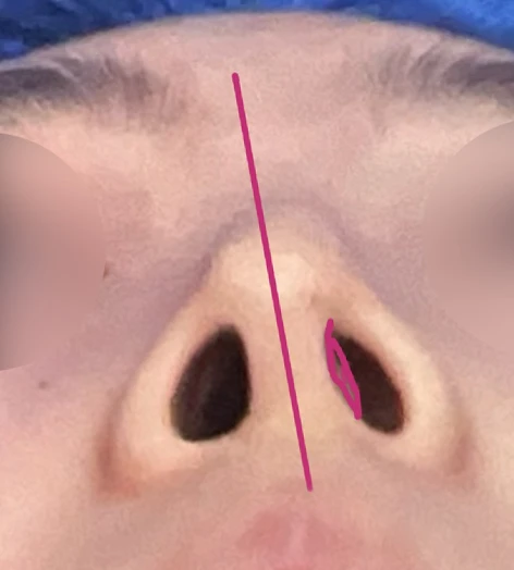
Preoperative design basal (nostril) view
On the preoperative CT scan, the septal cartilage (red line) can be seen deviating to the left, significantly narrowing the internal nasal cavity on the left side (black area).
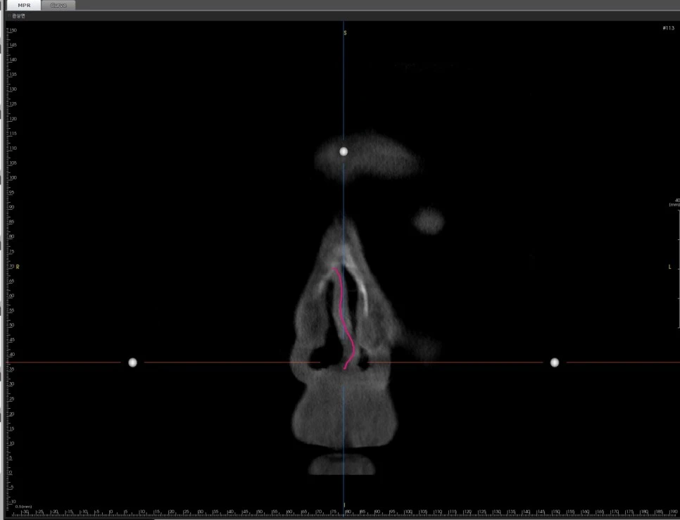
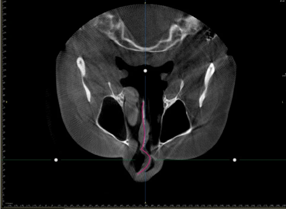
The CT scan taken at our clinic shows that the septal cartilage (red line) is severely deviated to the left, obstructing the internal space of the left nasal cavity.
When the septal cartilage is severely deviated, the most directly affected area is the nasal tip, including the columella. As seen here, the direction in which the septal cartilage curves corresponds with the direction of the columellar tilt, and the rightward deviation of the nasal tip is likely related as well. In particular, a tilted columella is accompanied by nostril asymmetry.
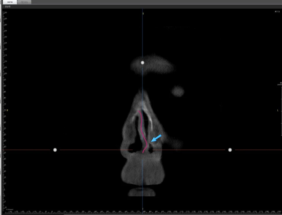
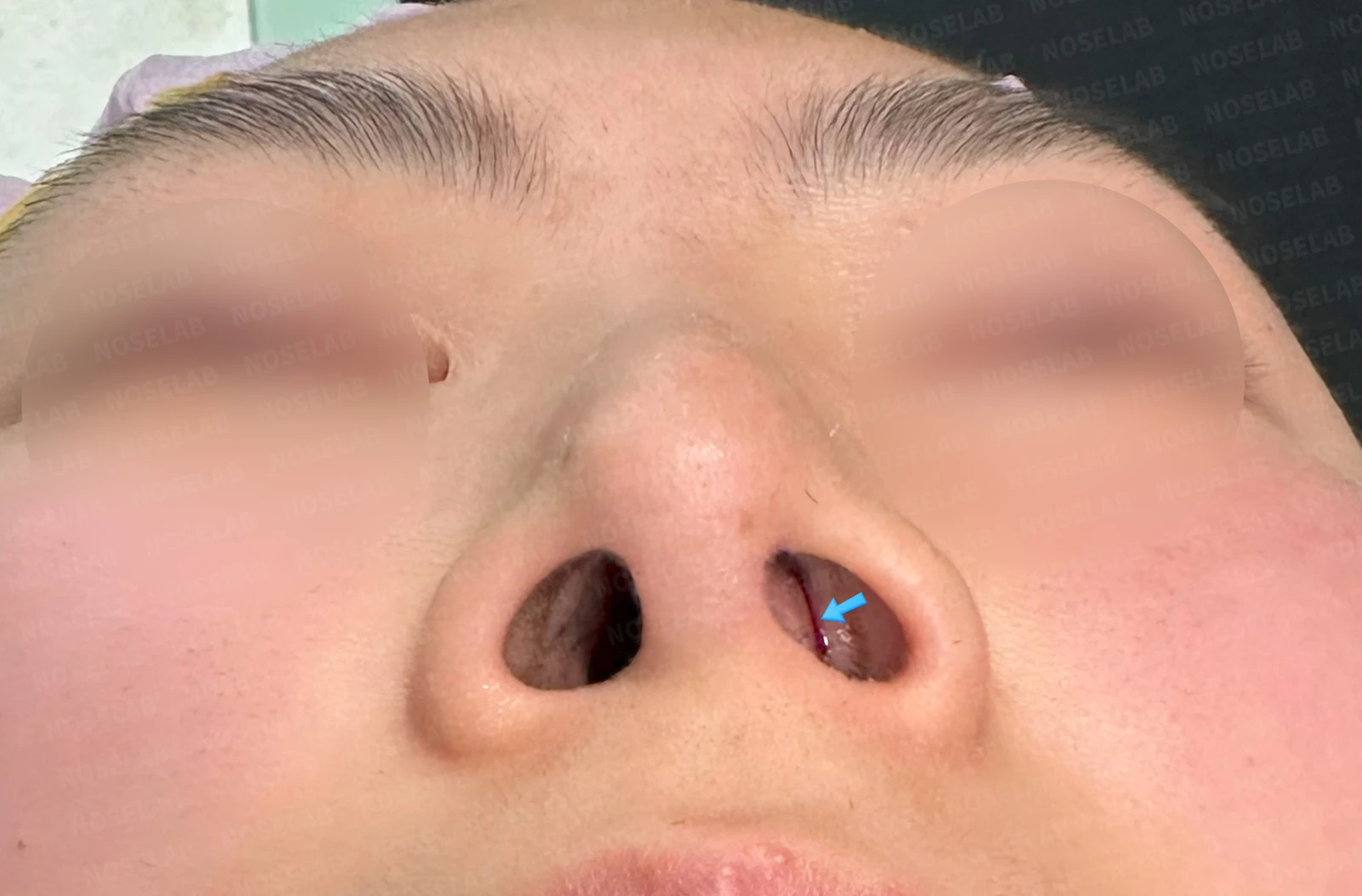
The CT scan shows a lack of visible airway space in the left nostril, and the basal nostril view demonstrates that the internal passage of the left nasal cavity is almost completely obstructed.
What was the surgical plan?
The surgical plan for this patient was to straighten the deviated septum. There are several ways to correct a deviated septum. However, revision surgery for a deviated septum carries the risk of re-deviation and further structural weakening of the cartilage, requiring extra caution. Moreover, in this type of case, the procedure must refine the nasal tip shape simultaneously, which makes sturdy septal cartilage support even more essential. Therefore, the surgical plan was established to reconstruct the deviated septal cartilage using autologous rib cartilage.
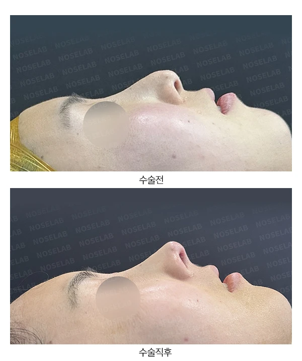
Preoperative vs. immediately after surgery
On the 90-degree profile view, the drooping nasal tip improved, and the retracted columella was brought up to a balanced position.
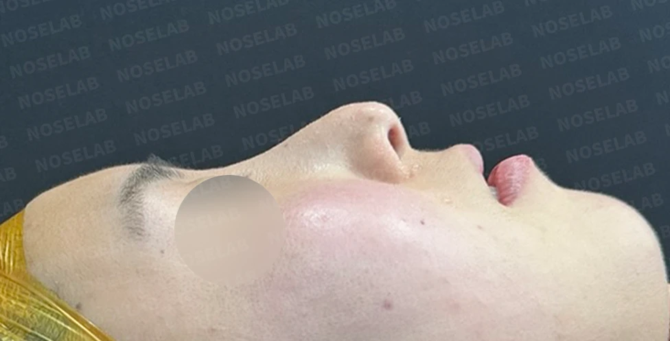
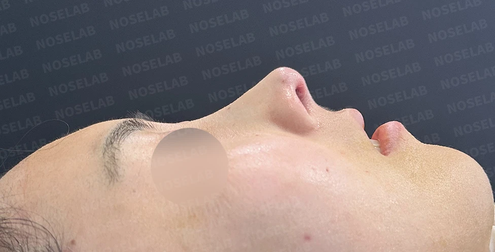
Before surgery / Day 0 immediately after surgery Photos taken immediately after surgery show swelling and may differ from the final result.
On the 45-degree oblique view, the previously blunt, drooping nasal tip changed into a more defined contour.
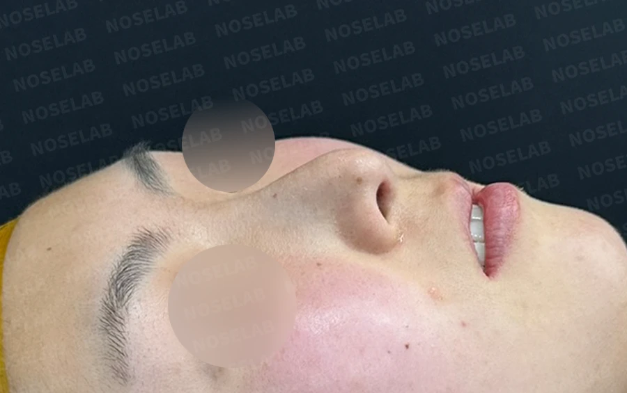
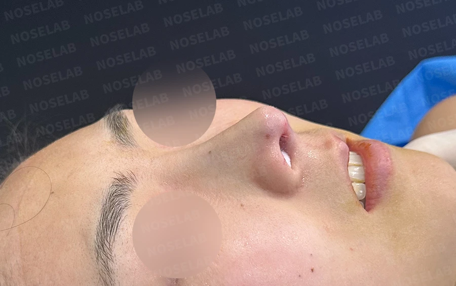
Before surgery / Day 0 immediately after surgery Photos taken immediately after surgery show swelling and may differ from the final result.
On the front view, the rightward deviation of the nasal tip improved noticeably, and the nostril asymmetry showed marked improvement.
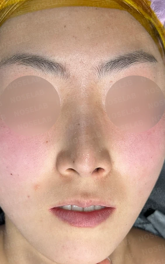
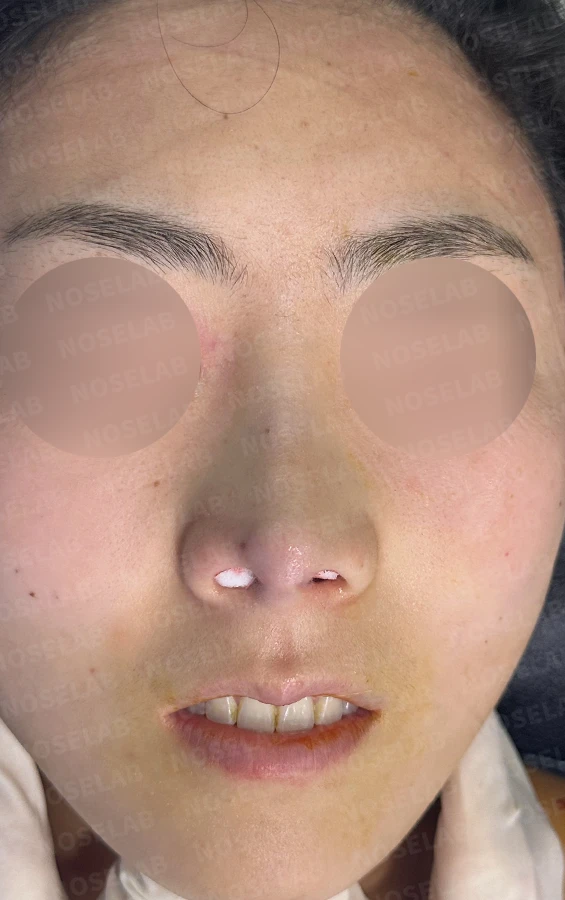
Before surgery / Day 0 immediately after surgery Photos taken immediately after surgery show swelling and may differ from the final result.
The most noticeable change appears in the basal nostril view. The columella, which had leaned to the right, was repositioned upright. Although internal cotton packing obscures the deep view, this image indicates that the septal cartilage has been straightened.
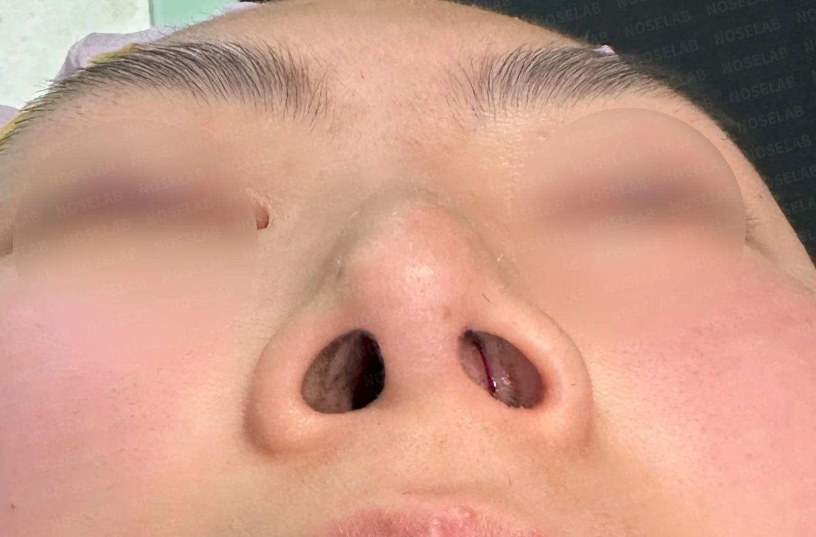
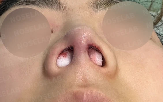
Before surgery / Day 0 immediately after surgery Photos taken immediately after surgery show swelling and may differ from the final result.
Photos immediately after surgery
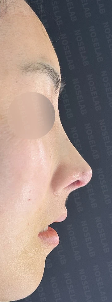
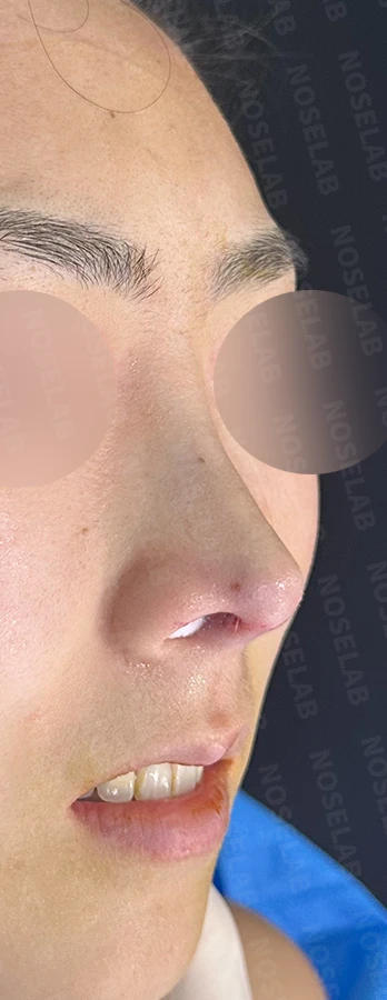
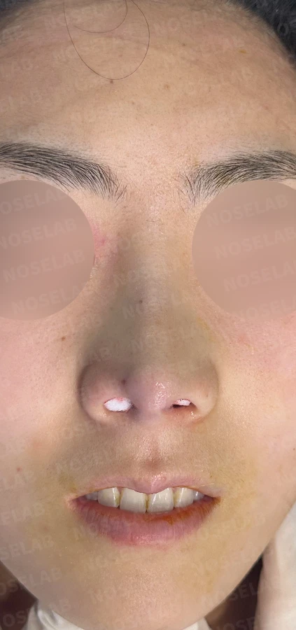
Day 0 immediately after surgery: 90-degree profile, 45-degree oblique, front view Immediate postoperative photos show swelling and may differ from the final result.
Day 0 immediately after surgery: nostril view Immediate postoperative photos show swelling and may differ from the final result.
My perspective on functional rhinoplasty
Functional rhinoplasty refers to performing surgery for nasal obstruction and aesthetic rhinoplasty at the same time. It is the procedure I am most focused on and perform most frequently. Aesthetic surgery is demanding because achieving symmetry is difficult, and aligning with each patient's expectations is equally challenging. Surgery for nasal obstruction involves evenly widening both visible passages and deeper internal nasal spaces. Opening the unseen pathways through which air flows is especially complex. Neither procedure can be called simple. Furthermore, as a board-certified otolaryngologist, performing functional rhinoplasty requires uniting clinical duty with aesthetic judgment, which brings a significant sense of responsibility.
I once brought up a question in a lighthearted conversation: if faced with a critical dilemma during nasal surgery where I had to choose between function and aesthetics, which would I pick? While the answer might seem clear, for a practitioner it can be a genuine consideration, since aesthetics are often prioritized in cosmetic surgery. My choice was function. I consider that core to my professional mission. It sounds straightforward in words, but to me it was a meaningful principle.
I check for cartilage that was damaged or depleted in previous operations and proceed with reconstruction.
"It may not look vastly different right now, but with time it will become sturdier, resist deformation, and allow smooth breathing."
That is what I continually remind myself throughout surgery.
I ended up speaking at length, but I hope this information is helpful to those seeking both relief from nasal obstruction and aesthetic improvement.
This was Dr. Peter C. Kang. Thank you.

Limitations and risks
When autologous rib cartilage is used, scarring and pain may remain at the donor site on the chest, and pneumothorax (pleural injury) can occur rarely. Over time, the grafted cartilage may warp or undergo partial resorption.
When the septum is corrected at the same time, nasal obstruction may persist or recur, and in rare cases, septal perforation or changes in smell may occur.
Common potential risks include infection, bleeding, asymmetry, scarring, skin necrosis, and insufficient improvement; results vary depending on individual skin condition and previous surgical history.