Hello. I am Dr. Peter C. Kang, a rhinoplasty surgeon and board-certified otolaryngologist (ENT specialist). This is a space where I provide accessible medical knowledge regarding nasal obstruction (blocked nose). While many patients visit our clinic (NOSELAB) seeking purely cosmetic procedures, an even greater number visit us for revision rhinoplasty due to complications from surgeries at other clinics, or because they are suffering from nasal obstruction.
First, let us look into CT scans, which are very commonly taken when planning rhinoplasty and visiting clinics for consultations. A CT scan can provide an immense amount of information, and for a board-certified otolaryngologist, head and neck and paranasal sinus CT scans are very familiar and yield crucial insights.
What does a CT scan, which is such a common test, allow us to see? Before exploring CT scans, let us first look at X-rays. The radiological examination we most commonly encounter is a chest X-ray. In this examination, radiation known as X-rays passes through our body and exposes photographic film, producing an image composed of black and white. Air allows 100% of the radiation to pass through, so it appears black, while bone is dense and does not readily permit radiation to pass through, appearing white. In an X-ray examination, structures appear on a spectrum from black to white depending on their density. A CT scan takes hundreds of these X-ray images from various angles, which a computer then processes into a format that is easy to view. Simply put, you can think of CT scans as being used mainly to evaluate bone-related structures, whereas MRI scans examine soft tissues other than bone in detail.
Therefore, in rhinoplasty and functional nasal surgery, a CT scan allows us to evaluate the approximate shape of internal nasal structures, assess how evenly air is distributed between the left and right sides of the nasal cavity, check for any obstructed areas, detect conditions such as sinusitis, and identify whether an implant is present in the nasal bridge (dorsum)—and if so, its size, length, and exact position.
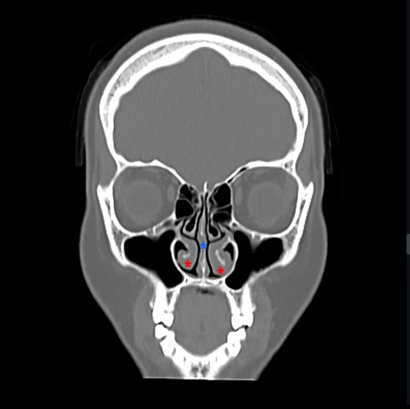
This image is a normal coronal CT slice viewed from the front. If you view it as a face, both eyes are visible, the brain is visible above, and the space in the center is the nasal cavity. The black areas are air, the white areas are bone, and the soft tissues appear gray. *marks where the septal cartilage is located, and *is called the inferior turbinate; undergoing surgery for rhinitis involves a procedure to reduce the size of the inferior turbinates. Cartilage itself cannot actually be visualized clearly on a CT scan. It is impossible to determine on a CT scan whether septal cartilage has been previously harvested or to measure cartilage thickness and size.
Today, we took a simple and accessible look at CT scans among the radiological imaging tests used to evaluate nasal function. A CT scan is an examination involving radiation exposure. Although the CT scanners in most clinics are low-dose CT devices with reduced radiation output, frequent scanning can be harmful to the body. Because radiation exposure can be a risk factor for head and neck cancers, it is advisable to avoid overly frequent CT scans within a 6-month period.